
















Copyright: ©Author(s) 2027.
World J Gastroenterol. Jan 7, 2027; 33(1): 123681
Published online Jan 7, 2027. doi: 10.3748/wjg.123681
Published online Jan 7, 2027. doi: 10.3748/wjg.123681
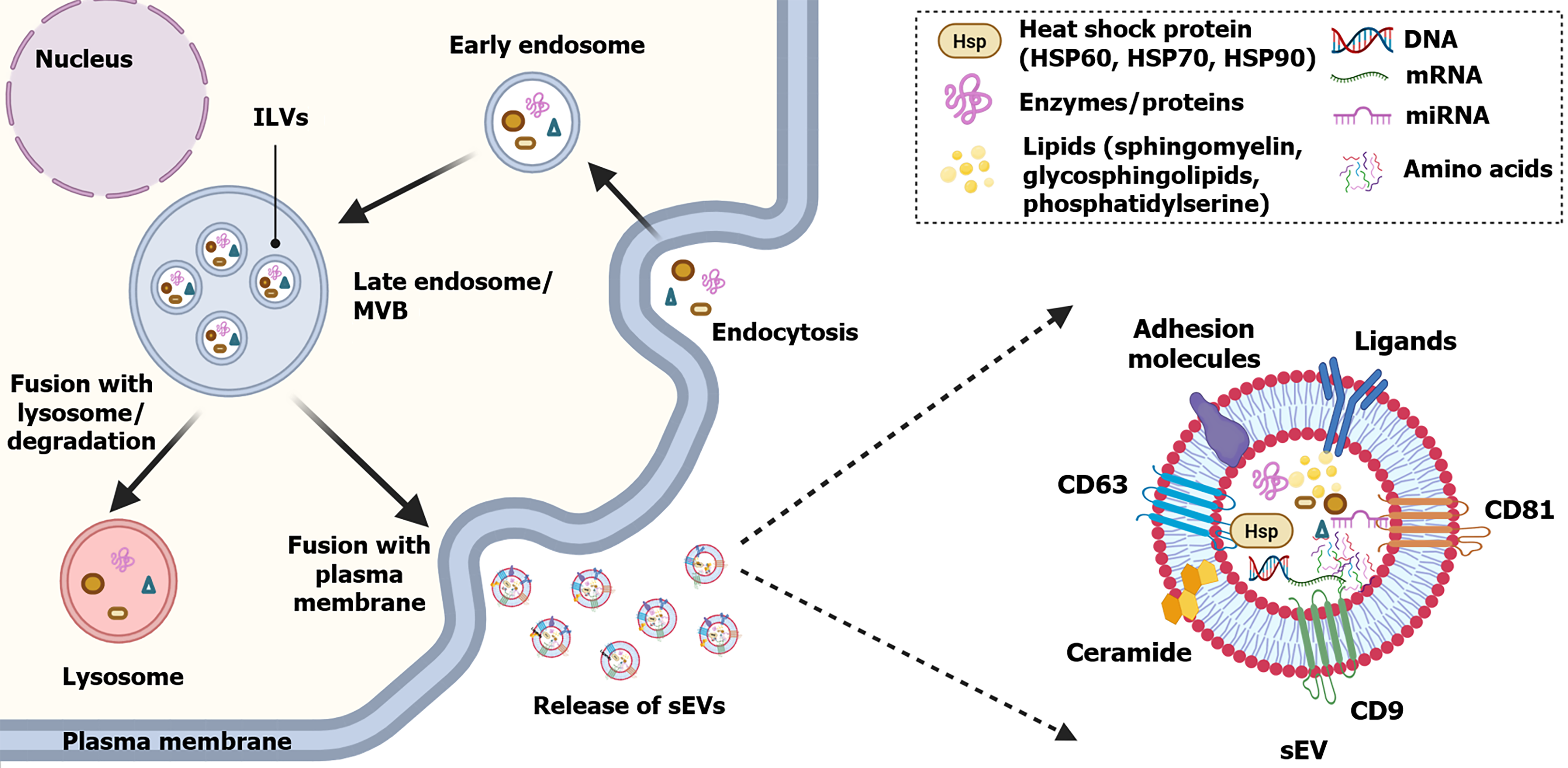
Figure 1 Conceptual schematic of small extracellular vesicles biogenesis and structure.
Endocytosis leads to the formation of early endosomes, followed by endosomal maturation and the generation of multivesicular bodies (MVBs). Intraluminal vesicles are formed by inward budding of the endosomal limiting membrane within MVBs. MVBs may either fuse with lysosomes for degradation or fuse with the plasma membrane, resulting in the extracellular release of intraluminal vesicles as small extracellular vesicles. Small extracellular vesicles contain diverse cargos, including nucleic acids, proteins, lipids, and enzymes, and may display extracellular vesicles (EV)-associated tetraspanins such as CD9, CD63, and CD81. These proteins are commonly used for EV characterization but are not universal or definitive markers of a specific EV subtype; multiparametric characterization is recommended. ILVs: Intraluminal vesicles; MVB: Multivesicular body; sEVs: Small extracellular vesicles; HSP: Heat shock protein.
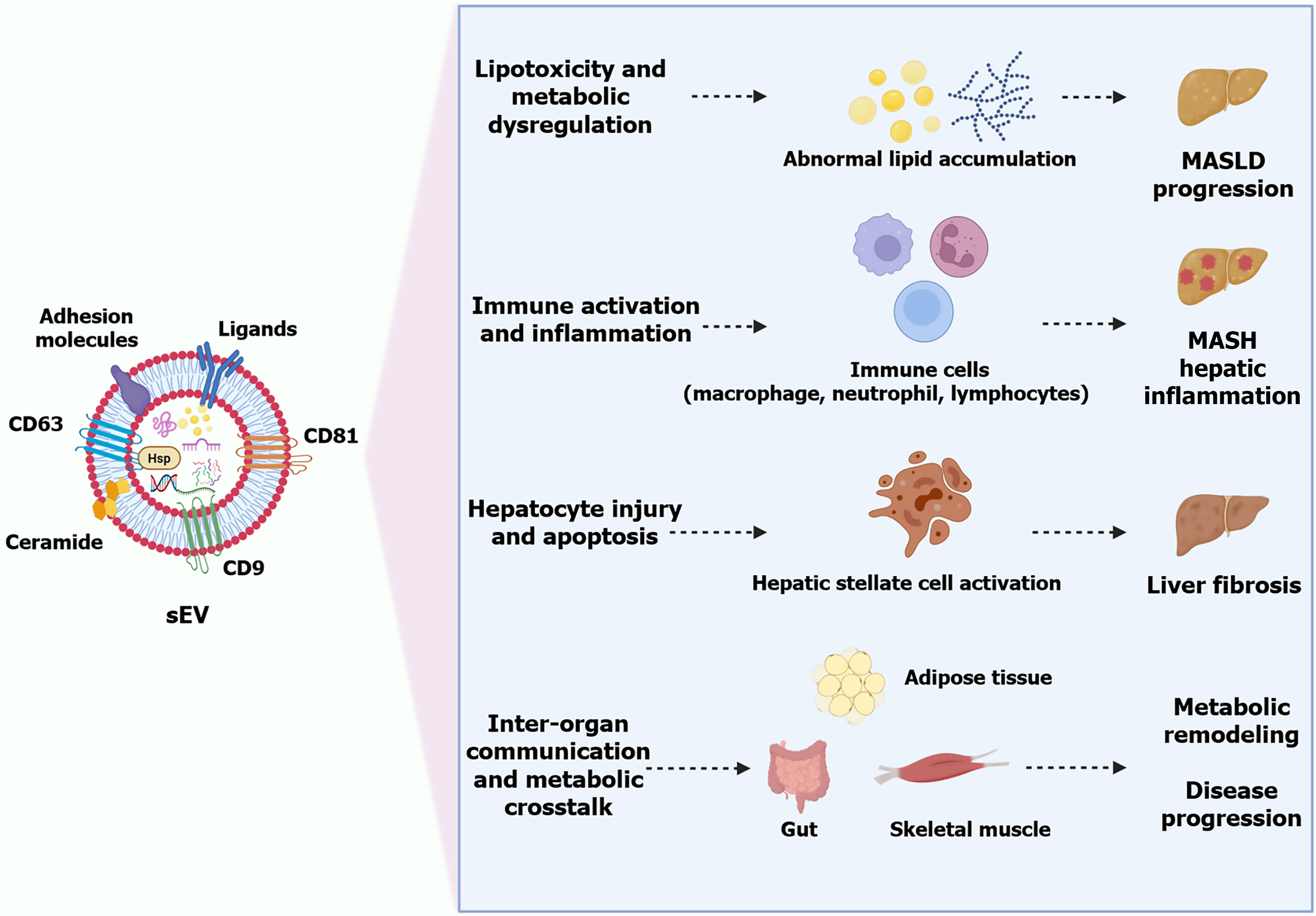
Figure 2 Conceptual schematic of pathological roles of small extracellular vesicles in metabolic dysfunction-associated steatotic liver disease progression.
Small extracellular vesicle-mediated signaling contributes to major pathological processes in metabolic dysfunction-associated steatotic liver disease, including lipotoxicity and metabolic dysregulation, immune activation and inflammation, hepatocyte injury and apoptosis, fibrogenic remodeling, and inter-organ metabolic crosstalk. These processes are associated with abnormal lipid accumulation, activation of hepatic immune cells and hepatic stellate cells, and metabolic communication involving the gut, adipose tissue, and skeletal muscle, collectively contributing to metabolic dysfunction-associated steatohepatitis, liver fibrosis, metabolic remodeling, and disease progression. The depicted mechanisms are primarily supported by preclinical and experimental evidence. MASLD: Metabolic dysfunction-associated steatotic liver disease; MASH: Metabolic dysfunction-associated steatohepatitis; sEV: Small extracellular vesicle.
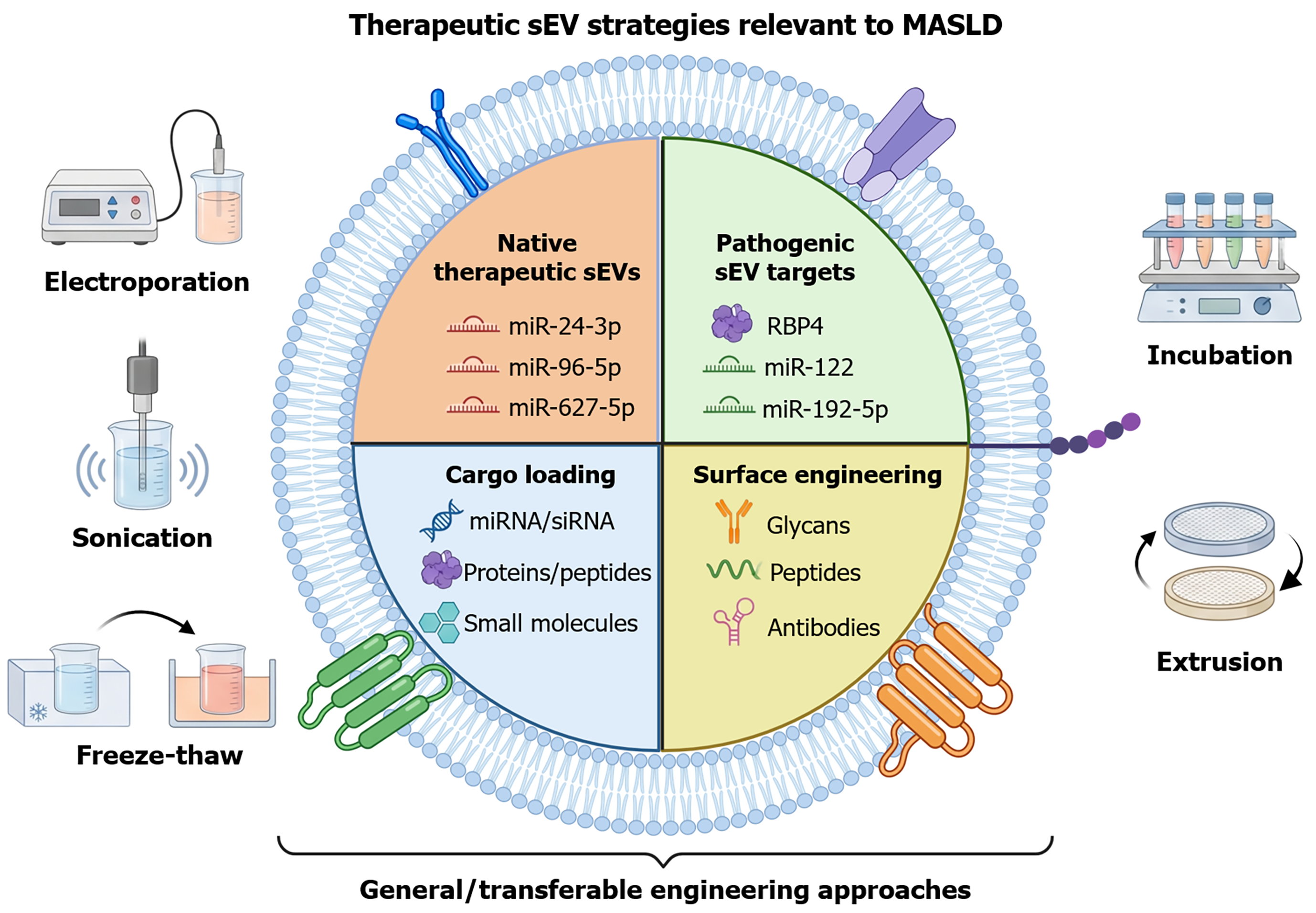
Figure 3 Conceptual schematic of therapeutic roles and engineering strategies of small extracellular vesicles relevant to metabolic dysfunction-associated steatotic liver disease.
The upper panels summarize metabolic dysfunction-associated steatotic liver disease (MASLD)-related evidence for native therapeutic small extracellular vesicles and pathogenic small extracellular vesicle-associated targets. The lower panels summarize general or transferable extracellular vesicle-engineering approaches, including cargo loading and surface engineering. Electroporation, sonication, freeze-thaw cycling, passive incubation, and extrusion represent general cargo-loading methods and should not be interpreted as established MASLD-specific therapeutic strategies unless validated directly in nonalcoholic fatty liver disease/MASLD/metabolic dysfunction-associated steatohepatitis models. Evidence categories and disease-model relevance for representative therapeutic studies are detailed in Table 2. sEV: Small extracellular vesicle; MASLD: Metabolic dysfunction-associated steatotic liver disease.
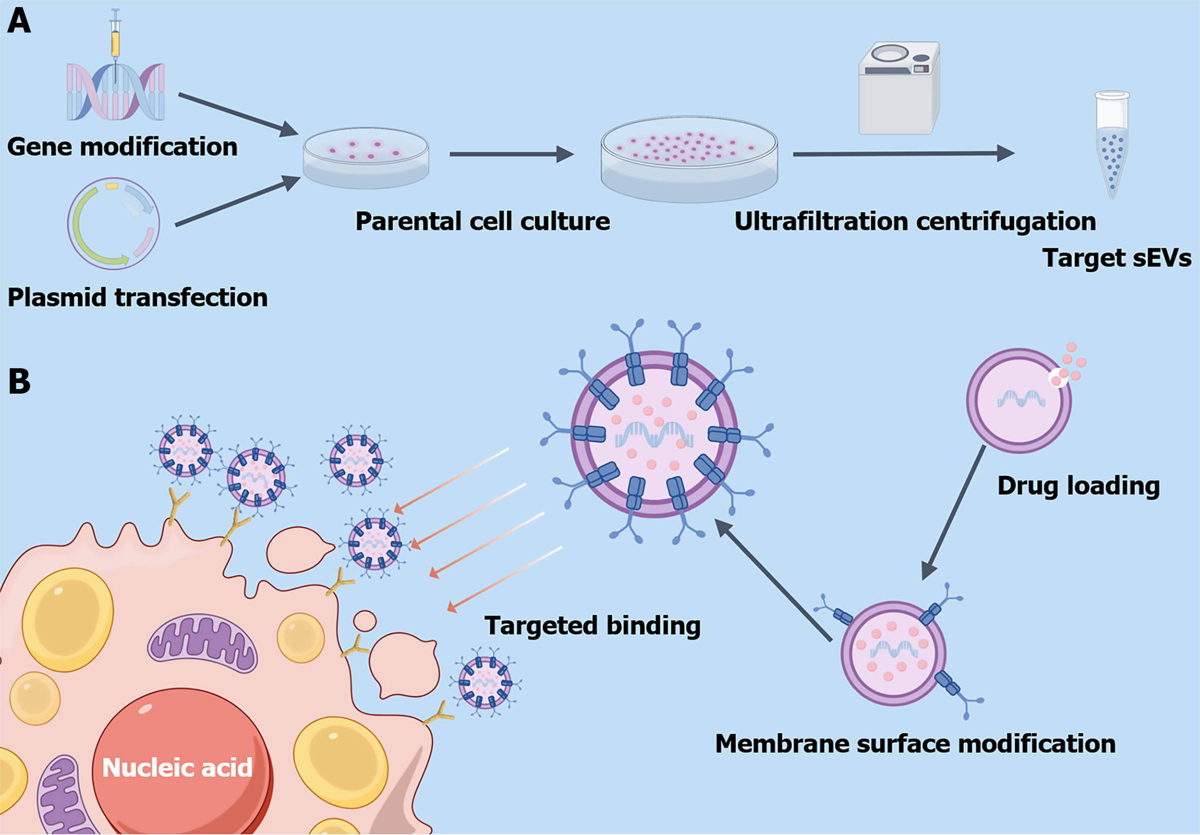
Figure 4 Conceptual schematic of major engineering strategies for small extracellular vesicles.
This conceptual schematic illustrates two major approaches for engineering small extracellular vesicles. A: Parental cells can be engineered through approaches such as genetic modification or plasmid transfection, enabling the resulting small extracellular vesicles to acquire desired therapeutic cargos or functions; B: After isolation, small extracellular vesicles can be further engineered through cargo loading and membrane surface modification. Therapeutic agents may be introduced by transiently altering membrane permeability, while targeting ligands can be conjugated to the vesicle membrane to enhance delivery specificity. sEV: Small extracellular vesicle.
- Citation: Ding LL, Liu H, Yu Y, Liu XC, Zhang SQ, Qian H, Hao DL. Small extracellular vesicles as liquid biopsy targets and precision therapeutics for metabolic dysfunction-associated steatotic liver disease. World J Gastroenterol 2027; 33(1): 123681
- URL: https://www.wjgnet.com/1007-9327/full/v33/i1/123681.htm
- DOI: https://dx.doi.org/10.3748/wjg.123681