
















Copyright: ©Author(s) 2026.
World J Gastroenterol. Dec 28, 2026; 32(48): 123215
Published online Dec 28, 2026. doi: 10.3748/wjg.123215
Published online Dec 28, 2026. doi: 10.3748/wjg.123215
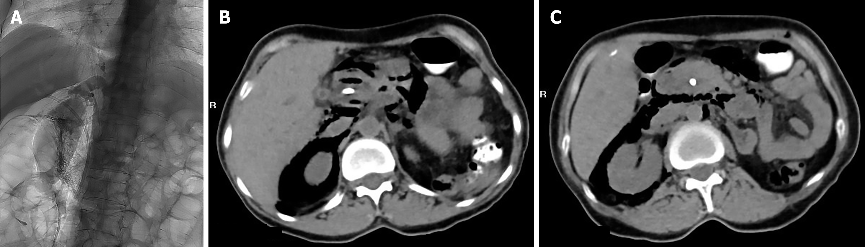
Figure 1 Early suspected retroperitoneal perforation after endoscopic retrograde cholangiopancreatography.
A: Fluoroscopic image during endoscopic retrograde cholangiopancreatography showing dilatation of the intra- and extrahepatic bile ducts, with an approximately 1.2 cm filling defect in the mid-to-upper common bile duct. During gallstone extraction, contrast extravasation was observed and the stone could no longer be clearly delineated; B and C: Axial computed tomography images obtained the following day showing pneumobilia and extensive retroperitoneal extraluminal gas with mediastinal extension, raising suspicion of early retroperitoneal perforation.
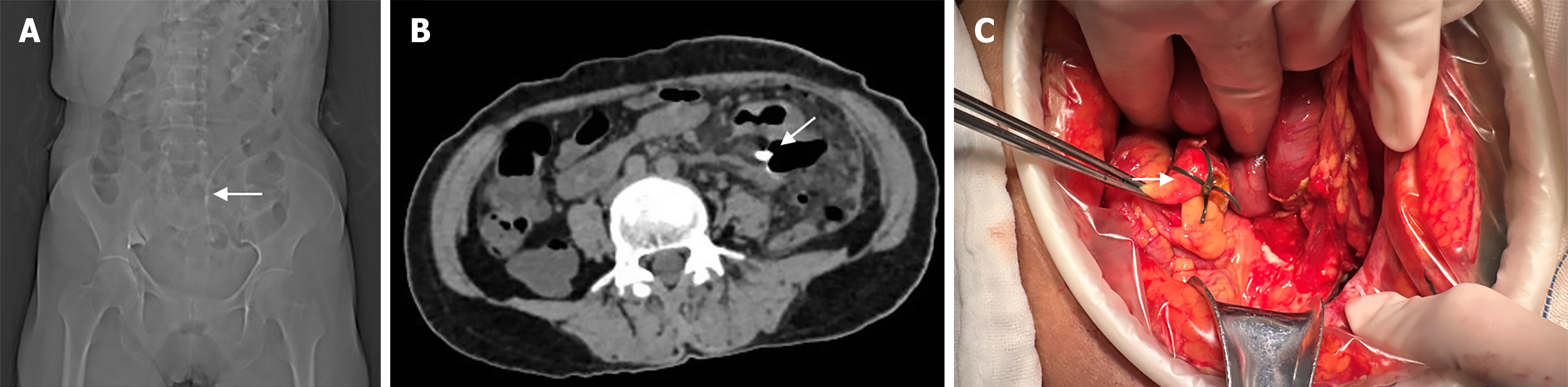
Figure 2 Delayed sigmoid perforation due to biliary stent migration.
A: Plain abdominal radiograph showing distal migration of the biliary stent to the sigmoid colon; B: Axial CT image showing the migrated stent, with one portion remaining within the lumen of the sigmoid colon and the other extending beyond the bowel wall into the abdominal cavity, accompanied by multiple free intraperitoneal air collections; C: Intraoperative photograph showing a perforation of the sigmoid colon, with part of the biliary stent protruding outside the bowel lumen.
- Citation: Danzeng SL, Chen YH, Zha X, Wang Q. Two-stage complications after endoscopic retrograde cholangiopancreatography comprising early retroperitoneal perforation and delayed biliary stent migration: A case report. World J Gastroenterol 2026; 32(48): 123215
- URL: https://www.wjgnet.com/1007-9327/full/v32/i48/123215.htm
- DOI: https://dx.doi.org/10.3748/wjg.123215