
















Copyright: ©Author(s) 2026.
World J Gastroenterol. Dec 21, 2026; 32(47): 122949
Published online Dec 21, 2026. doi: 10.3748/wjg.122949
Published online Dec 21, 2026. doi: 10.3748/wjg.122949
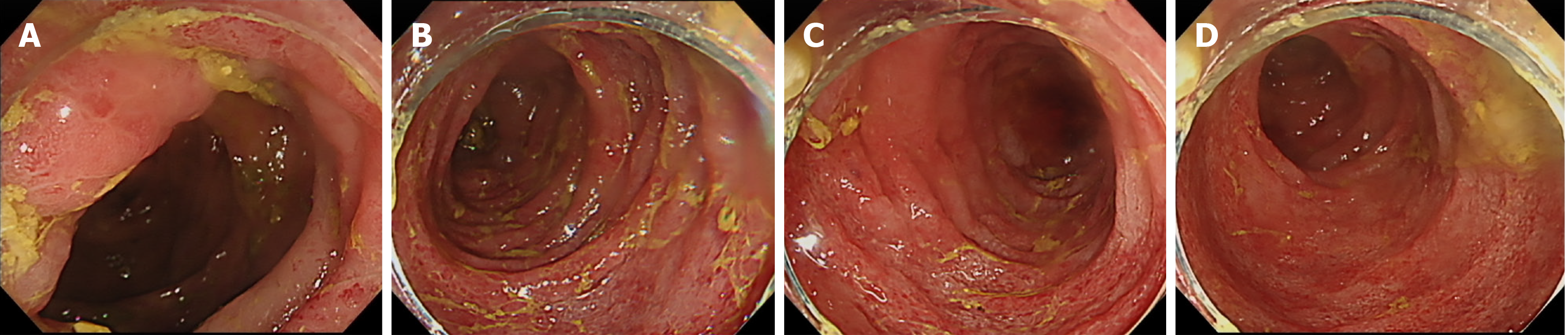
Figure 1 Initial colonoscopic findings.
Colonoscopy at presentation revealed continuous mucosal inflammation characterized by edema, erythema, and friability extending from the transverse colon through the descending and sigmoid colon to the rectum. No definite deep ulceration was identified, and a relatively sharp demarcation from adjacent normal mucosa was noted at the proximal extent of involvement. A: Transverse colon; B: Descending colon; C: Sigmoid colon; D: Rectum.
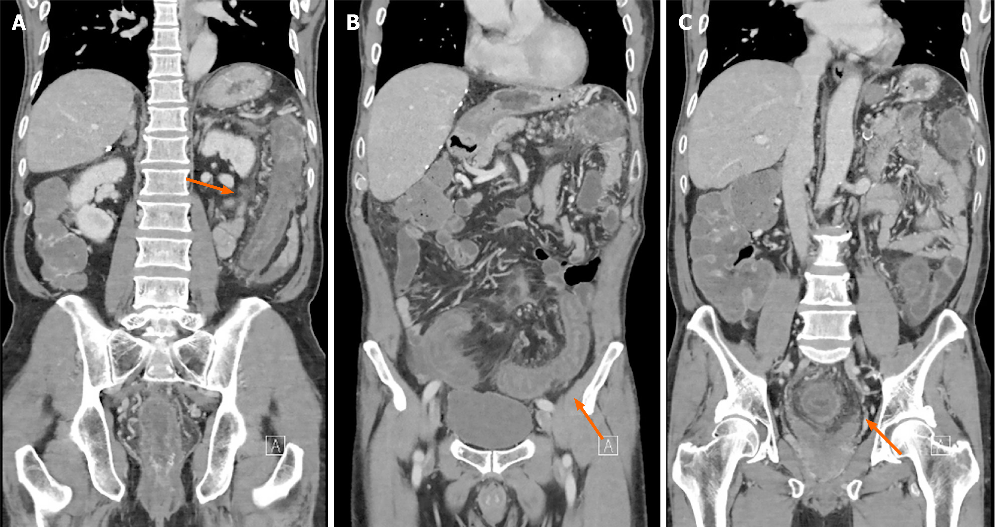
Figure 2 Abdominal computed tomography findings.
Contrast-enhanced abdominal computed tomography demonstrated diffuse colonic wall thickening with pericolonic fat stranding involving the descending colon, sigmoid colon, and rectum (arrows). These findings corresponded to the distribution of inflammation observed on colonoscopy. A: Descending colon; B: Sigmoid colon; C: Rectum.
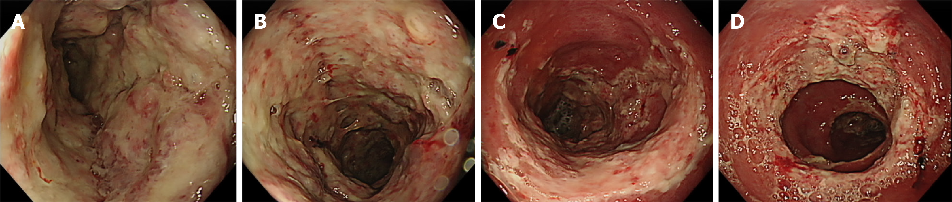
Figure 3 Follow-up sigmoidoscopy demonstrating marked progression of ulcerative lesions.
Follow-up sigmoidoscopy performed for hematochezia showed marked worsening of colonic inflammation despite ongoing medical therapy. The transverse colon and descending colon demonstrated extensive mucosal ulceration with irregular, deep ulcers covered by thick exudate. Similar findings were observed in the sigmoid colon and rectum, where ulcerations appeared more extensive and deeper, with areas of mucosal denudation and suspected vascular exposure. These findings indicate severe progression of disease activity despite corticosteroid and subsequent filgotinib therapy. A: Transverse colon; B: Descending colon; C: Sigmoid colon; D: Rectum.
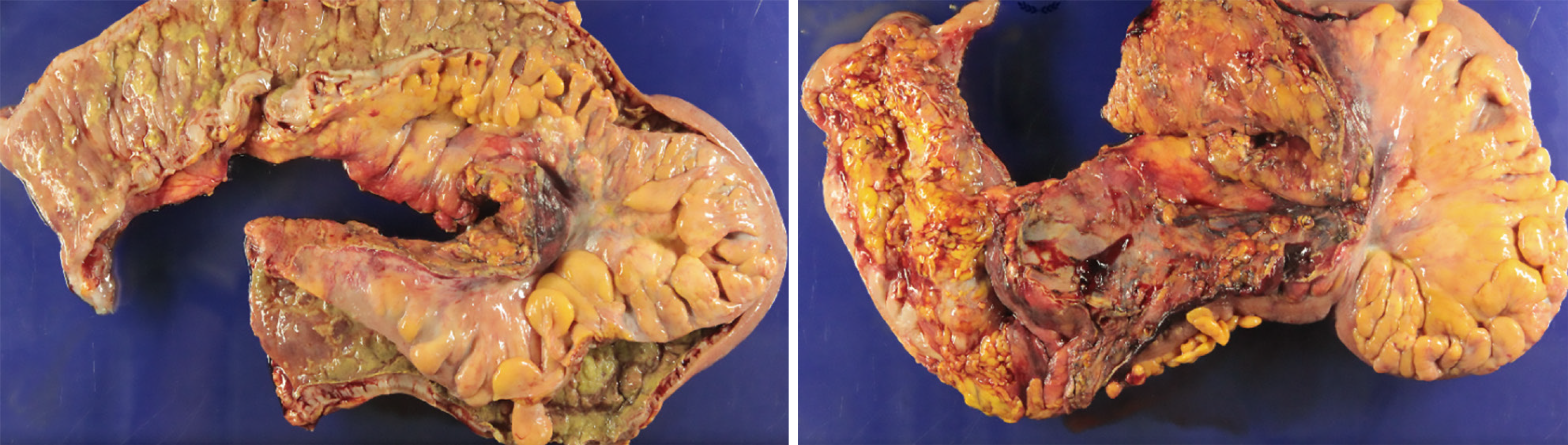
Figure 4 Gross findings of the resected colon.
Gross examination of the resected specimen demonstrated diffuse inflammatory changes extending from the mid-transverse colon to the rectum. The mucosa showed extensive ulceration, edema, and areas of hemorrhage, with particularly severe involvement of the sigmoid colon, where near-necrotizing changes were observed. These findings are consistent with fulminant colitis.
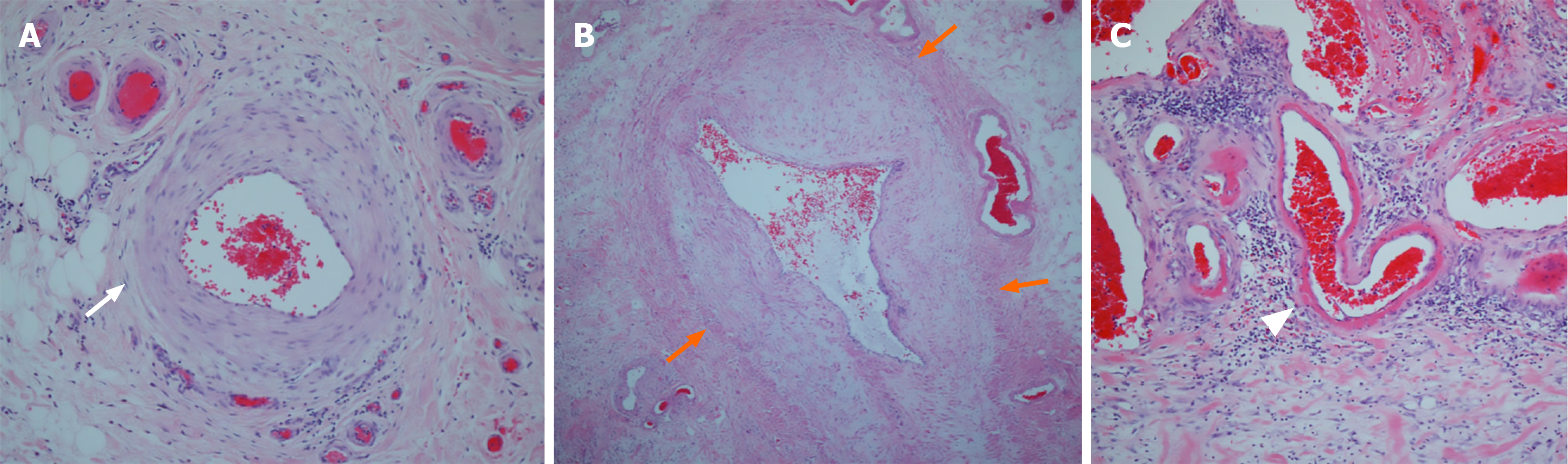
Figure 5 Histopathologic findings of the resected colon consistent with idiopathic myointimal hyperplasia of mesenteric veins.
A: A mesenteric artery showing a preserved normal wall architecture without intimal thickening (white arrow), × 100; B: A mesenteric vein demonstrating marked myointimal hyperplasia with near-complete luminal obliteration (orange arrows), × 100; C: Small venules exhibiting subendothelial fibrin deposition, seen as eosinophilic material beneath the endothelium (arrowhead), × 40. These findings are characteristic of idiopathic myointimal hyperplasia of mesenteric veins, highlighting selective venous involvement with arterial sparing.
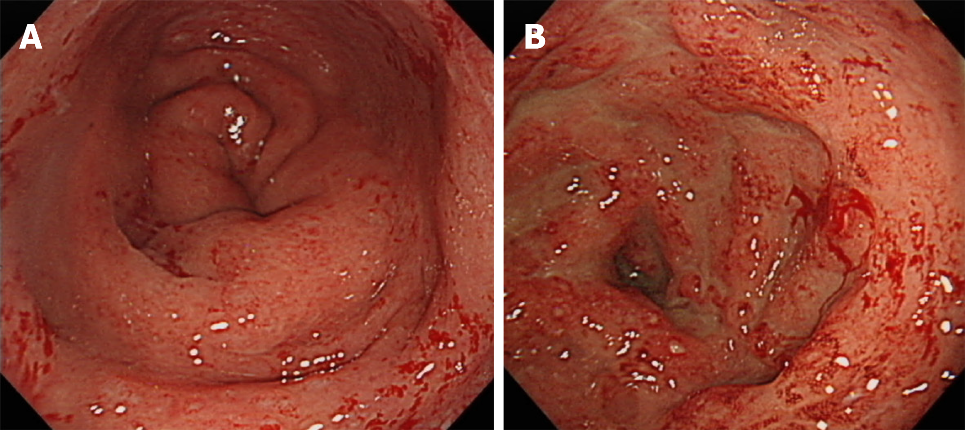
Figure 6 Postoperative follow-up sigmoidoscopy of the rectal stump at 3 months and 12 months after surgery.
A: At 3 months after surgery, the rectal stump showed mild erythema and friability without definite ulceration; B: At 12 months after surgery, the rectal stump demonstrated recurrent mucosal inflammation with erythema, friability, and newly developed shallow ulcerations. These findings indicate interval worsening of rectal inflammation over time.
- Citation: Lee JS, Kim EY, Kwon JG, Bae JH, Kim M, Yang CS, Yu SH, Cho CH, Jo HH. Idiopathic myointimal hyperplasia of mesenteric veins mimicking steroid-refractory ulcerative colitis: A case report. World J Gastroenterol 2026; 32(47): 122949
- URL: https://www.wjgnet.com/1007-9327/full/v32/i47/122949.htm
- DOI: https://dx.doi.org/10.3748/wjg.122949