
















Copyright: ©Author(s) 2026.
World J Gastroenterol. Dec 14, 2026; 32(46): 122970
Published online Dec 14, 2026. doi: 10.3748/wjg.122970
Published online Dec 14, 2026. doi: 10.3748/wjg.122970
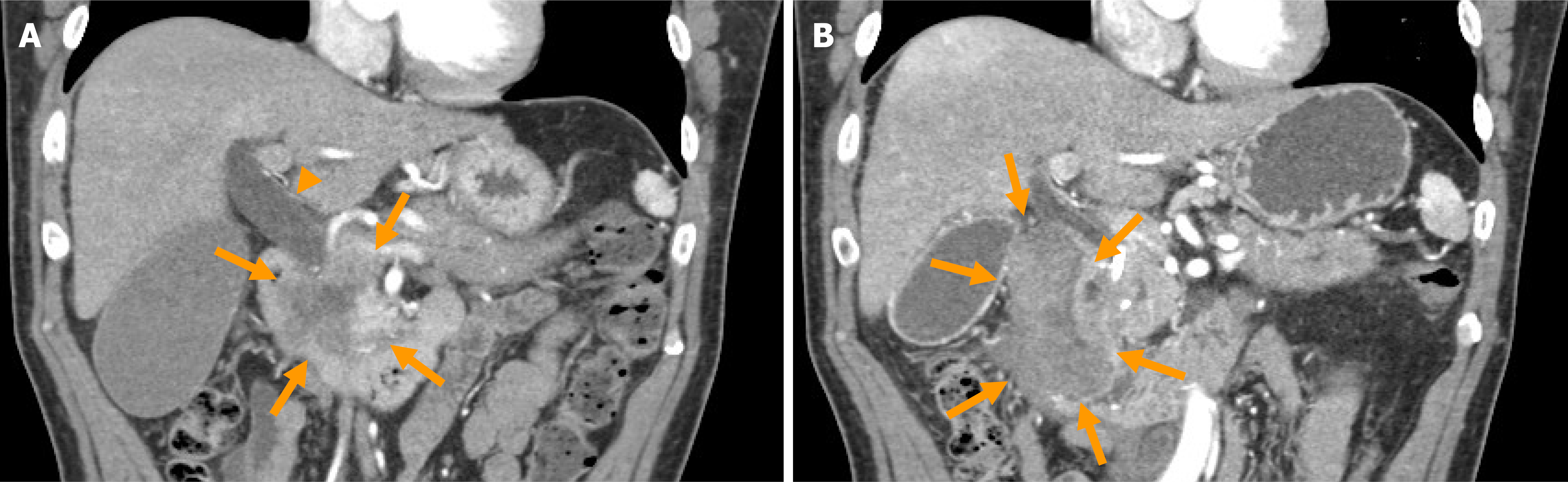
Figure 1 Abdominal contrast computed tomography.
A: Abdominal contrast computed tomography in the coronal plane showing an ill-defined 40 mm hypovascular mass at the head of the pancreas (arrows), together with dilatation of the common bile duct (arrowhead); B: Abdominal contrast computed tomography in the coronal plane revealing a high-density mass in the second part of the duodenum (arrows), consistent with a hematoma in the wall of the duodenum.
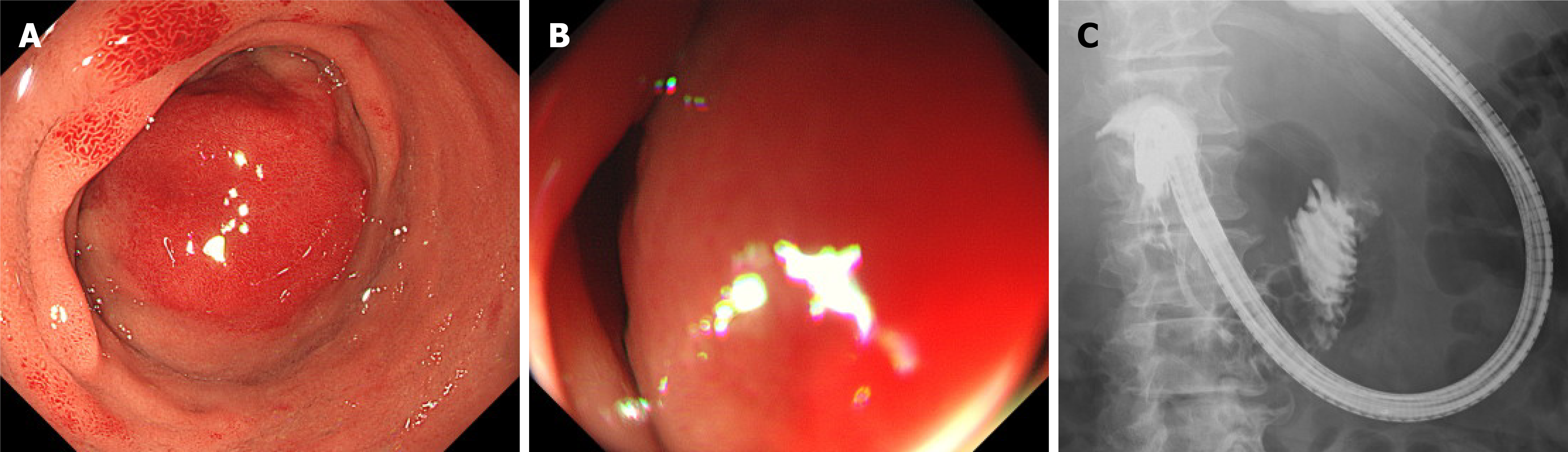
Figure 2 Upper gastrointestinal endoscopy.
A: Upper gastrointestinal endoscopy showing a smooth red swelling arising from the subepithelial layer from the superior duodenal angle on the opposite side of the ampulla of Vater, consistent with an intramural duodenal hematoma; B: The duodenal lumen was narrow because of the subepithelial swelling; C: The contrast was able to pass without significant impedance to the jejunum.
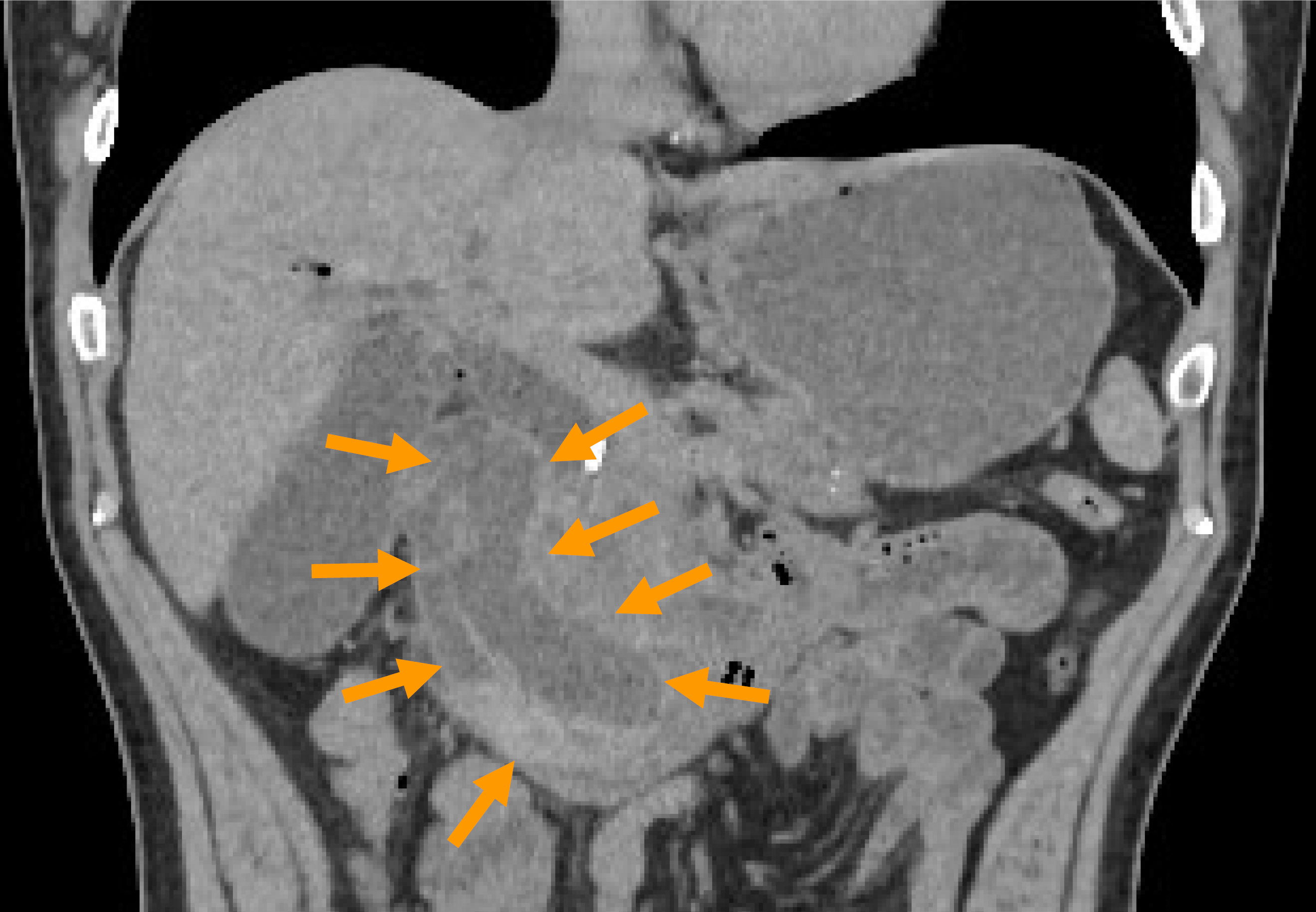
Figure 3
Abdominal computed tomography in the coronal plane showed the enlargement of the intramural duodenal hematoma, consistent with a rebleed (arrows).
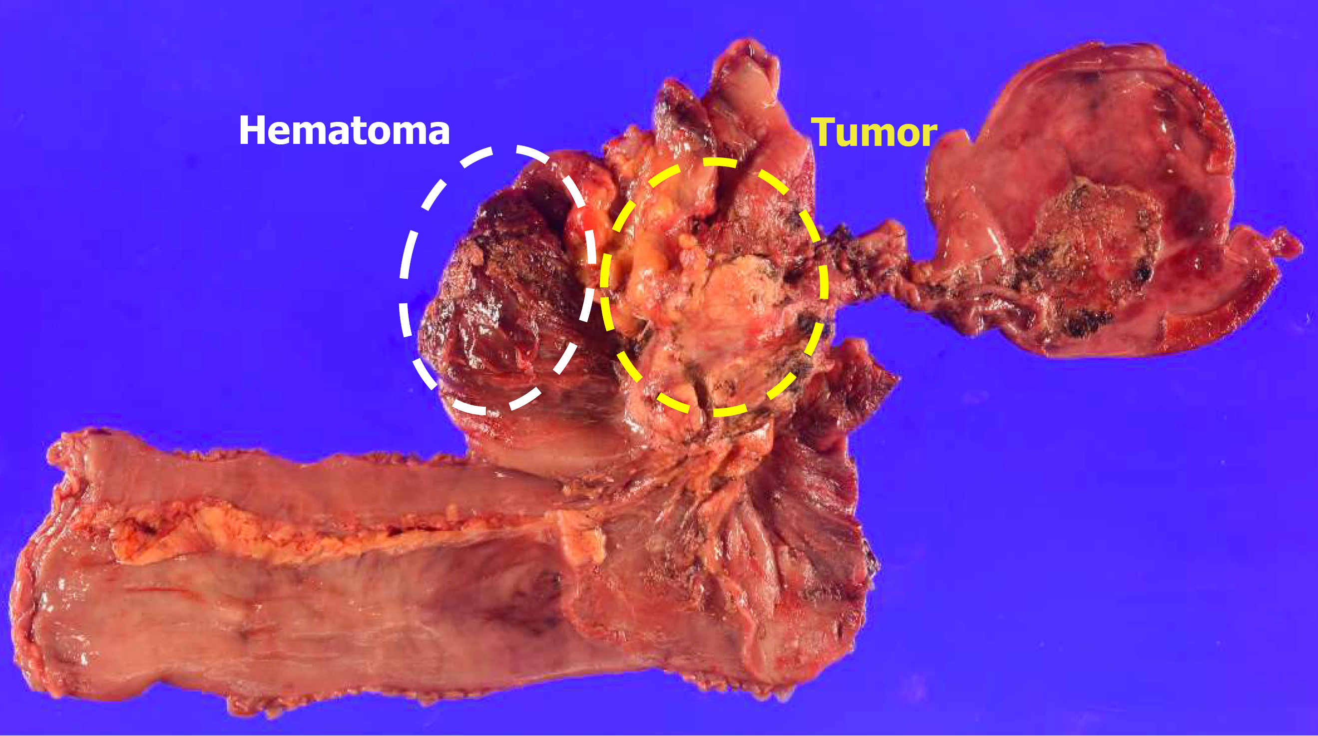
Figure 4
In the resected specimen, the hematoma (white circle) was separate from the tumor at the head of the pancreas (yellow circle) and the ampulla of Vater.
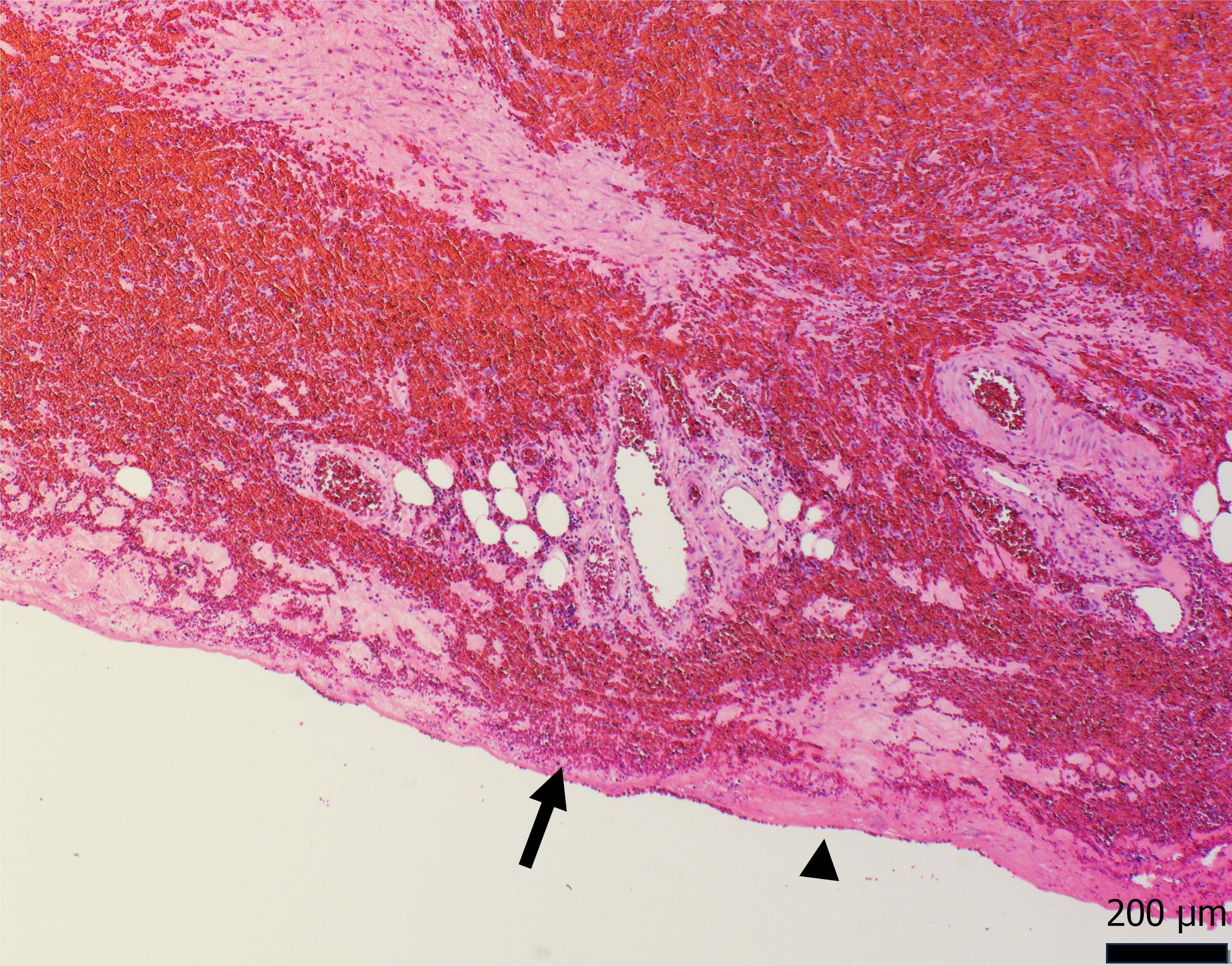
Figure 5
Histopathology of the resected specimen showed that the hematoma was located mainly within the muscularis propria, but in one area, there was only the subserosa (arrowhead) covering the hematoma (arrow).
- Citation: Colvin HS, Wato M, Sakakihara I, Akashi K, Okamoto K, Sui K, Nakamura S, Inaba T. Pancreatic adenocarcinoma complicated by intramural duodenal hematoma: A case report and review of literature. World J Gastroenterol 2026; 32(46): 122970
- URL: https://www.wjgnet.com/1007-9327/full/v32/i46/122970.htm
- DOI: https://dx.doi.org/10.3748/wjg.122970