
















Copyright: ©Author(s) 2026.
World J Gastroenterol. Nov 21, 2026; 32(43): 121298
Published online Nov 21, 2026. doi: 10.3748/wjg.121298
Published online Nov 21, 2026. doi: 10.3748/wjg.121298
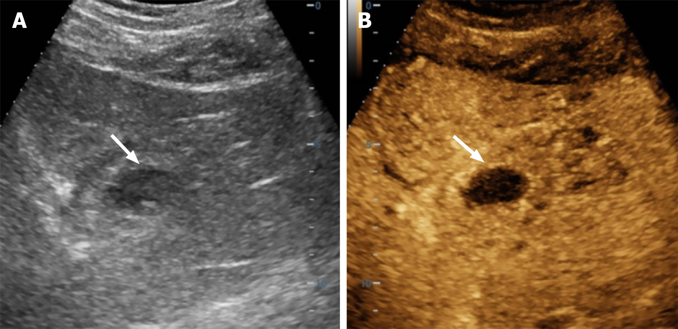
Figure 1 Liver ultrasound images of the liver lesion.
A: Routine liver ultrasound identified a 7 cm hypoechoic lesion in the left lobe; B: Contrast-enhanced ultrasound revealed a complex hepatic mass (7.8 cm × 7.7 cm) with < 50% liquefaction, suggesting evolving abscess formation.
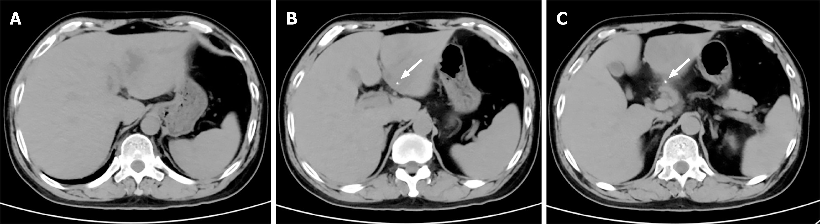
Figure 2 Results of abdomen enhanced computed tomography.
A: An irregular hypodense mass shadow in the left lateral lobe of the liver; B and C: A strip-shaped high-density shadow in the left lateral lobe of the liver, with local invasion of the pyloric canal wall.
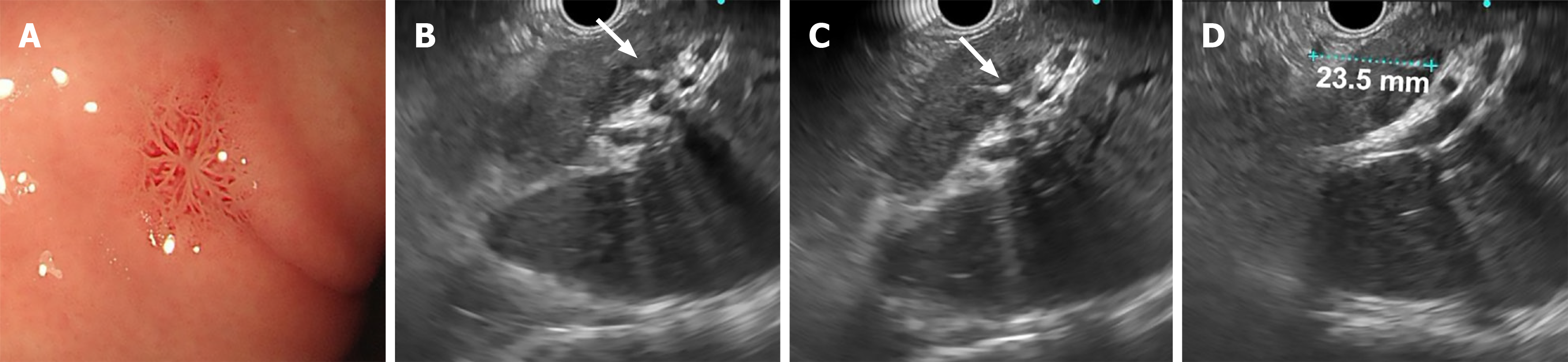
Figure 3 Results of simultaneous upper endoscopy and endoscopic ultrasonography.
A: A red scar was seen in the gastric corpus; B-D: A 23.5 mm strip-shaped lesion within the liver mass, invading the hepatic capsule with extrahepatic extension.
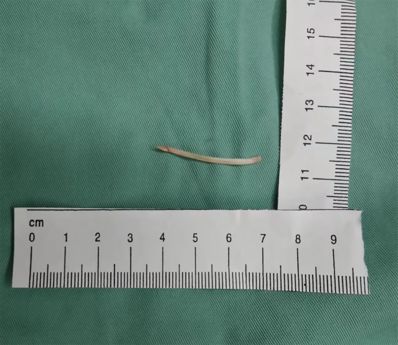
Figure 4
A 3 cm fish bone was removed from the liver mass.
- Citation: Zhang YF, Xu LL, Tang GM. Liver abscess caused by Parvimonas micra originating from a fish bone: A case report. World J Gastroenterol 2026; 32(43): 121298
- URL: https://www.wjgnet.com/1007-9327/full/v32/i43/121298.htm
- DOI: https://dx.doi.org/10.3748/wjg.121298