
















Copyright: ©Author(s) 2026.
World J Gastroenterol. Oct 7, 2026; 32(37): 120384
Published online Oct 7, 2026. doi: 10.3748/wjg.120384
Published online Oct 7, 2026. doi: 10.3748/wjg.120384
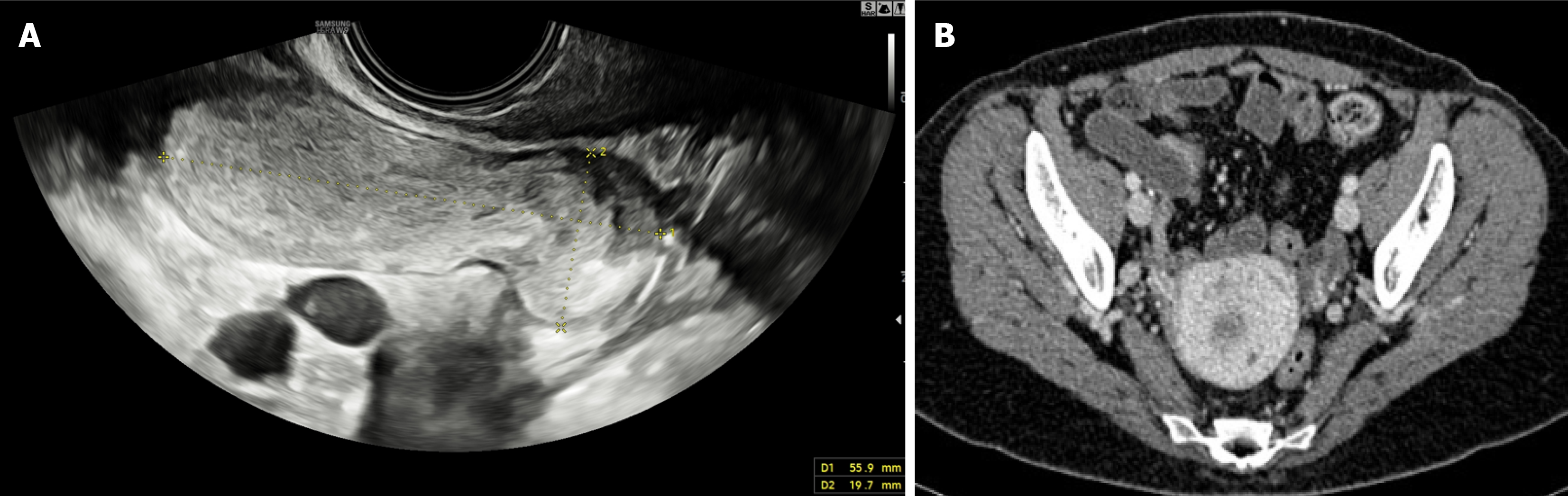
Figure 1 Contrast-enhanced computed tomography images of a 56-year-old woman with low-grade appendiceal mucinous neoplasms, diagnosed on histopathological examination.
A: Abdominal ultrasound shows a tubular, appendiceal lesion measuring approximately 55 mm × 20 mm, with hyperechoic to anechoic content and thickened walls; B: Axial contrast-enhanced computed tomography scan demonstrating appendiceal dilatation with low-attenuation mucinous content and smooth walls.
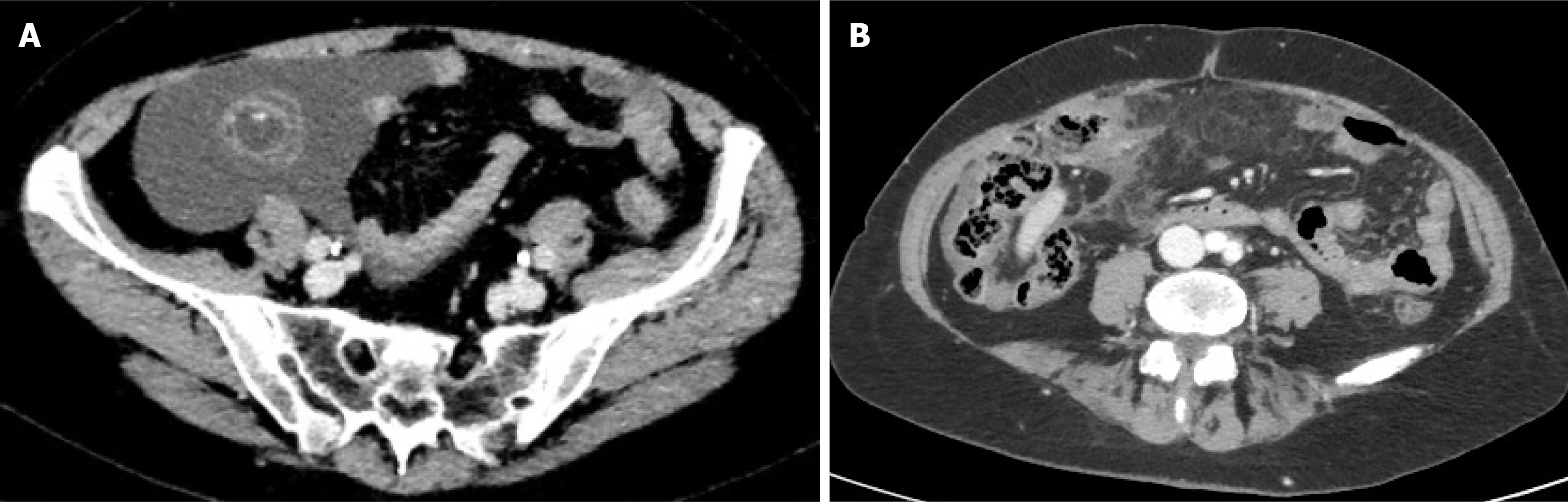
Figure 2 Contrast-enhanced computed tomography images of a 68-year-old male patient.
A: Axial image showing a high-grade appendiceal mucinous neoplasms, presenting as a well-defined, tubular appendiceal mass with low-attenuation mucinous content and mild wall enhancement; B: Axial image demonstrating pseudomyxoma peritonei, characterized by low-attenuation mucinous ascites, and mild peritoneal thickening, consistent with mucinous peritoneal spread.
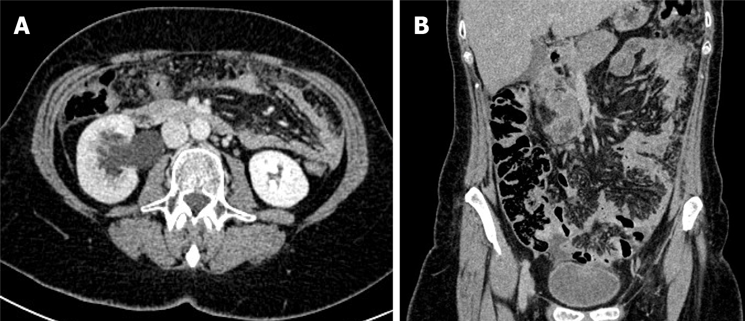
Figure 3 Contrast-enhanced computed tomography images in a 73-year-old man with mucinous appendiceal signet ring cell carcinoma.
A: Axial image demonstrating grade II hydronephrosis secondary to extrinsic ureteral compression by the tumor, associated with diffuse increased attenuation and stranding of the visceral abdominal fat, consistent with pseudomyxoma peritonei; B: Coronal reconstruction showing a poorly marginated appendiceal mass lesion; associated stranding of the abdominal visceral fat is also appreciable.
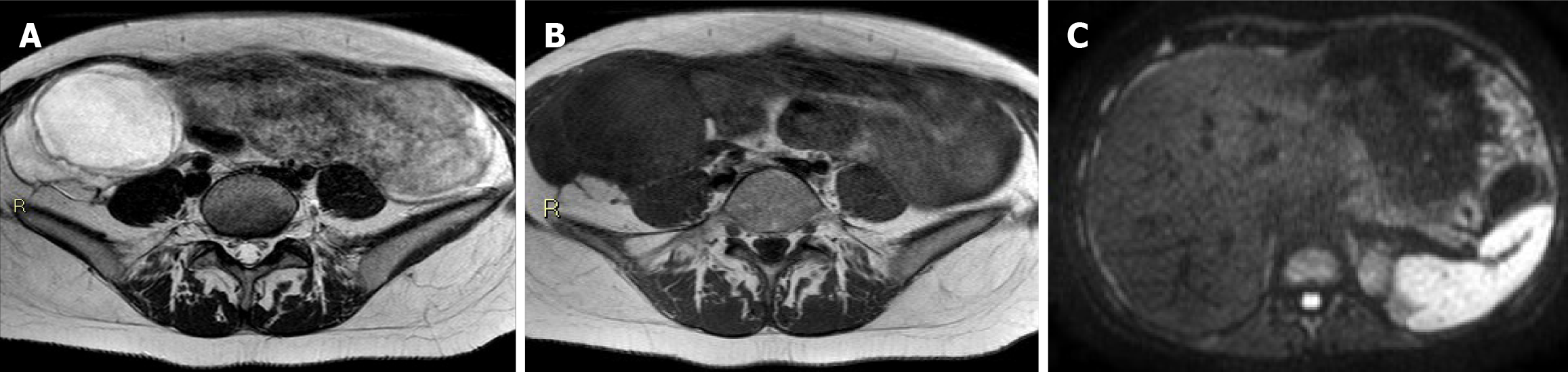
Figure 4 Magnetic resonance imaging of a 62-year-old man with low-grade appendiceal mucinous neoplasms.
A: Axial T2-weighted image demonstrating the appendiceal tumor, characterized by high signal intensity consistent with its mucinous content; B: Axial T1-weighted image showing the same lesion, with relatively low signal intensity compared to adjacent soft tissues; C: Diffusion-weighted imaging (b = 800 second/mm2) depicting Glissonian peritoneal implants with high signal intensity, consistent with peritoneal dissemination.
- Citation: Morelli C, Villanova I, Lorusso G, Cozzi M, Greco S, Calbi R, Maggialetti N, Calabrese A. Imaging of appendiceal mucinous epithelial neoplasms: A narrative review. World J Gastroenterol 2026; 32(37): 120384
- URL: https://www.wjgnet.com/1007-9327/full/v32/i37/120384.htm
- DOI: https://dx.doi.org/10.3748/wjg.120384