
















Copyright: ©Author(s) 2026.
World J Gastroenterol. Sep 7, 2026; 32(33): 118587
Published online Sep 7, 2026. doi: 10.3748/wjg.118587
Published online Sep 7, 2026. doi: 10.3748/wjg.118587
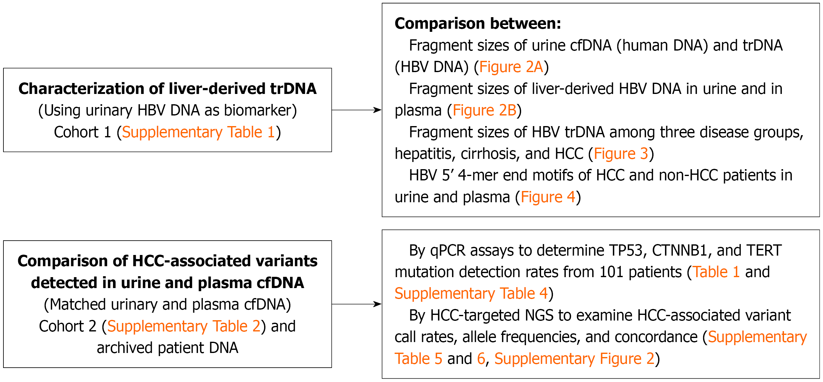
Figure 1 The study outline.
trDNA: Transrenal DNA; HBV: Hepatitis B virus; cfDNA: Cell-free DNA; HCC: Hepatocellular carcinoma; qPCR: Quantitative polymerase chain reaction; NGS: Next-generation sequencing.
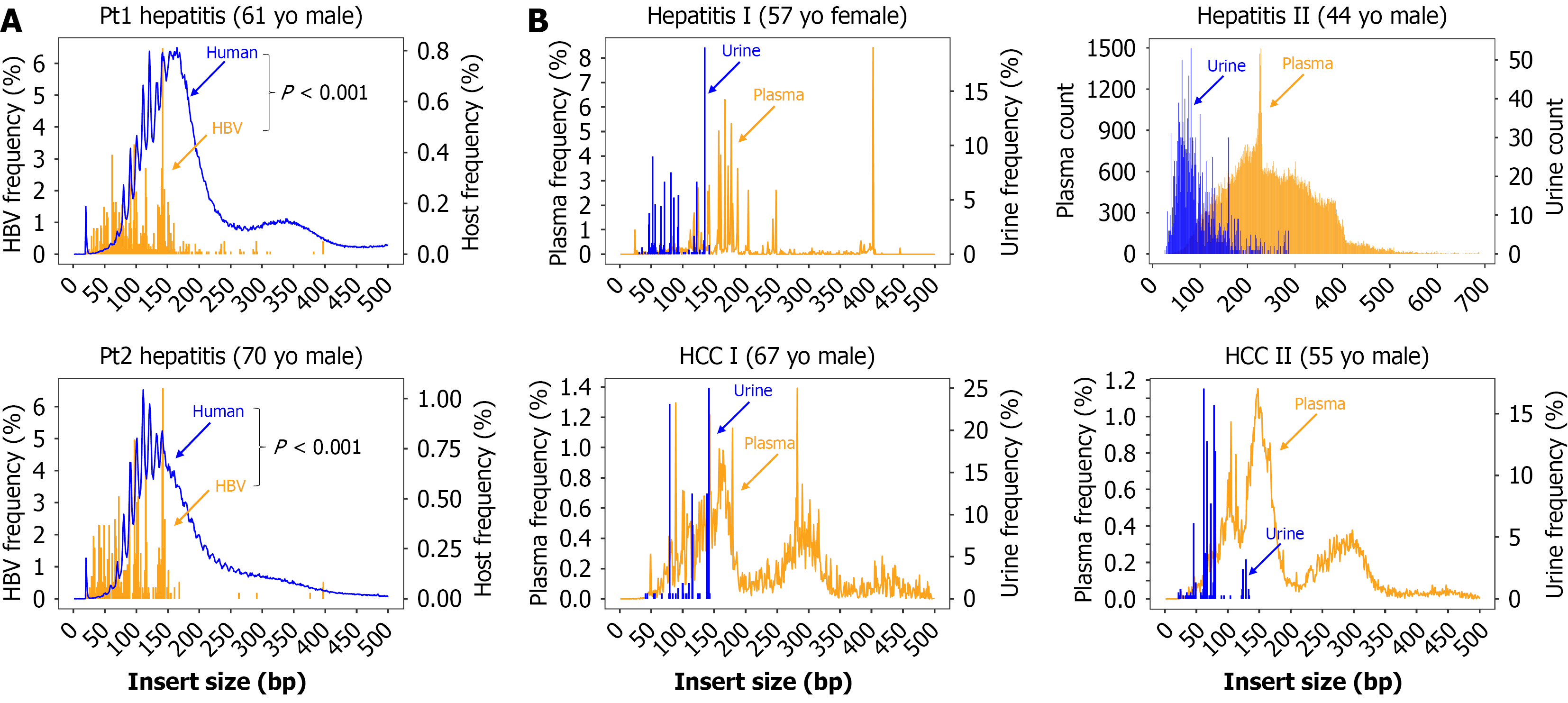
Figure 2 Hepatitis B virus transrenal DNA fragment size distributions compared to human cell-free DNA in urine and liver-derived hepatitis B virus cell-free DNA in plasma.
A: Comparisons of DNA fragment sizes of transrenal DNA (hepatitis B virus DNA; orange) and urine cell-free DNA (human DNA; blue) from two hepatitis B patients, Pt 1 and Pt 2, with serum viral load undetectable at the time of specimen collection; B: Comparisons of hepatitis B virus DNA fragment sizes between matched urine (transrenal DNA) and plasma (cell-free DNA) from two hepatitis B patients, hepatitis I and hepatitis II, and two hepatocellular carcinoma (HCC) patients, HCC I and HCC II. Hepatitis II had a 6.9 Log IU/mL serum viral load at the time of specimen collection. The other three patients had undetectable viral load at the time of specimen collection. HBV: Hepatitis B virus; HCC: Hepatocellular carcinoma.
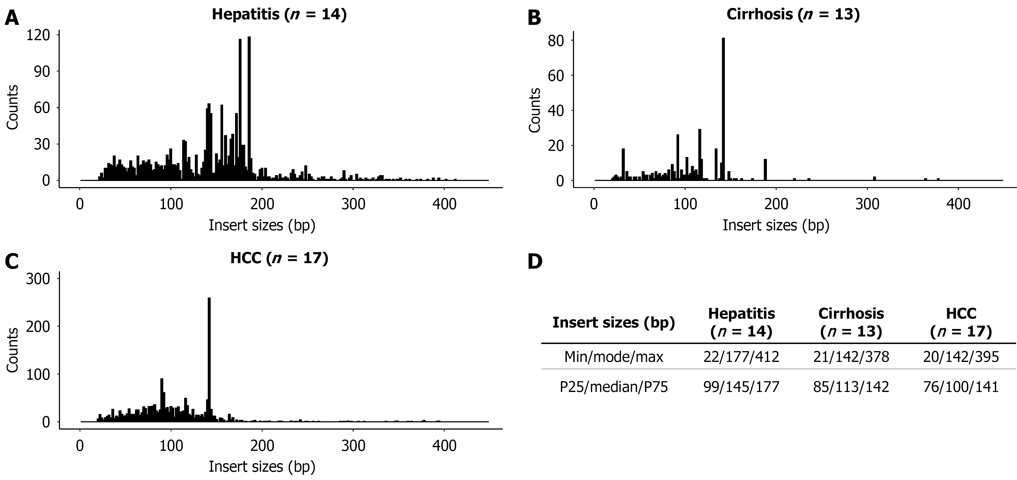
Figure 3 Comparison of hepatitis B virus DNA fragment sizes in urine from 14 hepatitis, 13 cirrhosis, and 17 hepatocellular carcinoma patients.
A-C: Distribution of hepatitis B virus DNA insert size from three disease groups, hepatitis (A), cirrhosis (B), and hepatocellular carcinoma (C), as listed in Supplementary Table 1, are plotted for each group; D The insert size statistics. HCC: Hepatocellular carcinoma.
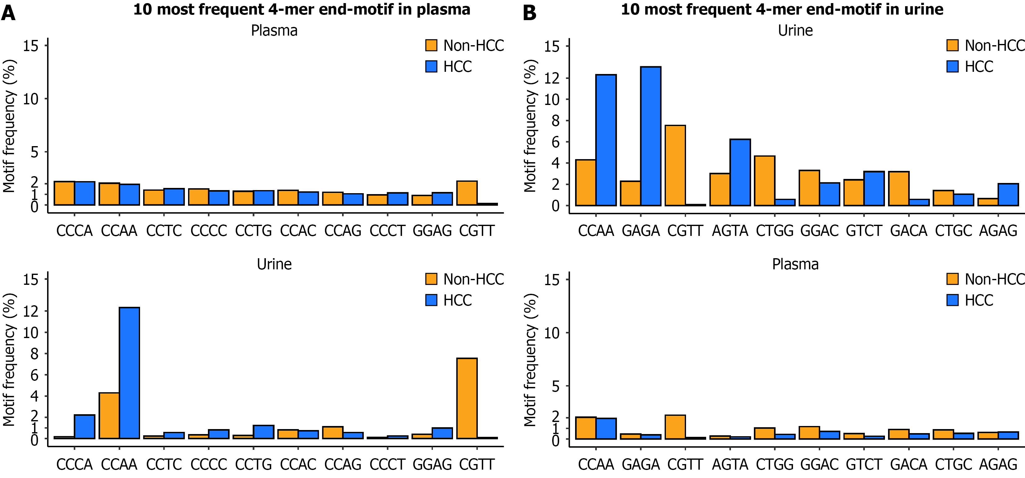
Figure 4 Comparison of the 10 most frequent 5’ 4-mer end motifs of hepatitis B virus DNA fragments in plasma and in urine between hepatocellular carcinoma and non-hepatocellular carcinoma hepatitis B patients.
A and B: The 10 most frequent 5’ 4-mer end motifs identified in plasma (A) and in urine (B) (top panel) and in the corresponding matched urine and plasma (bottom panel) from hepatocellular carcinoma (blue) and non-hepatocellular carcinoma (orange) patients are compared. Motifs are sorted by motif frequencies in the top panel. HCC: Hepatocellular carcinoma.
- Citation: Kim AK, Lin SY, Liu HN, Jain S, Gade TP, Shieh FS, Hamilton J, Hann HW, Chang TT, Goryunov D, Wang Z, Su YH. Urinary cell-free DNA as a noninvasive liquid biopsy for hepatocellular carcinoma. World J Gastroenterol 2026; 32(33): 118587
- URL: https://www.wjgnet.com/1007-9327/full/v32/i33/118587.htm
- DOI: https://dx.doi.org/10.3748/wjg.118587