
















Copyright: ©Author(s) 2026.
World J Gastroenterol. Aug 14, 2026; 32(30): 122191
Published online Aug 14, 2026. doi: 10.3748/wjg.122191
Published online Aug 14, 2026. doi: 10.3748/wjg.122191
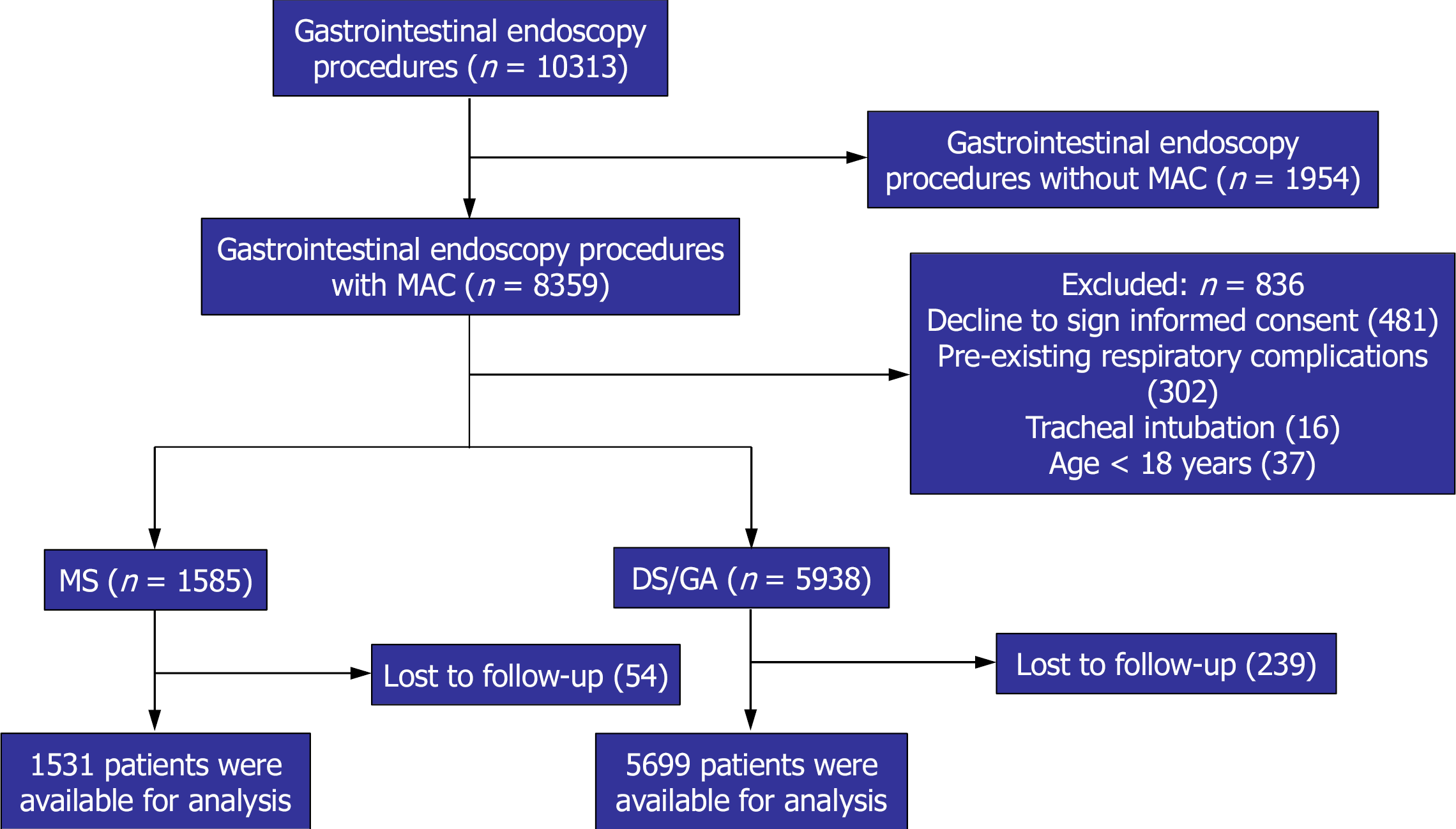
Figure 1 Flow diagram showing patient eligibility.
GA: General anesthesia; MS: Moderate sedation; DS: Deep sedation; MAC: Monitored anesthesia care.
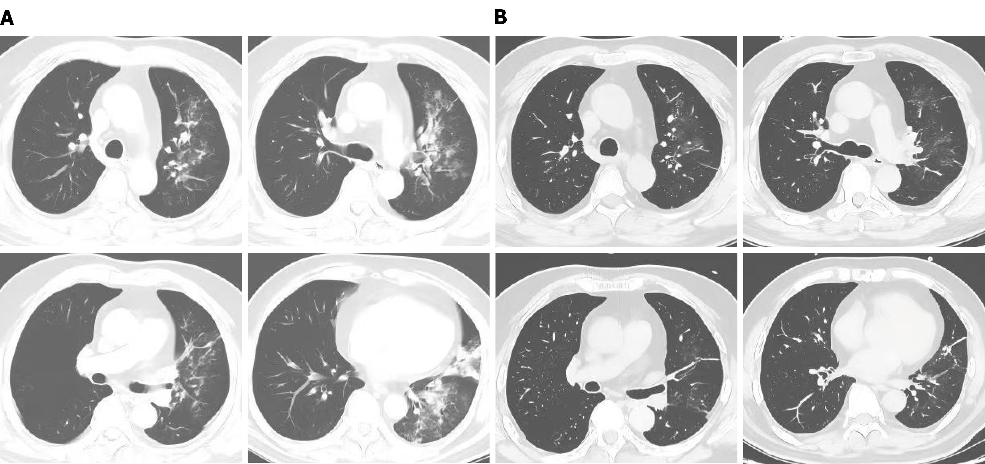
Figure 2 Chest computed tomography findings before and after treatment.
A: Chest computed tomography image obtained 5 hours post-operatively, showing left lung infiltration; B: Chest computed tomography image obtained after 7 days of treatment, showing marked resolution of the infiltration.
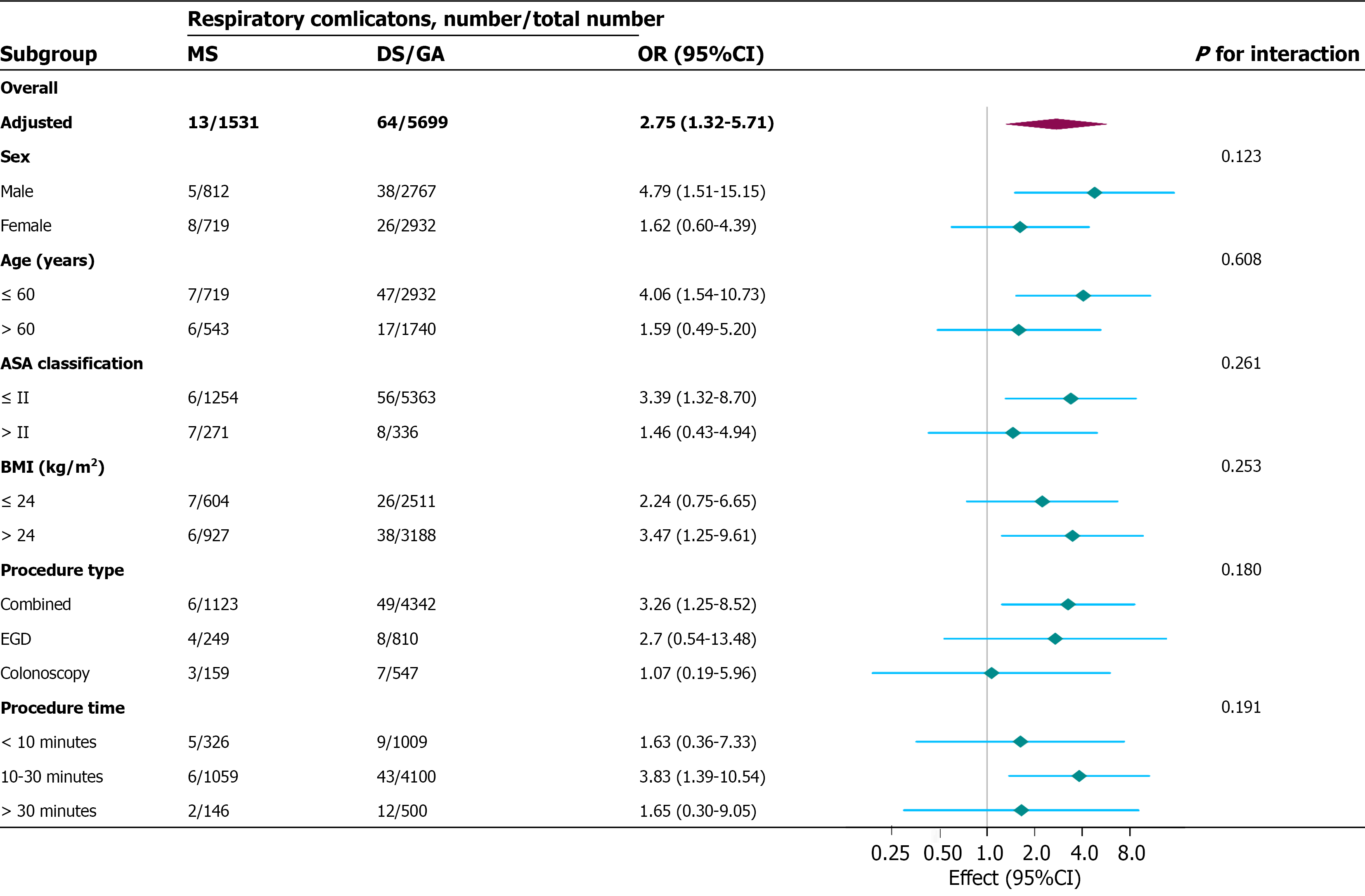
Figure 3 Relationships between sedation and respiratory complications according to general characteristics.
In addition to the stratification component itself, each stratification factor was adjusted for all other variables (American Society of Anesthesiologists classification, heart disease, asthma, hiatus hernia, gastrointestinal ulcer, gastroesophageal reflux disease, history of upper gastrointestinal and oral-pharyngeal surgery, and regurgitation and aspiration). ASA: American Society of Anesthesiologists; EGD: Esophagogastroduodenoscopy; GA: General anesthesia; BMI: Body mass index; MS: Moderate sedation; DS: Deep sedation; OR: Odds ratio.
- Citation: Ma LX, Zhang LN, Zhu YZ, Wang MH, Du XB, Qi LJ, Sun J, Yi XL, Wang P, Zhou ZG, Ji XY, Zhang L. Risk of respiratory complications following gastrointestinal endoscopy under monitored anesthesia care: A prospective cohort study. World J Gastroenterol 2026; 32(30): 122191
- URL: https://www.wjgnet.com/1007-9327/full/v32/i30/122191.htm
- DOI: https://dx.doi.org/10.3748/wjg.122191