
















Copyright: ©Author(s) 2026.
World J Gastroenterol. Aug 7, 2026; 32(29): 120498
Published online Aug 7, 2026. doi: 10.3748/wjg.120498
Published online Aug 7, 2026. doi: 10.3748/wjg.120498
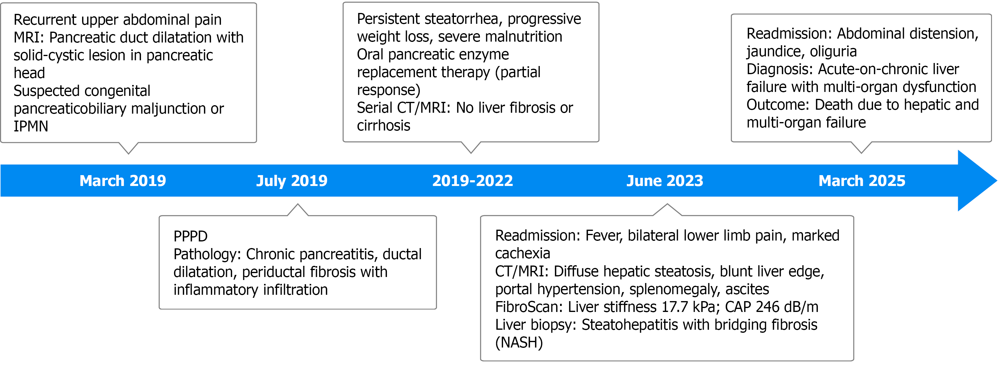
Figure 1 Chronological progression of the patient’s clinical course is summarized in the timeline.
PPPD: Pylorus-preserving pancreatoduo
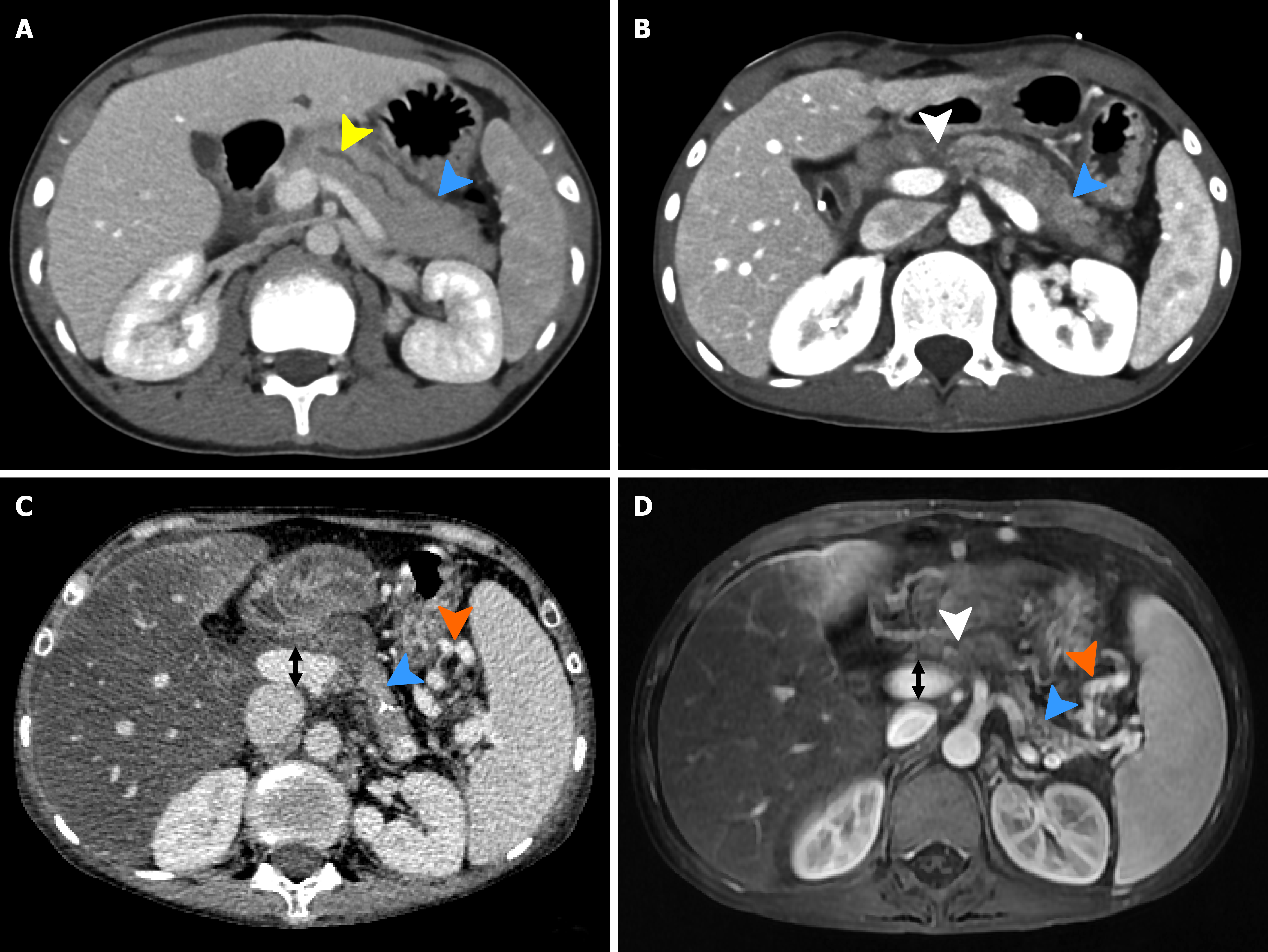
Figure 2 Serial abdominal imaging findings at different time points.
A: Preoperative contrast-enhanced abdominal computed tomography (CT) showing dilatation of the main pancreatic duct (yellow arrow) and a swollen pancreas (blue arrow). The liver parenchyma demonstrates homogeneous attenuation without evidence of hepatic steatosis or cirrhosis; B: Early postoperative contrast-enhanced abdominal CT demonstrating the pancreatic remnant at the surgical margin (white arrow) and the residual pancreatic body and tail (blue arrow). The liver parenchyma remains homogeneous, with no radiological features suggestive of hepatic steatosis or cirrhosis; C: Contrast-enhanced abdominal CT at the 4-year postoperative follow-up. Marked atrophy of the residual pancreatic body and tail is observed (blue arrow). Multiple dilated and tortuous vessels around the splenic vein and gastric fundus are present (orange arrow), and the main portal vein is widened to 16 mm (black arrow). The liver shows an irregular contour, altered lobar proportions, heterogeneous perfusion, and intrahepatic fat deposition, accompanied by splenomegaly; D: Contrast-enhanced abdominal magnetic resonance imaging at the 4-year postoperative follow-up demonstrating the pancreatic remnant (white arrow) and severe atrophy of the residual pancreatic body and tail (blue arrow). Prominent dilated collateral vessels around the splenic vein and gastric fundus are again noted (orange arrow), with enlargement of the main portal vein (black arrow, 16 mm). The liver exhibits an irregular surface, distorted lobar architecture, heterogeneous enhancement, abnormal hepatic fat metabolism, and splenomegaly.
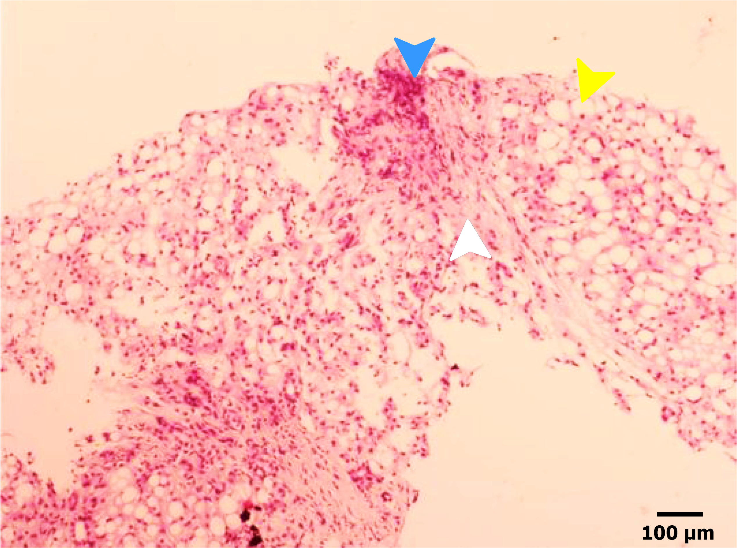
Figure 3 Histopathological examination of the liver biopsy specimen obtained at the 4-year postoperative follow-up (hematoxylin and eosin staining).
The liver tissue shows diffuse macrovesicular steatosis of hepatocytes (yellow arrow), accompanied by inflammatory cell infiltration predominantly composed of lymphocytes (blue arrow). Marked fibrous tissue proliferation is observed within the lobules and portal areas (white arrow), resulting in blurring of the normal lobular architecture. In addition, increased proliferation of small bile ducts is noted within hepatocytes and portal tracts, indicating advanced chronic liver injury with fibrotic remodeling.
- Citation: Hu H, Liu YJ, Li MC, Li Y, Lin SD. Rapid progression to liver failure following pancreatoduodenectomy in an adolescent: A case report. World J Gastroenterol 2026; 32(29): 120498
- URL: https://www.wjgnet.com/1007-9327/full/v32/i29/120498.htm
- DOI: https://dx.doi.org/10.3748/wjg.120498