
















Copyright: ©Author(s) 2026.
World J Gastroenterol. Aug 7, 2026; 32(29): 119374
Published online Aug 7, 2026. doi: 10.3748/wjg.119374
Published online Aug 7, 2026. doi: 10.3748/wjg.119374
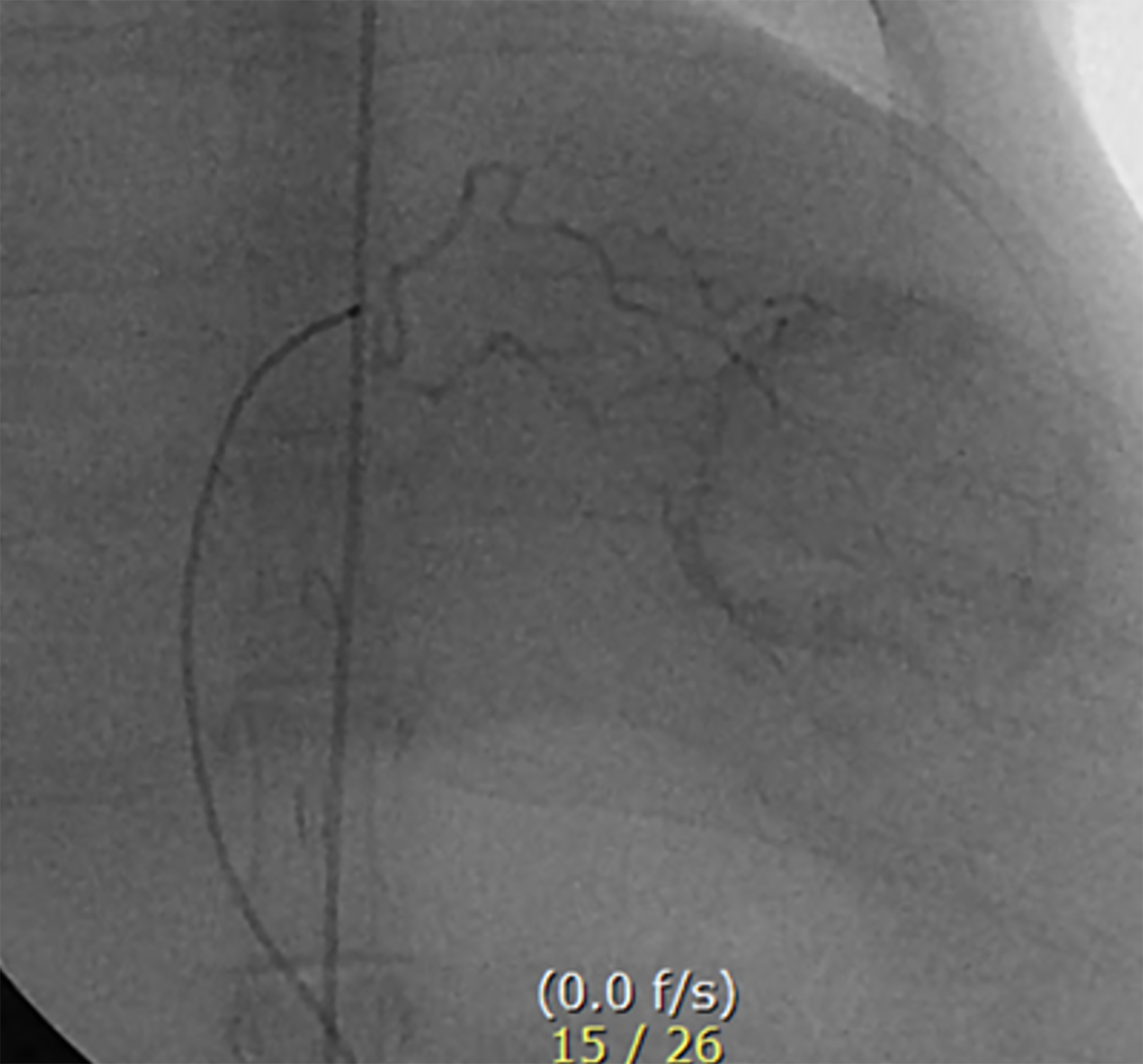
Figure 1 Angiography of VX2 tumor via the hepatic artery in a rabbit liver.
The hepatic angiogram demonstrates tumor staining in the left lobe of the liver. The catheter tip is positioned in the left hepatic artery through the celiac axis using the trans-auricular approach. Triolein emulsion or normal saline, along with doxorubicin, were administered through a microcatheter.
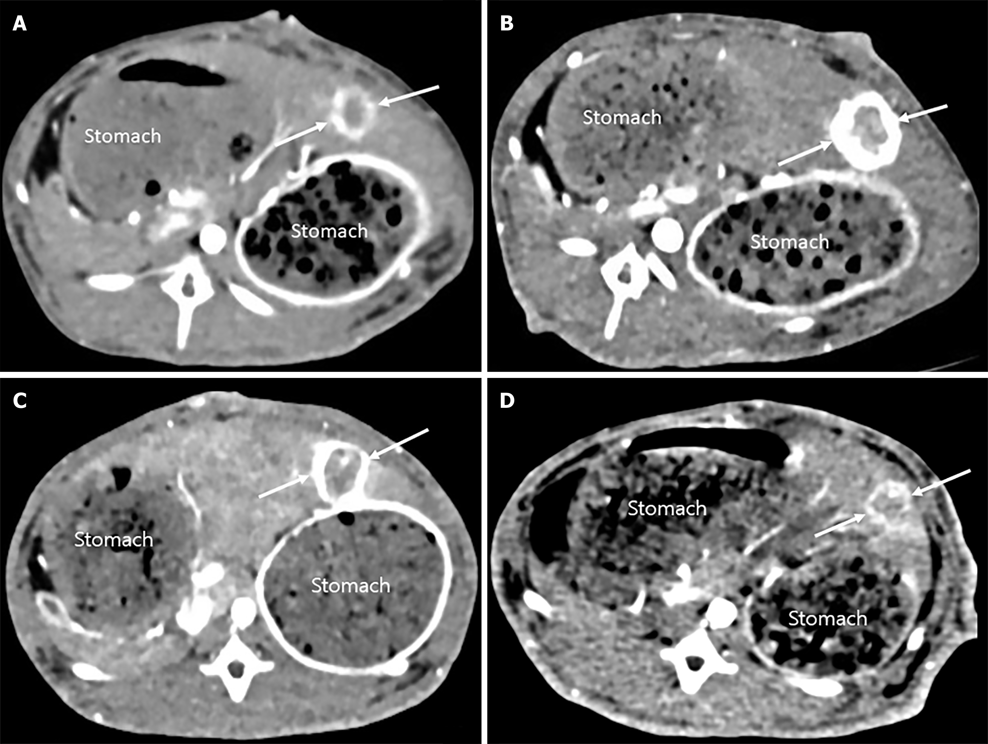
Figure 2 Representative computed tomography images of VX2 liver tumors before and after transcatheter arterial chemoembolization.
A and B: In the normal saline with doxorubicin group, pre-transcatheter arterial chemoembolization (TACE) (A) and post-TACE (B) images show a marked enlargement in tumor volume (arrows) and a significantly higher tumor growth ratio; C and D: In the triolein emulsion with doxorubicin group, pre-TACE (C) and post-TACE (D) images demonstrate a smaller reduction in tumor volume (arrows) and a significantly lower tumor growth ratio. All panels depict the largest tumor diameter slice for accurate volume estimation, and the tumor locations are consistent across pre- and post-TACE images.
- Citation: Kim YW, Kim HJ, Choi SH, Yun MS. Enhancing delivery of doxorubicin using triolein emulsion in rabbit VX2 liver tumor model. World J Gastroenterol 2026; 32(29): 119374
- URL: https://www.wjgnet.com/1007-9327/full/v32/i29/119374.htm
- DOI: https://dx.doi.org/10.3748/wjg.119374