
















Copyright: ©Author(s) 2026.
World J Gastroenterol. Aug 7, 2026; 32(29): 116126
Published online Aug 7, 2026. doi: 10.3748/wjg.116126
Published online Aug 7, 2026. doi: 10.3748/wjg.116126
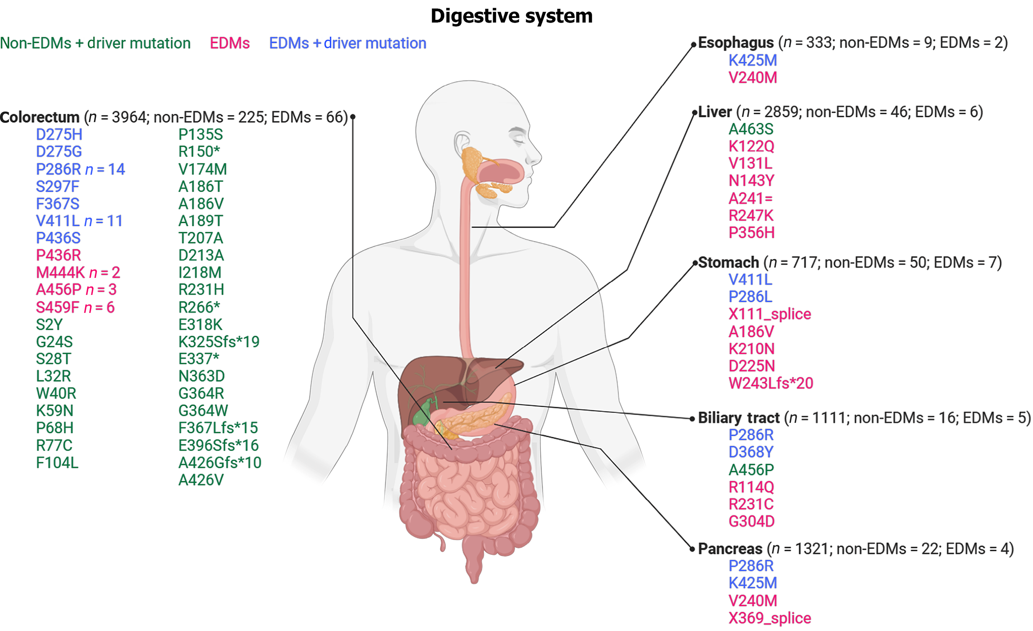
Figure 1 Pan-gastrointestinal landscape of somatic DNA polymerase epsilon mutations.
Schematic overview of the frequency and classification of somatic polymerase epsilon gene mutations across major digestive system cancers. For each organ, the figure displays the total number of samples analyzed (n). Mutations are classified based on their location relative to the proofreading region of the gene: Those located within this region are termed exonuclease domain mutations (EDMs), while those located elsewhere are termed non-EDMs. The esophagus data specifically represents esophageal adenocarcinoma. Specific variants are color-coded by their classification: Non-EDMs (green), EDMs (red), and driver EDMs (blue). Data were summarized from a comprehensive cancer genomics database. EDMs: Exonuclease domain mutations.
- Citation: Zou LF, Yang SQ, Yu SD, Wang CL. Letter to the Editor: From colorectal to pan-gastrointestinal cancer: Unlocking the therapeutic potential of DNA polymerase epsilon mutations. World J Gastroenterol 2026; 32(29): 116126
- URL: https://www.wjgnet.com/1007-9327/full/v32/i29/116126.htm
- DOI: https://dx.doi.org/10.3748/wjg.116126