
















Copyright: ©Author(s) 2026.
World J Gastroenterol. Jun 7, 2026; 32(21): 117828
Published online Jun 7, 2026. doi: 10.3748/wjg.v32.i21.117828
Published online Jun 7, 2026. doi: 10.3748/wjg.v32.i21.117828
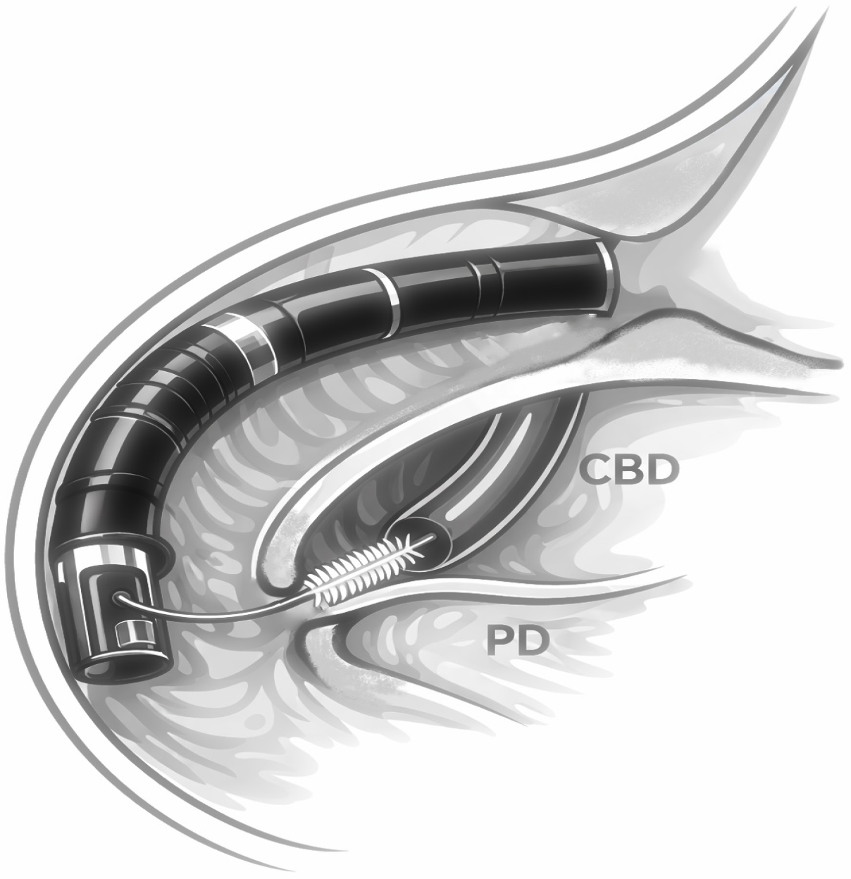
Figure 1 Schematic illustration of endoscopic retrograde cholangiopancreatography brush cytology technique for sampling biliary strictures.
Endoscopic retrograde cholangiopancreatography showing a cytology brush advanced through the endoscope into the common bile duct for tissue sampling at the site of the biliary stricture. The pancreatic duct is shown for anatomical reference. This schematic demonstrates the brushing technique used to obtain cellular material for cytological and molecular analysis. Illustration created by the authors. CBD: Common bile duct; PD: Pancreatic duct.
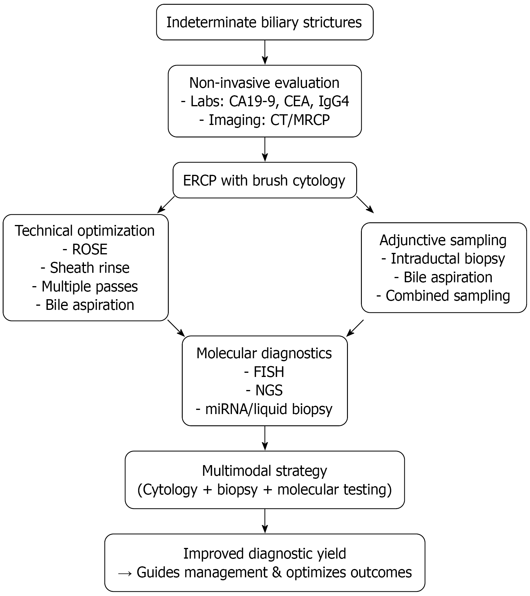
Figure 2 Diagnostic algorithm for the evaluation of indeterminate biliary strictures.
Schematic diagnostic algorithm for indeterminate biliary strictures showing the role of endoscopic retrograde cholangiopancreatography brush cytology, refinements, biopsy, and molecular diagnostics, culminating in a multimodal strategy to improve outcomes (in cases in which endoscopic retrograde cholangiopancreatography with brush cytology is not diagnostic, spyglass cholangioscopy is recommended). CA19-9: Carbohydrate antigen 19-9; CEA: Carcinoembryonic antigen; MRCP: Magnetic resonance cholangiopancreatography; ERCP: Endoscopic retrograde cholangiopancreatography; ROSE: Rapid on-site evaluation; FISH: Fluorescence in situ hybridization; NGS: Next-generation sequencing; miRNA: MicroRNA.
- Citation: Kordeni K, Vezakis A, Voulgaris T, Sotirianakou A, Politi E, Gazouli M. Methods improving endoscopic retrograde cholangiopancreatography brush cytology in malignant biliary strictures. World J Gastroenterol 2026; 32(21): 117828
- URL: https://www.wjgnet.com/1007-9327/full/v32/i21/117828.htm
- DOI: https://dx.doi.org/10.3748/wjg.v32.i21.117828