
















Copyright: ©Author(s) 2026.
World J Gastroenterol. May 28, 2026; 32(20): 117044
Published online May 28, 2026. doi: 10.3748/wjg.v32.i20.117044
Published online May 28, 2026. doi: 10.3748/wjg.v32.i20.117044
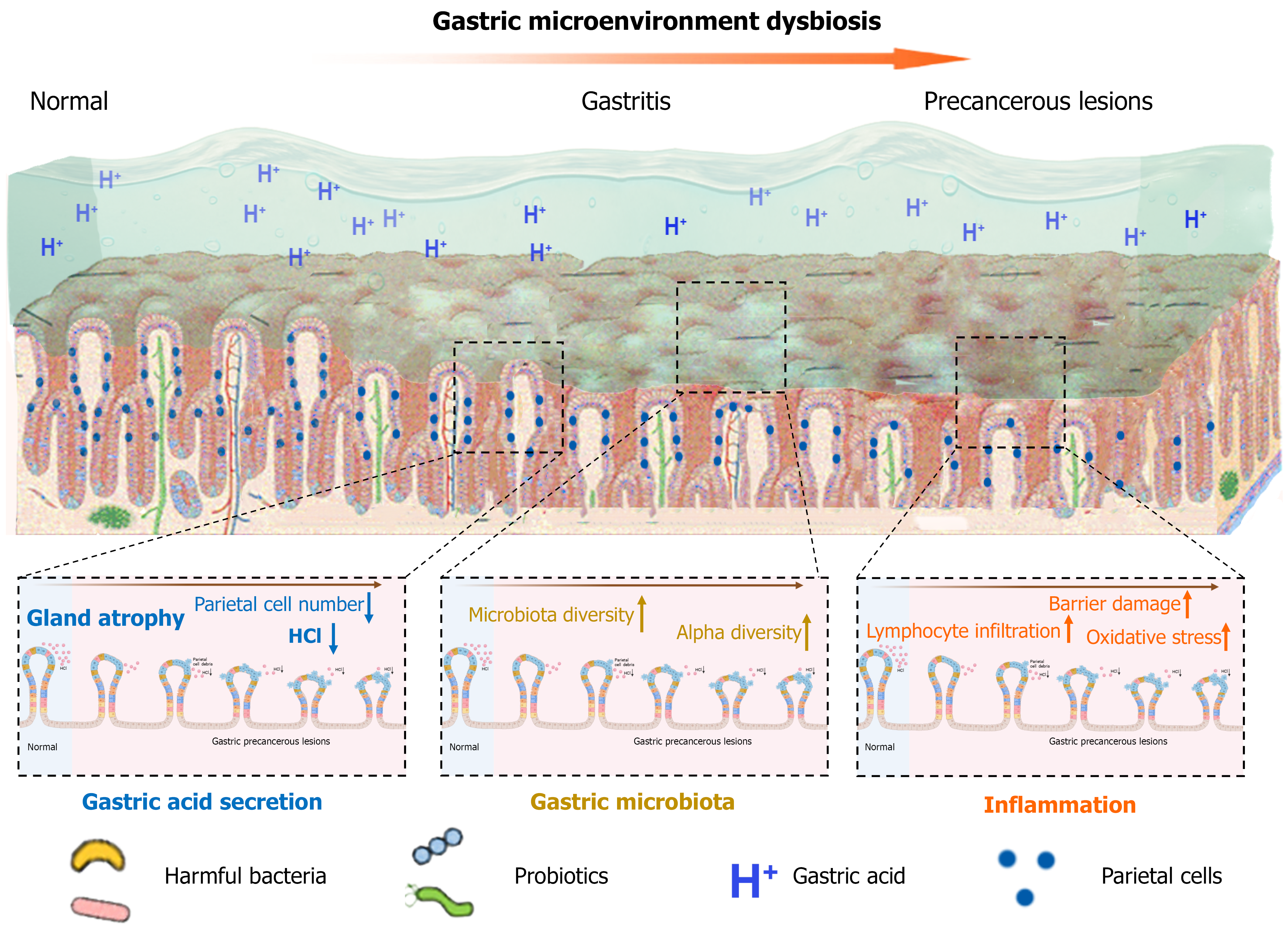
Figure 1 Illustration of dysbiosis of the gastric microenvironment.
This schematic figure depicts the altered gastric microenvironment under dysbiotic conditions, highlighting changes in gastric acidity, microbial composition, epithelial integrity, and inflammatory status that are associated with gastric carcinogenesis.
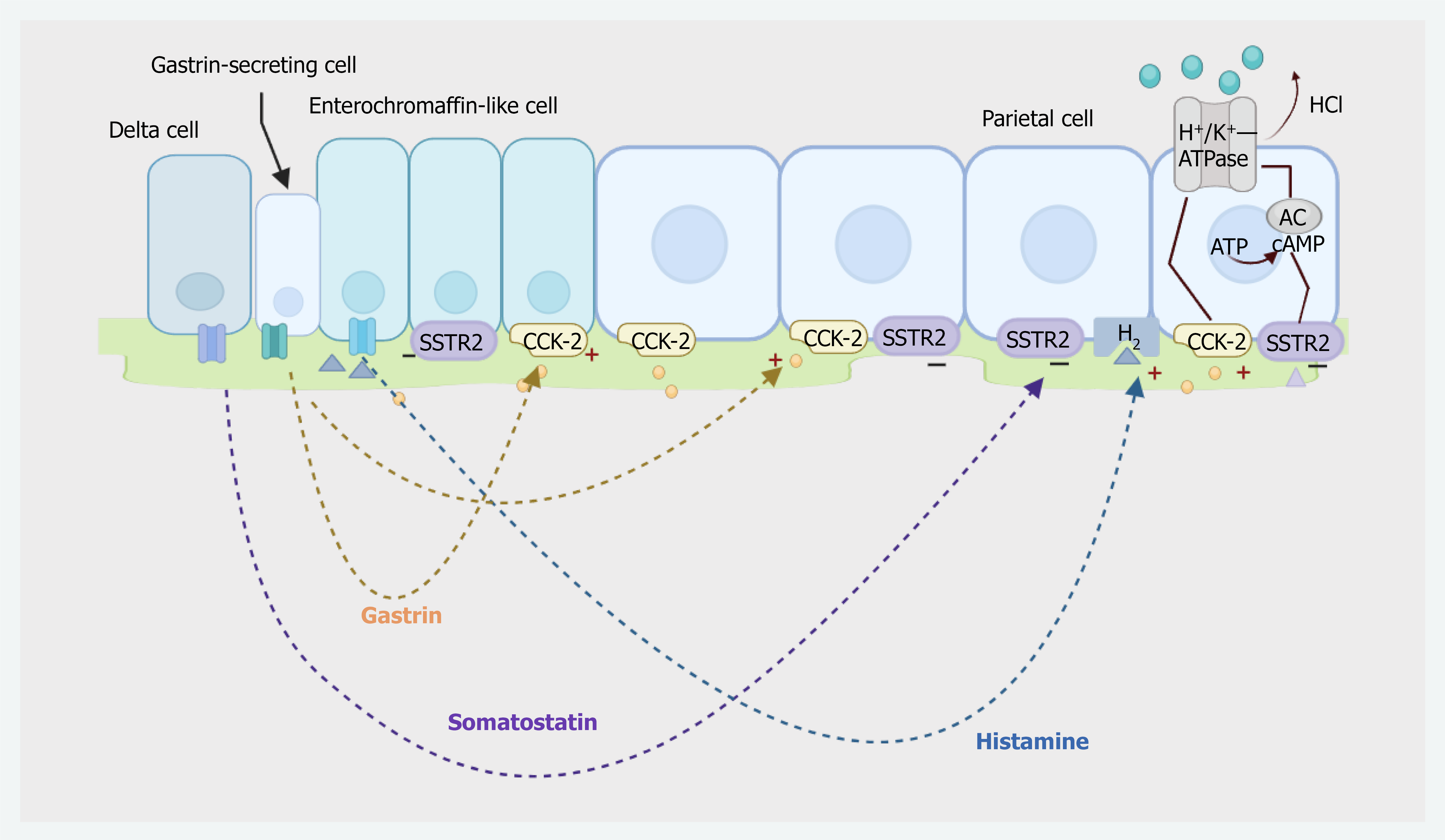
Figure 2 Illustration of the coordinated control of parietal cell acid secretion by gastrin, somatostatin, and histamine.
Gastrin stimulates enterochromaffin-like cells via CCK-2 receptors to release histamine, which activates parietal cells through H2 receptors. Somatostatin from delta cells exerts inhibitory effects through SSTR2 on gastrin cells, ECL cells, and parietal cells, maintaining feedback regulation of gastric acid output.
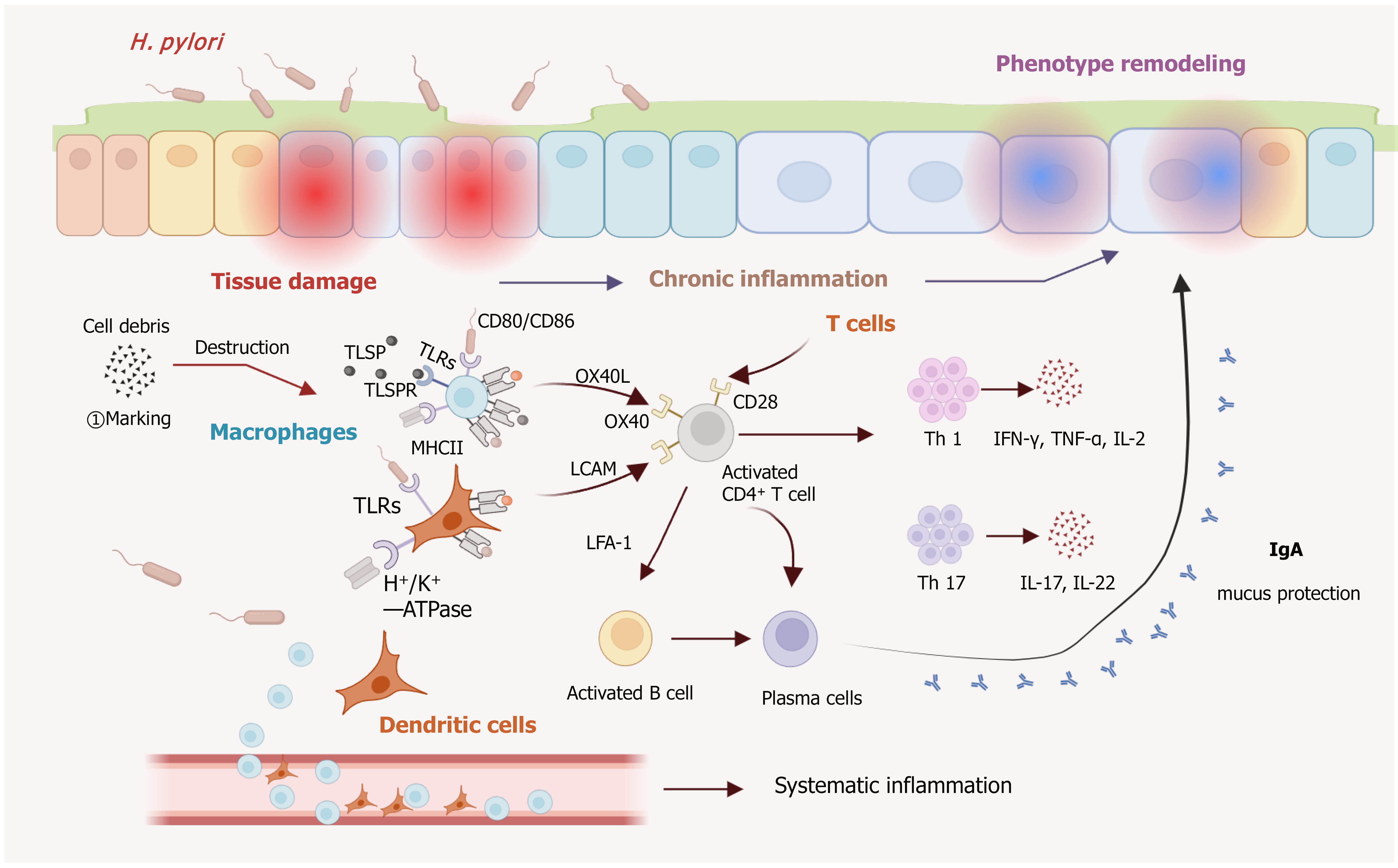
Figure 3 Helicobacter pylori-induced epithelial injury triggers the release and clearance of cell debris by macrophages, activating innate immune signaling and antigen presentation.
Dendritic cells and macrophages promote CD4+ T-cell activation through co-stimulatory pathways, driving Th1 and Th17 differentiation. The resulting cytokines (e.g., interferon-γ, tumor necrosis factor-α, interleukin-17) amplify chronic inflammation and contribute to remodeling of the epithelial phenotype. B cells activated through T-cell help differentiate into plasma cells and secrete IgA to support mucosal protection. Together, these interactions link local tissue damage with chronic inflammation and downstream epithelial changes. H. pylori: Helicobacter pylori; IL: Interleukin; IFN: Interferon; TNF-α: Tumor necrosis factor-α.
- Citation: Zheng HC, Zhou T, Sun L, Ding X. Gastric microenvironment and gastric cancer: Interplay of acid, microbiota, and inflammation. World J Gastroenterol 2026; 32(20): 117044
- URL: https://www.wjgnet.com/1007-9327/full/v32/i20/117044.htm
- DOI: https://dx.doi.org/10.3748/wjg.v32.i20.117044