Copyright
©The Author(s) 2025.
World J Gastroenterol. Oct 7, 2025; 31(37): 111081
Published online Oct 7, 2025. doi: 10.3748/wjg.v31.i37.111081
Published online Oct 7, 2025. doi: 10.3748/wjg.v31.i37.111081
Figure 1 Series of endoscopic image of endoscopic submucosal dissection.
A: A laterally spreading tumor in the sigmoid colon; B: Injecting methylene blue into the surrounding tumor before endoscopic submucosal dissection; C: Post-endoscopic submucosal dissection changes in the bowel.
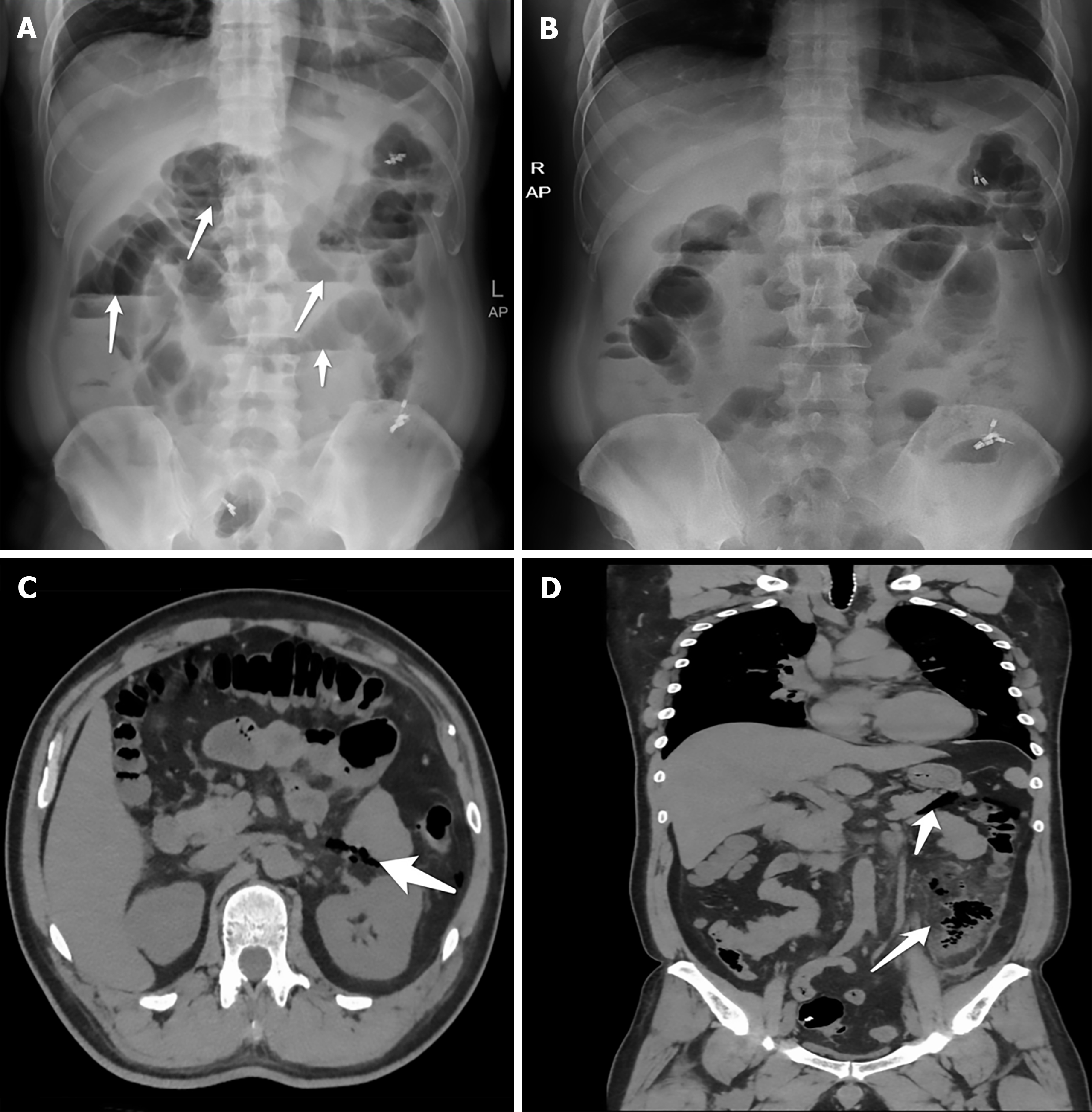
Figure 2 Initial and post treatment abdominal plain film and computed tomography scan.
A: The dilated intestinal tubes and air-fluid levels as the arrows shown after endoscopic submucosal dissection; B: Improvement of obstruction after treatment; C and D: After treatment and clip closure, computed tomography images demonstrate further the improvement of obstruction and confirm the etiology. The arrows indicated gas accumulation in the descending colon and surrounding exudation.
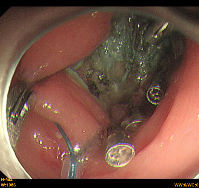
Figure 3
Endoscopic image of clip closure.
- Citation: Zhang Q, Hong D, Zhou YC, He GX, Jiang T. Acute intestinal obstruction caused by endoscopic submucosal dissection: A case report. World J Gastroenterol 2025; 31(37): 111081
- URL: https://www.wjgnet.com/1007-9327/full/v31/i37/111081.htm
- DOI: https://dx.doi.org/10.3748/wjg.v31.i37.111081


