©The Author(s) 2025.
World J Gastroenterol. Sep 28, 2025; 31(36): 111557
Published online Sep 28, 2025. doi: 10.3748/wjg.v31.i36.111557
Published online Sep 28, 2025. doi: 10.3748/wjg.v31.i36.111557
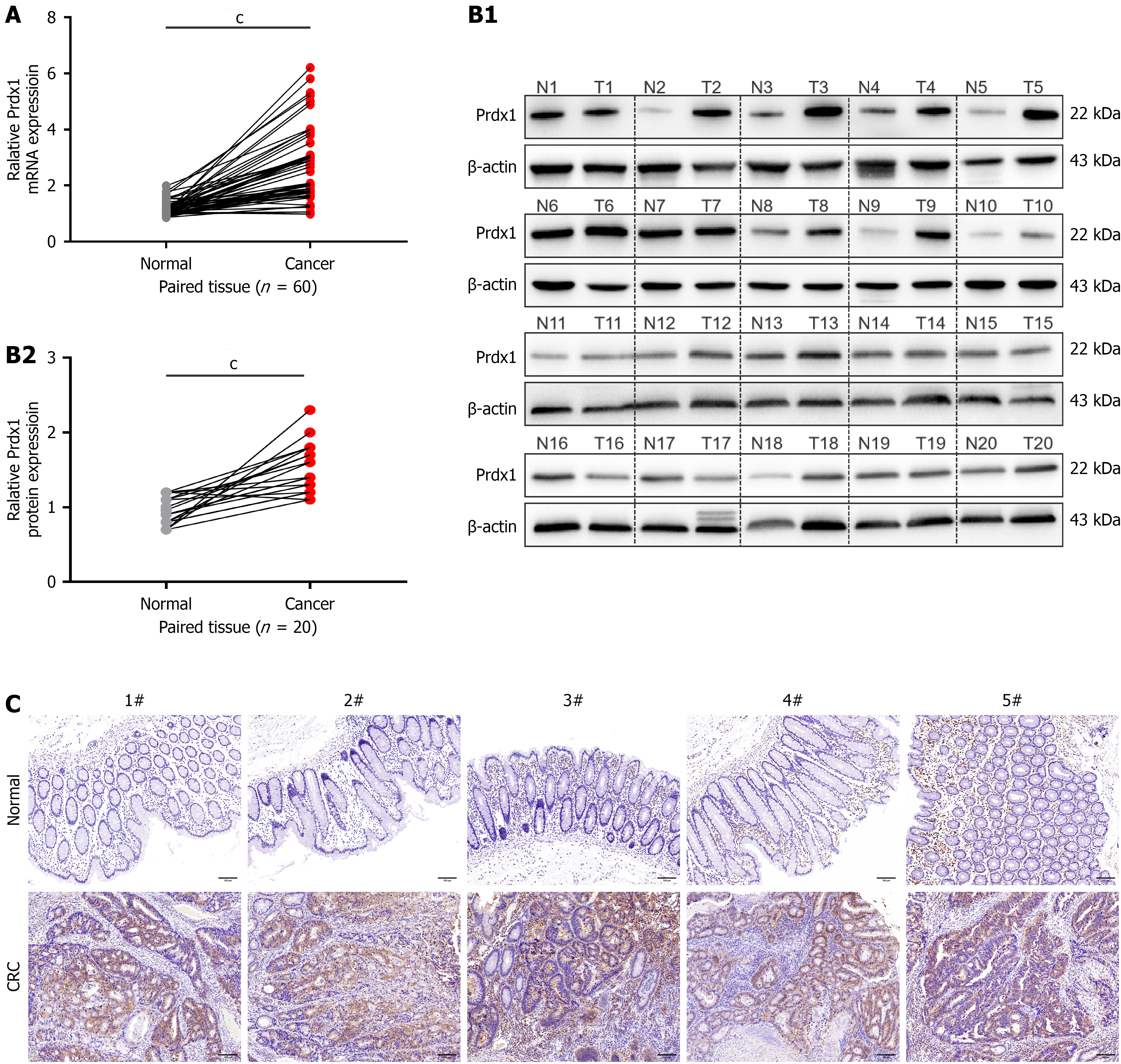
Figure 1 Peroxiredoxin 1 is up-regulated in colorectal cancer tissues.
A: The mRNA expression of peroxiredoxin 1 (Prdx1) was detected by quantitative real-time polymerase chain reaction in tumor tissues and adjacent normal tissues from patients with colorectal cancer (CRC), n = 60/group; B: The protein expression of Prdx1 was detected by western blotting in tumor tissues and adjacent normal tissues from patients with CRC, n = 20/group; C: Immunohistochemistry staining for Prdx1 in tumor tissues and adjacent normal tissues from patients with CRC, each representative image is from one sample (n = 20/group), scale bar: 100 μm (200 ×). Data are expressed as the mean ± SD; cP < 0.001. Prdx1: Peroxiredoxin 1; CRC: Colorectal cancer.
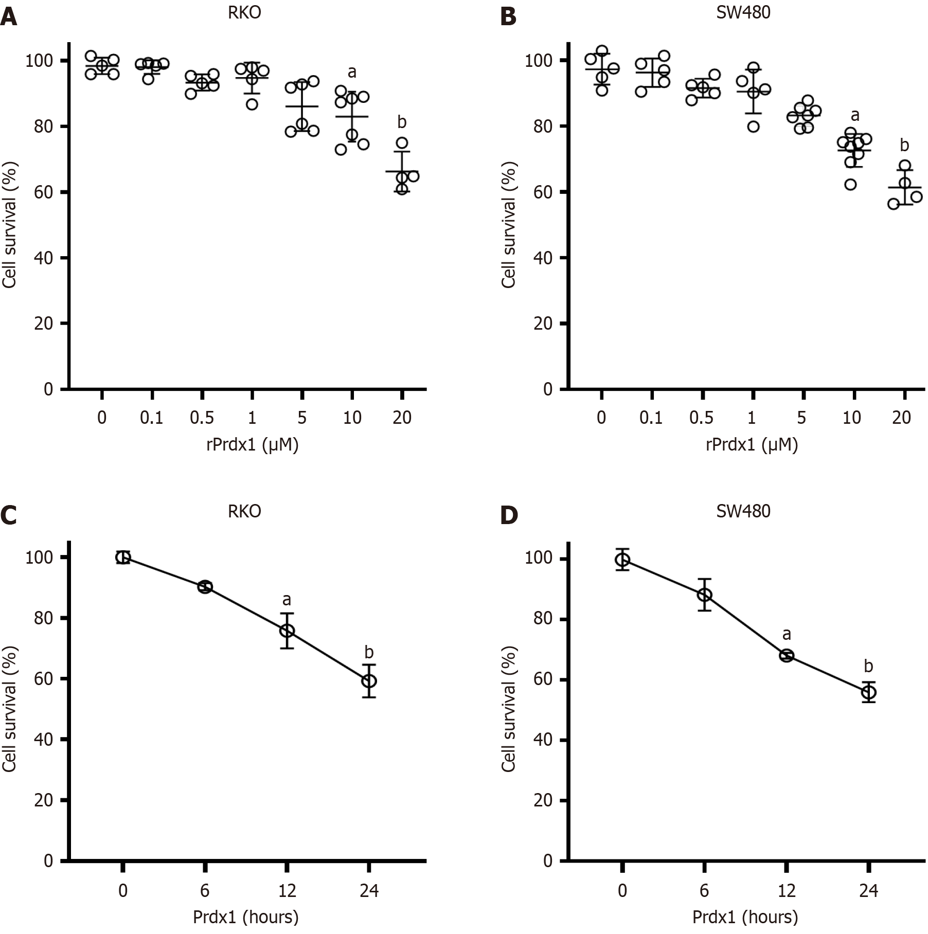
Figure 2 Recombinant peroxiredoxin 1 inhibits the viability of RKO and SW480 cells.
A and B: Survival of RKO and SW480 cells detected by MTT under recombinant peroxiredoxin 1 (rPrdx1) stimulation for 24 hours at different doses; C and D: Survival of RKO and SW480 cells detected by MTT under 20 μM rPrdx1 stimulation for different time periods. This experiment was repeated three times in independent experiments. Data are expressed as the mean ± SD; aP < 0.05 vs control group (without rPrdx1); bP < 0.01 vs control group (without rPrdx1). Prdx1: Peroxiredoxin 1; rPrdx1: Recombinant peroxiredoxin 1.
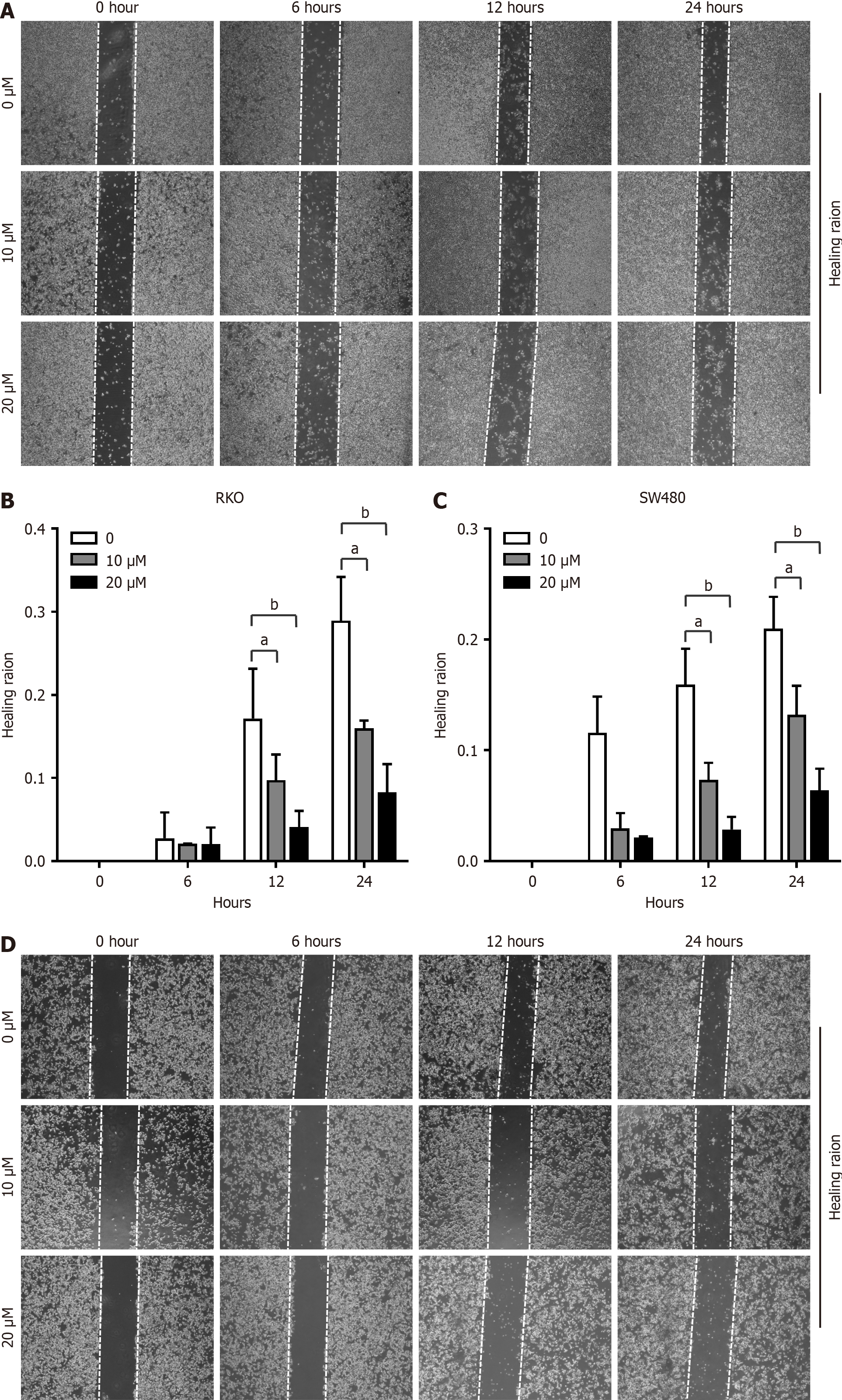
Figure 3 Recombinant peroxiredoxin 1 inhibits motility of RKO and SW480 cells.
A and B: Motility of RKO cells measured by the wound healing assay under different doses of recombinant peroxiredoxin 1 (rPrdx1) stimulation for 6, 12 and 24 hours, respectively; C and D: Motility of SW480 cells measured by the wound healing assay under different doses of rPrdx1 stimulation for 6, 12 and 24 hours, respectively. These experiments were repeated three times in independent experiments. Data are expressed as the mean ± SD. aP < 0.05 vs the control group (without rPrdx1); bP < 0.01 vs the control group (without rPrdx1).
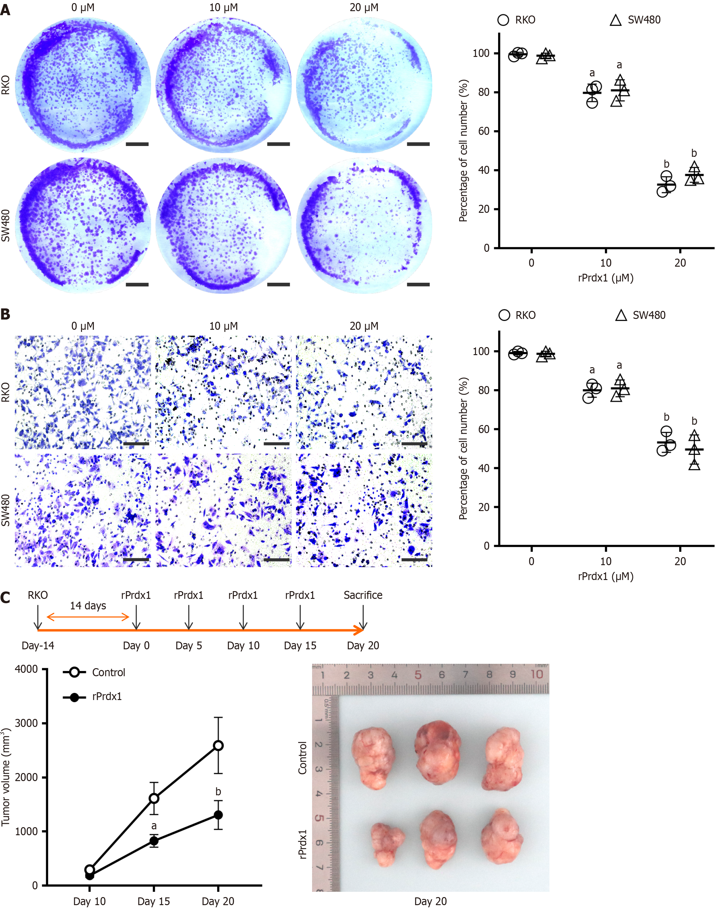
Figure 4 Recombinant peroxiredoxin 1 inhibits proliferation and invasion of RKO and SW480 cells.
A: Representative images of proliferation ability detected by the colony formation assay under stimulation by 10 μM or 20 μM recombinant peroxiredoxin 1 (rPrdx1) for 24 hours, scale bar: 1 cm; B: Representative images of invasion ability detected by the Transwell assay with stimulation by 10 μM or 20 μM rPrdx1 for 24 hours, scale bar: 200 μm (200 ×); the experiments for the colony formation assay and Transwell assay were repeated three times in independent experiments; C: Tumor volume analyses showed the inhibitory effect of intravenous injection of rPrdx1 on subcutaneous tumor formation in nude mice (n = 6/group). Bonferroni correction was used to adjust the significance for multiple comparisons after two-way RM ANOVA. Data are expressed as the mean ± SD. aP < 0.05 vs control group (without rPrdx1); bP < 0.01 vs control group (without rPrdx1). rPrdx1: Recombinant peroxiredoxin 1.
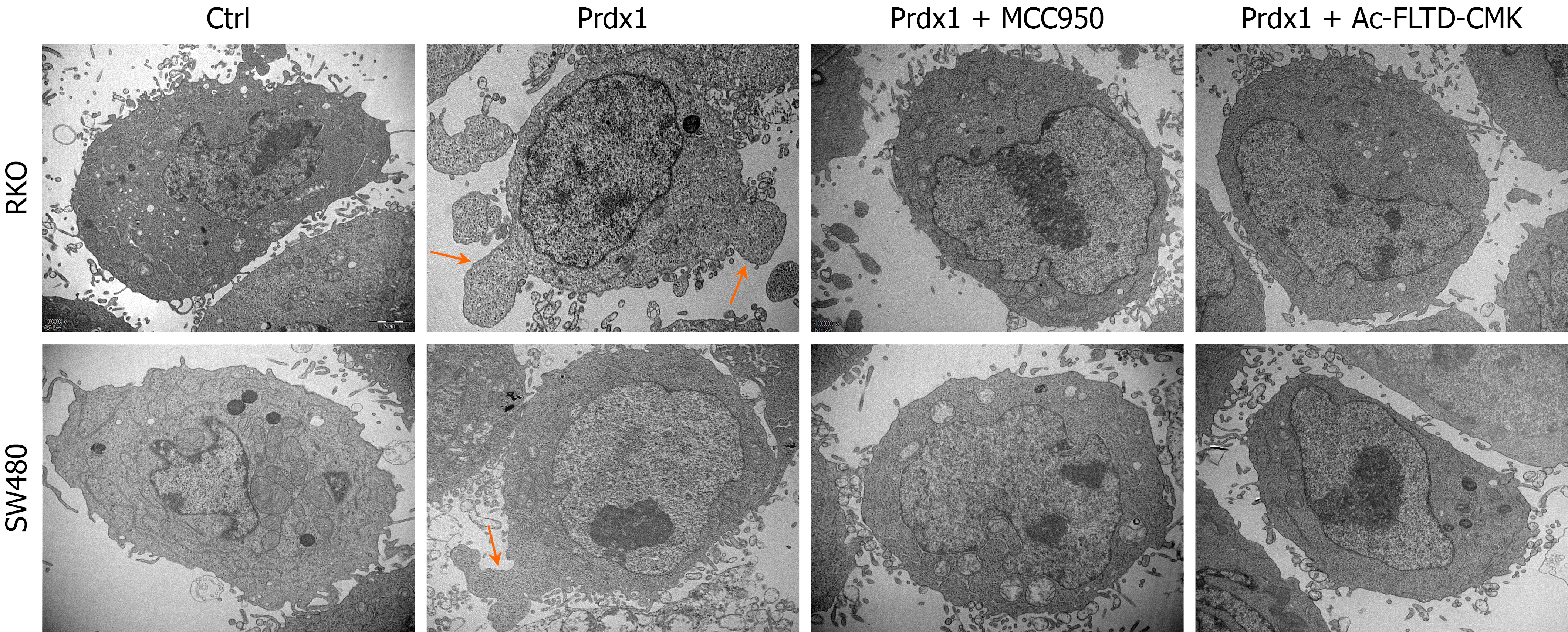
Figure 5 Recombinant peroxiredoxin 1 induces pyroptosis of RKO and SW480 cells.
Representative images of pyroptosis induced by recombinant peroxiredoxin 1 (rPrdx1) without or with NLRP3 inflammasome inhibitor (MCC950) and GSDMD inhibitor (Ac-FLTD-CMK) treatment (n = 3 per group), scale bar: 20 μm (× 10000). rPrdx1: 20 μM for 24 hours. MCC950: 10 μM for 24 hours. Ac-FLTD-CMK: 10 μM for 24 hours. Prdx1: Peroxiredoxin 1.
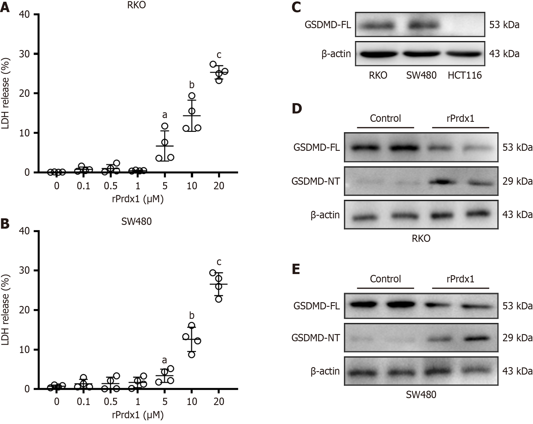
Figure 6 Recombinant peroxiredoxin 1 induces LDH release and up-regulation of GSDMD-NT in RKO and SW480 cells.
A: LDH release from RKO cells induced by different doses of recombinant peroxiredoxin 1 (rPrdx1) stimulation for 24 hours; B: LDH release from SW480 cells induced by different doses of rPrdx1 stimulation for 24 hours; C: GSDMD full-length (FL) expression in RKO, SW480 and HCT-116 cells detected by western blotting; D: GSDMD-FL and GSDMD N-terminal (NT) expression detected by western blotting in RKO cells under 20 μM rPrdx1 stimulation for 24 hours; E: GSDMD-FL and GSDMD-NT expression detected by western blotting in SW480 cells under 20 μM rPrdx1 stimulation for 24 hours. All these experiments were repeated three or four times in independent experiments. Data are expressed as the mean ± SD; aP < 0.05 vs control group (without rPrdx1); bP < 0.01 vs control group (without rPrdx1); cP < 0.001 vs control group (without rPrdx1). rPrdx1: Recombinant peroxiredoxin 1.
Figure 7 Recombinant peroxiredoxin 1 activates the NLRP3/GSDMD pathway to induce pyroptosis of RKO and SW480 cells.
A: The expression of GSDMD-FL, GSDMD-NT, NLRP3 and Caspase-1 (p10) in RKO and SW480 cells detected by western blotting under recombinant peroxiredoxin 1 (rPrdx1) stimulation; B: Survival of RKO and SW480 cells detected by MTT under rPrdx1 stimulation; C: LDH release from RKO and SW480 cells induced by rPrdx1; D: IL-1β release from RKO and SW480 cells induced by rPrdx1; E: IL-18 release from RKO and SW480 cells induced by rPrdx1. rPrdx1: 20 μM for 24 hours. MCC950: 10 μM for 24 hours. Ac-FLTD-CMK: 10 μM for 24 hours. All these experiments were repeated three or four times in independent experiments. Data are expressed as the mean ± SD. aP < 0.05; NS: No significance; rPrdx1: Recombinant peroxiredoxin 1.
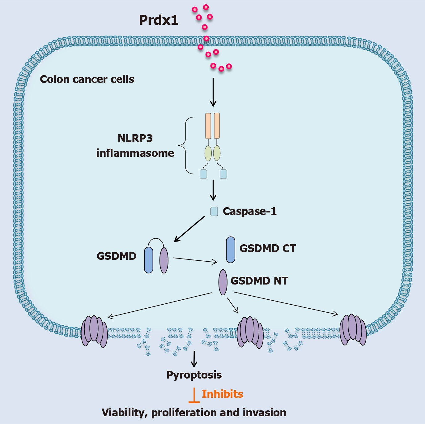
Figure 8 Schematic diagram illustrates peroxiredoxin 1 as a critical damage-associated molecular pattern to inhibit the malignant biological behavior of colon cancer cells.
Extracellular peroxiredoxin 1 induces pyroptosis of colon cancer cells by activating the NLRP3 inflamma
- Citation: He Y, Liu J, Zhou N, Xie LX, Jiang YF, Chen CL. Peroxiredoxin 1 inhibits tumorigenesis by activating the NLRP3/GSDMD pathway to induce pyroptosis of colorectal cancer cells. World J Gastroenterol 2025; 31(36): 111557
- URL: https://www.wjgnet.com/1007-9327/full/v31/i36/111557.htm
- DOI: https://dx.doi.org/10.3748/wjg.v31.i36.111557


