©The Author(s) 2025.
World J Gastroenterol. Mar 28, 2025; 31(12): 98174
Published online Mar 28, 2025. doi: 10.3748/wjg.v31.i12.98174
Published online Mar 28, 2025. doi: 10.3748/wjg.v31.i12.98174
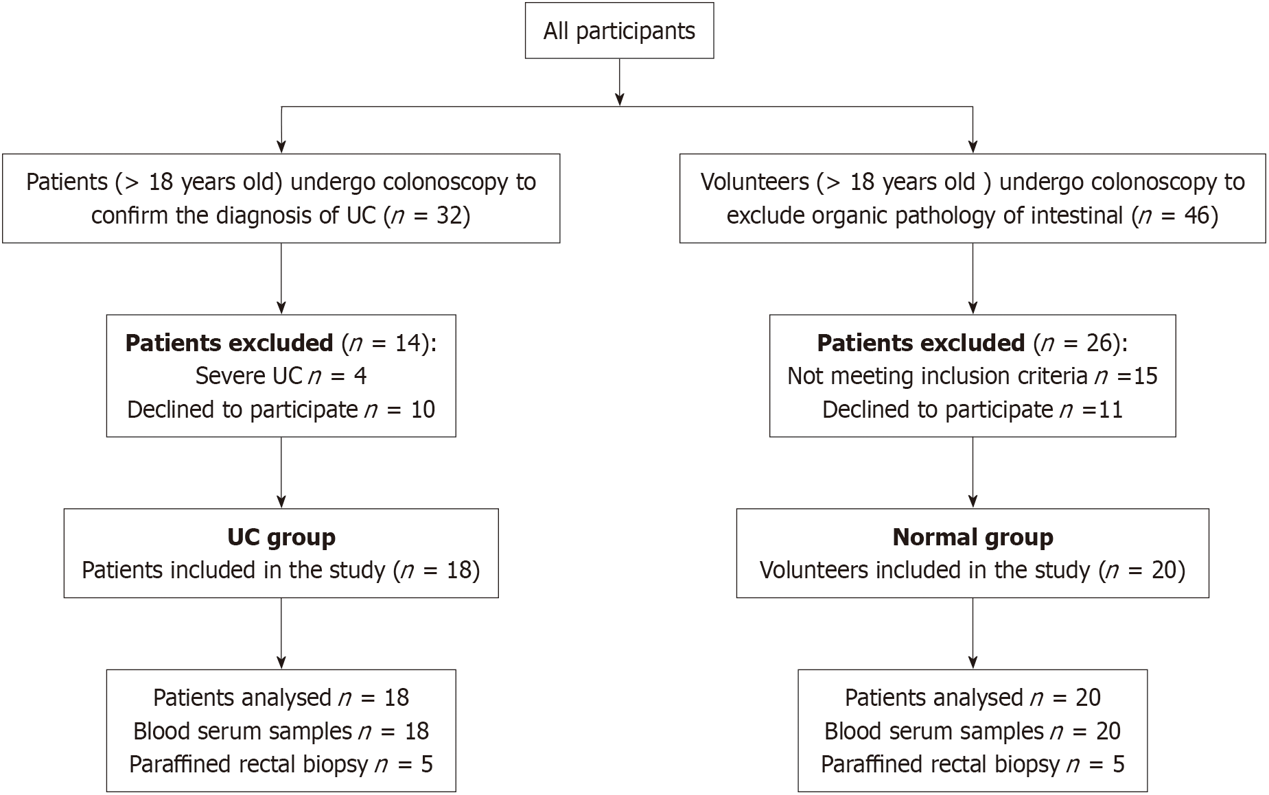
Figure 1 The screening process for participants in this study.
32 patients who met the diagnosis of ulcerative colitis were included, of which 4 patients belonged to severe ulcerative colitis and 10 patients refused to participate. Finally, total of 18 patients completed this study: 46 healthy volunteers were screened, of which 15 did not meet the inclusion criteria, 11 refused to participate, and 20 volunteers completed the experiment. UC: Ulcerative colitis.
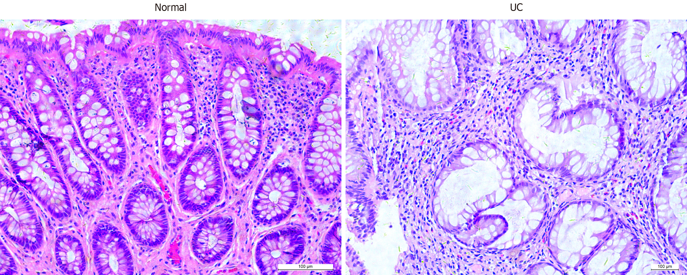
Figure 2 Histopathological manifestations of healthy controls and ulcerative colitis patients.
The number of glands in the colonic mucosal tissue of ulcerative colitis patients was reduced, crypt structures were distorted, twisted and branched, and the population of inflammatory cells in the lamina propria were increased compared with healthy control (magnification: 200 ×). UC: Ulcerative colitis.
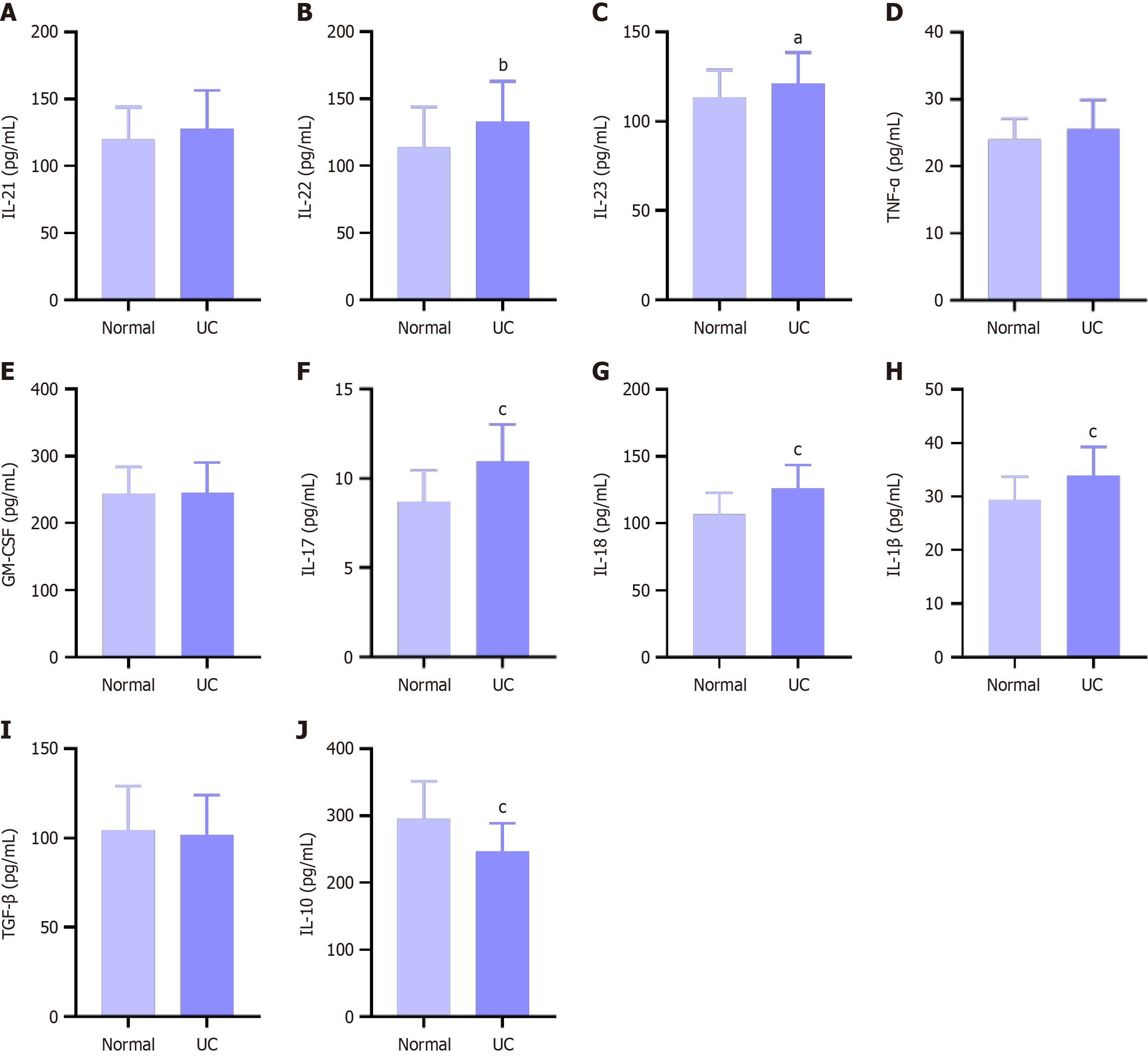
Figure 3 Levels of serum inflammatory factors in healthy controls and ulcerative colitis patients.
A: Interleukin-21 (IL-21); B: IL-22; C: IL-23 were significantly elevated; D: tumor necrosis factor α; E: Granulocyte-macrophage colony stimulating factor; F: IL-17; G: IL-18; H: IL-1β were significantly elevated; I: Anti-inflammatory cytokines transforming growth factor β were decreased; J: Anti-inflammatory cytokines IL-10 were decreased. aP < 0.05, bP < 0.01, cP < 0.001 vs healthy control. UC: Ulcerative colitis; IL: Interleukin; TNF-α: Tumor necrosis factor α; GM-CSF: Granulocyte-macrophage colony stimulating factor; TGF-β: Transforming growth factor β.
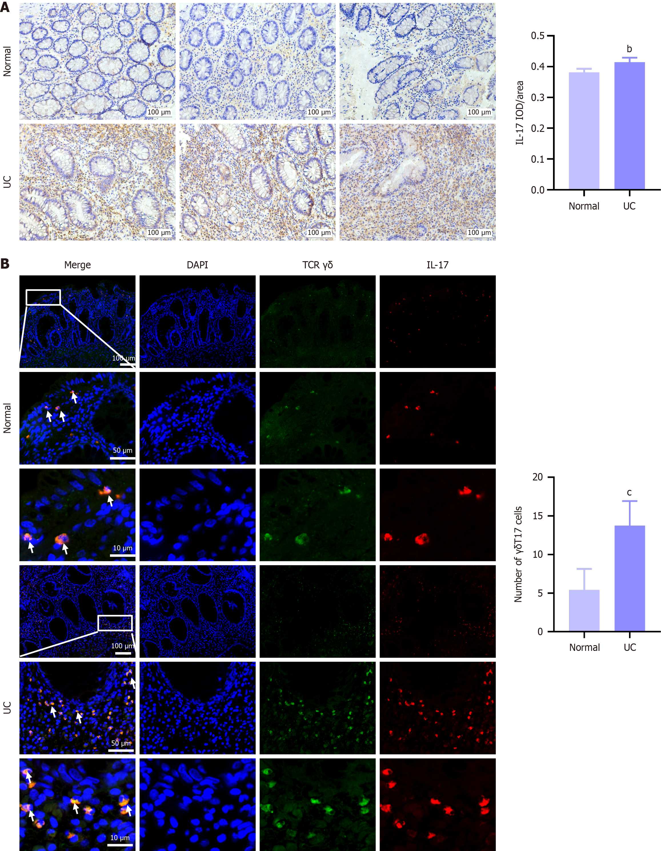
Figure 4 The expression levels of interleukin-17 and γδT17 cells in healthy controls and ulcerative colitis patients.
A: The expression level of interleukin-17 in colonic mucosal tissues of ulcerative colitis patients was markedly elevated. vs healthy control (magnification: 200 ×); B: The expression level of γδT17 cells in colonic mucosal tissues of ulcerative colitis patients was markedly elevated. bP < 0.01, cP < 0.001 vs healthy control (magnification: 40 ×, 200 ×, 400 ×). UC: Ulcerative colitis; IL: Interleukin; TCR: T cell receptor.
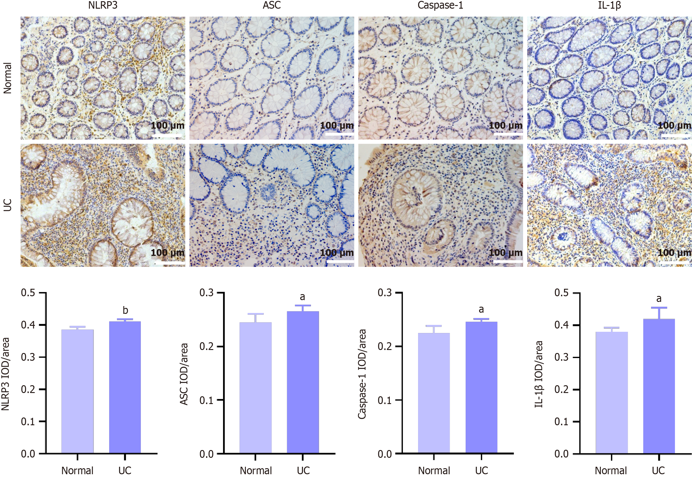
Figure 5 The expression levels of NLR family pyrin domain containing 3, apoptosis-associated speck-like protein, caspase-1 and interleukin-1β in colonic mucosal tissues.
NLR family pyrin domain containing 3, apoptosis-associated speck-like protein, caspase-1, and IL-1β are widely expressed in cytoplasm and their expression levels were elevated to different extents in colonic mucosal tissues of ulcerative colitis patients. aP < 0.05, bP < 0.01 vs healthy control (magnification: 200 ×). NLRP3: NLR family pyrin domain containing 3; ASC: Apoptosis-associated speck-like protein; IL: Interleukin; UC: Ulcerative colitis; IOD: Integrated optical density.
- Citation: Ma J, Wang FY, Tang XD. Involvement of the NLRP3/IL-1β pathway in activation and effector functions of γδT17 cells in patients with ulcerative colitis. World J Gastroenterol 2025; 31(12): 98174
- URL: https://www.wjgnet.com/1007-9327/full/v31/i12/98174.htm
- DOI: https://dx.doi.org/10.3748/wjg.v31.i12.98174


