©The Author(s) 2024.
World J Gastroenterol. Feb 14, 2024; 30(6): 599-606
Published online Feb 14, 2024. doi: 10.3748/wjg.v30.i6.599
Published online Feb 14, 2024. doi: 10.3748/wjg.v30.i6.599
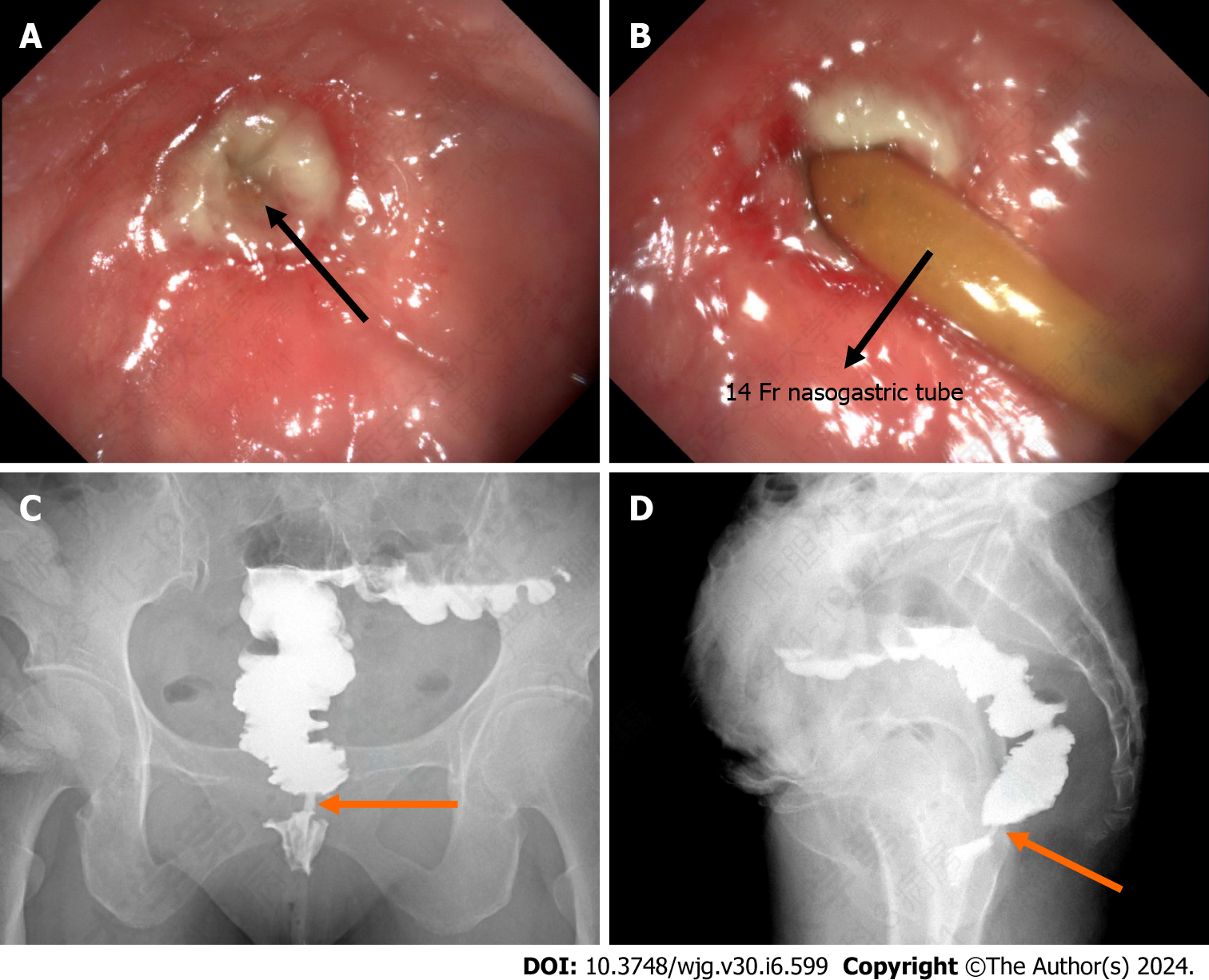
Figure 1 Colonoscopy and colography.
A: Rectal anastomosis stoma; B: Rectal stenosis can only be achieved through a 14 Fr nasogastric tube; C: Anteroposterior colography; D: Lateral colography.
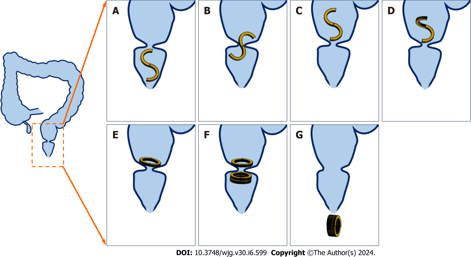
Figure 2 Operation plan.
A: An S-shaped magnetic ring is inserted through the anus; B: The S-shaped magnetic ring passes through the rectal stenosis; C: The S-shaped magnetic ring successfully passed the narrow rectum; D: Deformation of the S-shaped magnetic ring; E: The S-shaped magnetic ring deforms to an O-shape; F: The daughter ring and the parent ring attract each other and compress the narrow section of the rectum; G: Rectum stenosis recanalizes and the magnetic ring is discharged through the anus.
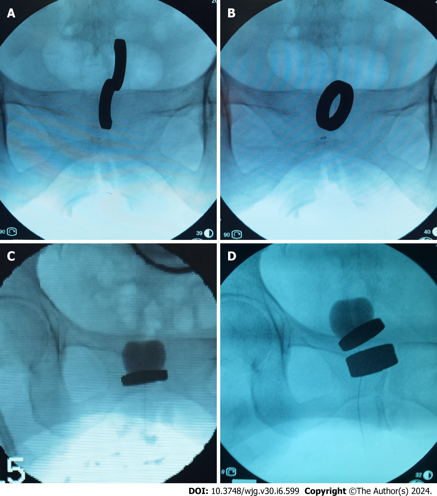
Figure 3 Surgical procedure.
A: The S-shaped magnetic ring passes the narrow rectum; B: The S-shaped magnetic ring becomes O-shaped; C: The catheter assists in adjusting the magnet position; D: The daughter ring and the parent ring attract each other and compress the narrow section of the rectum.
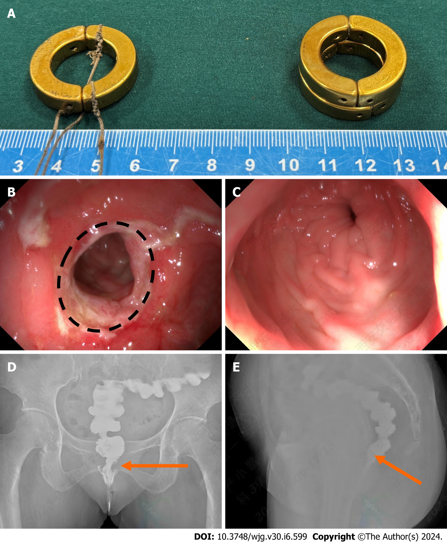
Figure 4 Postoperative anastomosis.
A: Daughter and parent magnetic rings expelled from the body; B: Rectal anastomosis stoma; C: The colonoscope smoothly passes the stenosis; D: Anteroposterior colography; E: Lateral colography.
- Citation: Zhang MM, Sha HC, Qin YF, Lyu Y, Yan XP. Y–Z deformable magnetic ring for the treatment of rectal stricture: A case report and review of literature. World J Gastroenterol 2024; 30(6): 599-606
- URL: https://www.wjgnet.com/1007-9327/full/v30/i6/599.htm
- DOI: https://dx.doi.org/10.3748/wjg.v30.i6.599


