Copyright
©The Author(s) 2024.
World J Gastroenterol. Jul 28, 2024; 30(28): 3393-3402
Published online Jul 28, 2024. doi: 10.3748/wjg.v30.i28.3393
Published online Jul 28, 2024. doi: 10.3748/wjg.v30.i28.3393
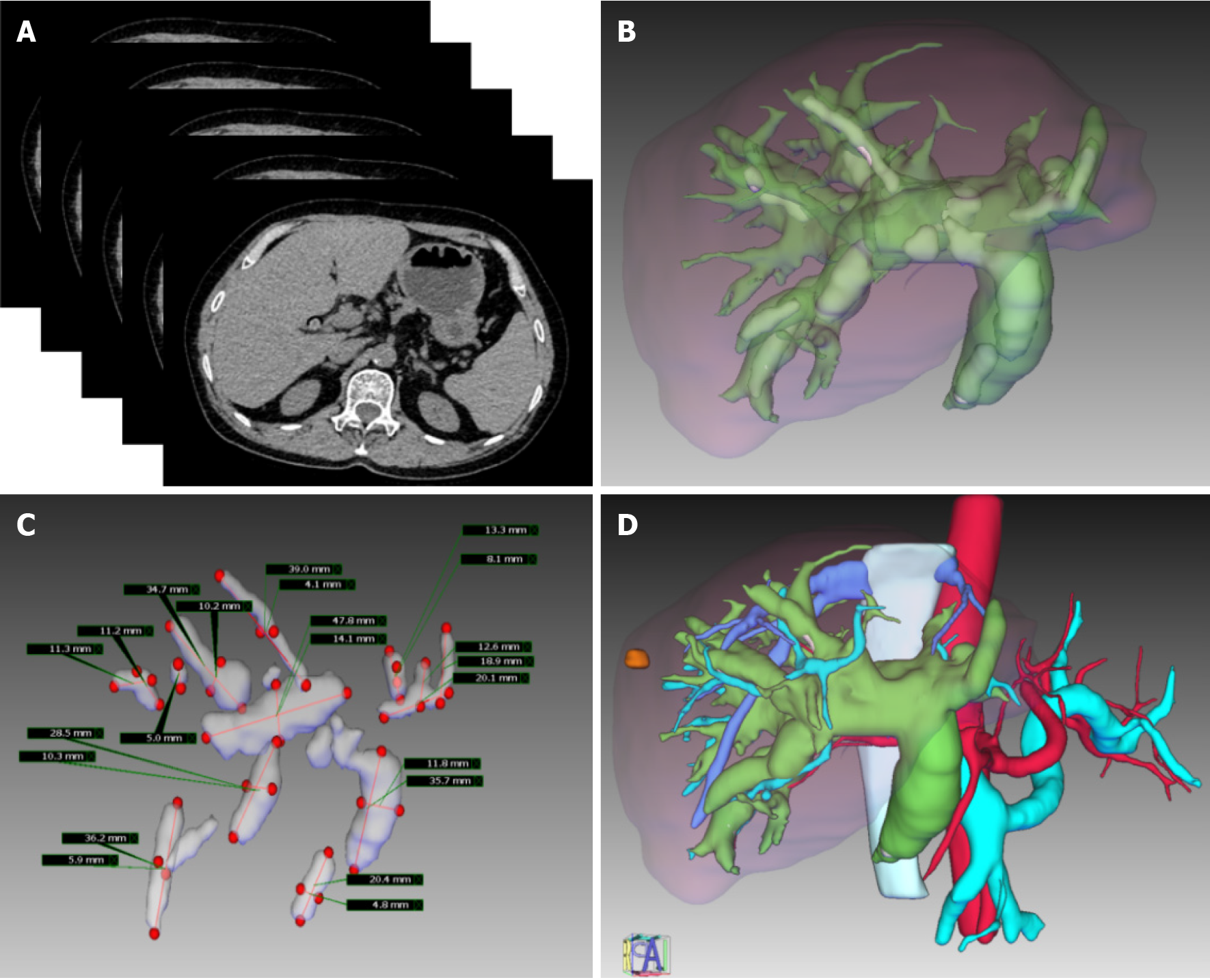
Figure 1 Details of the liver reconstruction model.
A: Iterative computed tomography images; B: The bile duct system involves the clearly visible hepatic bile duct segments, and the hepatolithiasis is labeled in yellow; C: Reconstruction of the location and size of the hepatolithiasis; D: A complete liver reconstruction model with intrahepatic bile duct stones.
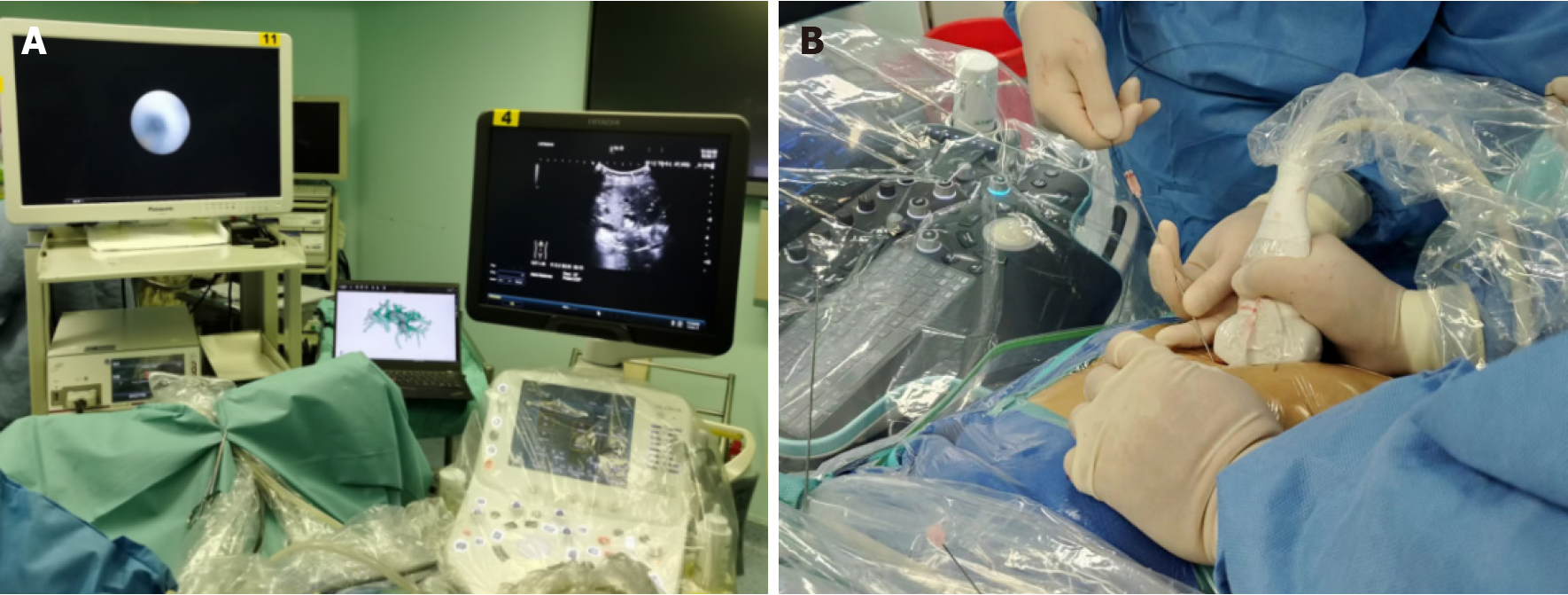
Figure 2 Three-dimensional model combined with real-time B-ultrasonic navigation.
A: Combination of three-dimensional reconstruction and B-ultrasound for guiding one-step percutaneous transhepatic cholangioscopic lithotripsy (PTCSL); B: Using the percutaneous transhepatic one-step biliary fistulation technique for one-step PTCSL.
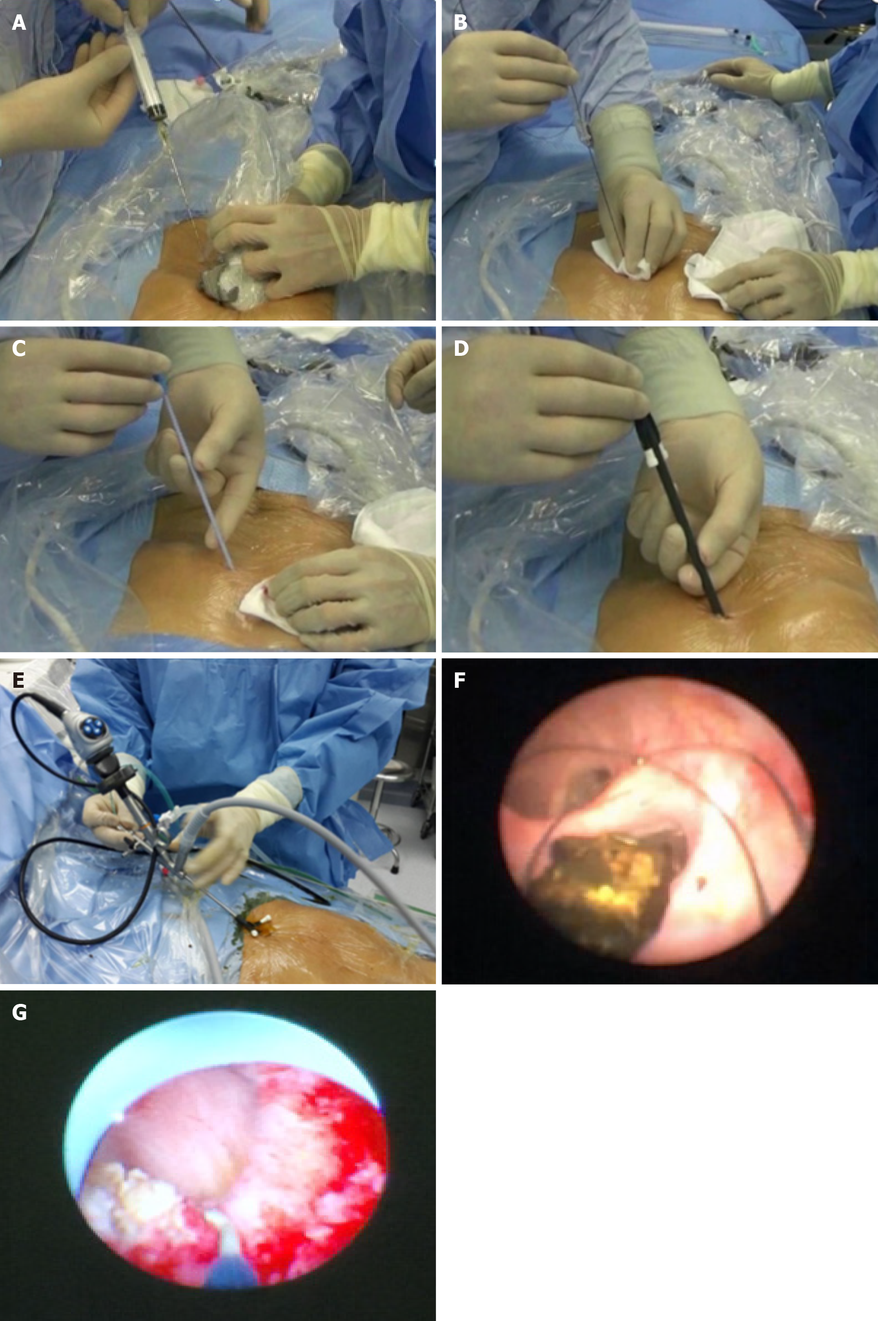
Figure 3 One-step percutaneous transhepatic cholangioscopic lithotripsy using the percutaneous transhepatic one-step biliary fistulation technique.
A: Ultrasound-guided puncture; B: Guidewire insertion; C: Fistula dilation D: Sheath insertion; E: Rigid choledochoscope exploration; F: Stone clearance; G: Stricture resolution using an electric knife.
- Citation: Ye YQ, Cao YW, Li RQ, Li EZ, Yan L, Ding ZW, Fan JM, Wang P, Wu YX. Three-dimensional visualization technology for guiding one-step percutaneous transhepatic cholangioscopic lithotripsy for the treatment of complex hepatolithiasis. World J Gastroenterol 2024; 30(28): 3393-3402
- URL: https://www.wjgnet.com/1007-9327/full/v30/i28/3393.htm
- DOI: https://dx.doi.org/10.3748/wjg.v30.i28.3393


