©The Author(s) 2024.
World J Gastroenterol. Jul 14, 2024; 30(26): 3247-3252
Published online Jul 14, 2024. doi: 10.3748/wjg.v30.i26.3247
Published online Jul 14, 2024. doi: 10.3748/wjg.v30.i26.3247
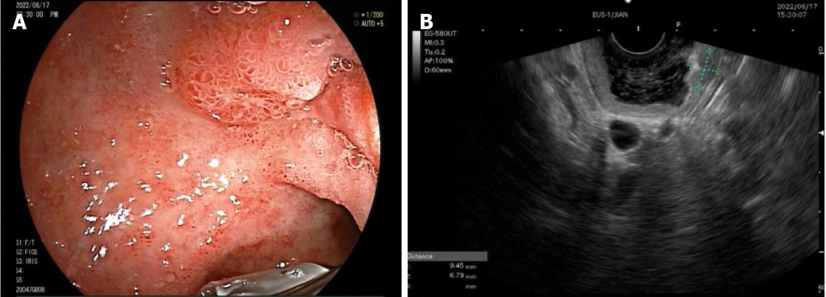
Figure 1 Imaging of lesions in the duodenal bulb via ultrasound endoscopy.
A: Ultrasound endoscopy revealed a hypoechoic focus in the duodenal bulb that was irregularly shaped; B: The lesion originated from the submucosa.
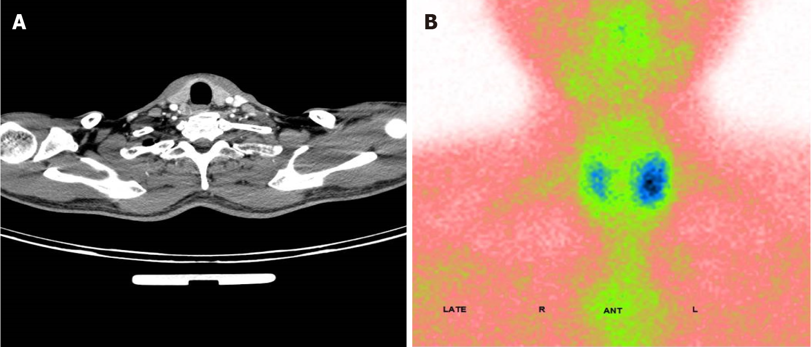
Figure 2 Imaging of lesions of the thyroid and parathyroid glands.
A: Enhanced computerized tomography image showing a blood-rich nodule in the posterior left lobe of the thyroid gland; B: Emission computed tomography revealed a hyperfunctional lesion change in the left lobe of the thyroid gland.
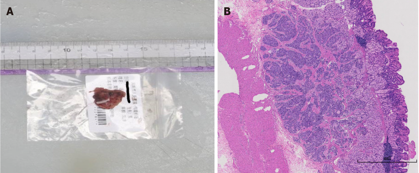
Figure 3 Postoperative pathological analysis of the duodenal bulbous lesion.
A: The postoperative duodenal bulb mass specimen measured approximately 2 cm × 2 cm × 0.7 cm; B: Histopathological analysis findings suggested that the lesion was a neuroendocrine tumor, stage G1.
- Citation: Yuan JH, Luo S, Zhang DG, Wang LS. Early detection of multiple endocrine neoplasia type 1: A case report. World J Gastroenterol 2024; 30(26): 3247-3252
- URL: https://www.wjgnet.com/1007-9327/full/v30/i26/3247.htm
- DOI: https://dx.doi.org/10.3748/wjg.v30.i26.3247


