©The Author(s) 2024.
World J Gastroenterol. Jun 14, 2024; 30(22): 2834-2838
Published online Jun 14, 2024. doi: 10.3748/wjg.v30.i22.2834
Published online Jun 14, 2024. doi: 10.3748/wjg.v30.i22.2834
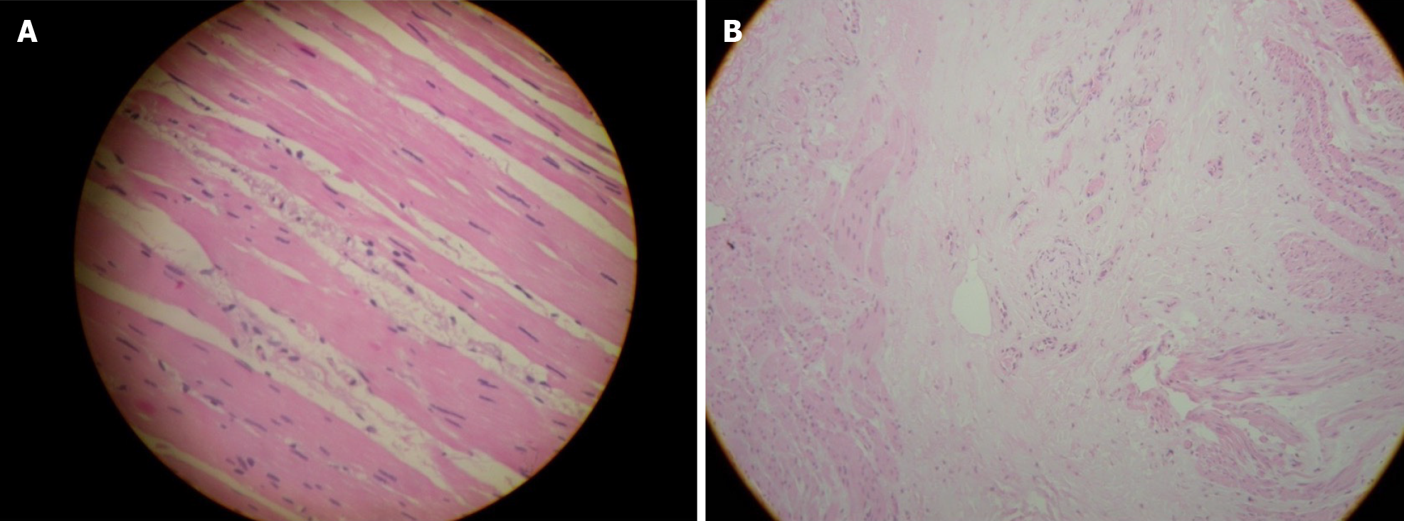
Figure 1 Mild fibrosis, in a patient with achalasia cardia.
A: Mild fibrosis, in a patient with early stage of achalasia cardia (AC); B: Severe fibrosis and muscle atrophy, in a patient with late stage of AC.
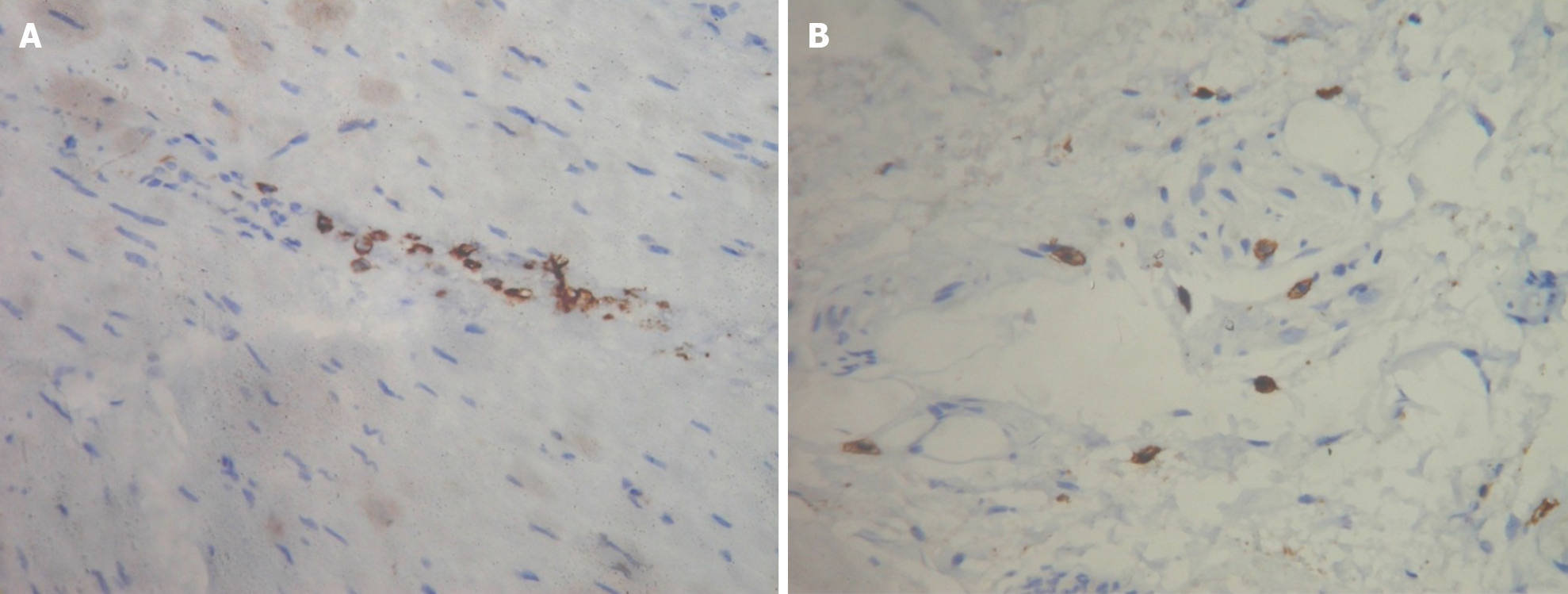
Figure 2 Immunohistochemistry.
A: Lymphocytes seen by CD3 immunohistochemistry; B: Mast cells seen on immunohistochemistry (CD117).
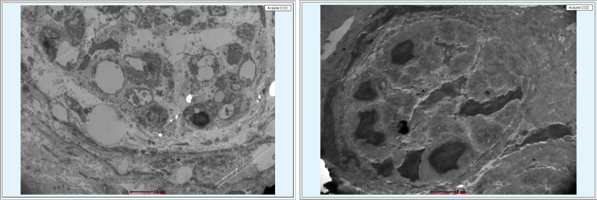
Figure 3 Degenerated pre and post ganglionic nerve cells on electron microscopy.
- Citation: Samarasam I, Joel RK, Pulimood AB. Gastroesophageal reflux following per-oral endoscopic myotomy: Can we improve outcomes? World J Gastroenterol 2024; 30(22): 2834-2838
- URL: https://www.wjgnet.com/1007-9327/full/v30/i22/2834.htm
- DOI: https://dx.doi.org/10.3748/wjg.v30.i22.2834


