Copyright
©The Author(s) 2024.
World J Gastroenterol. Jan 14, 2024; 30(2): 137-145
Published online Jan 14, 2024. doi: 10.3748/wjg.v30.i2.137
Published online Jan 14, 2024. doi: 10.3748/wjg.v30.i2.137
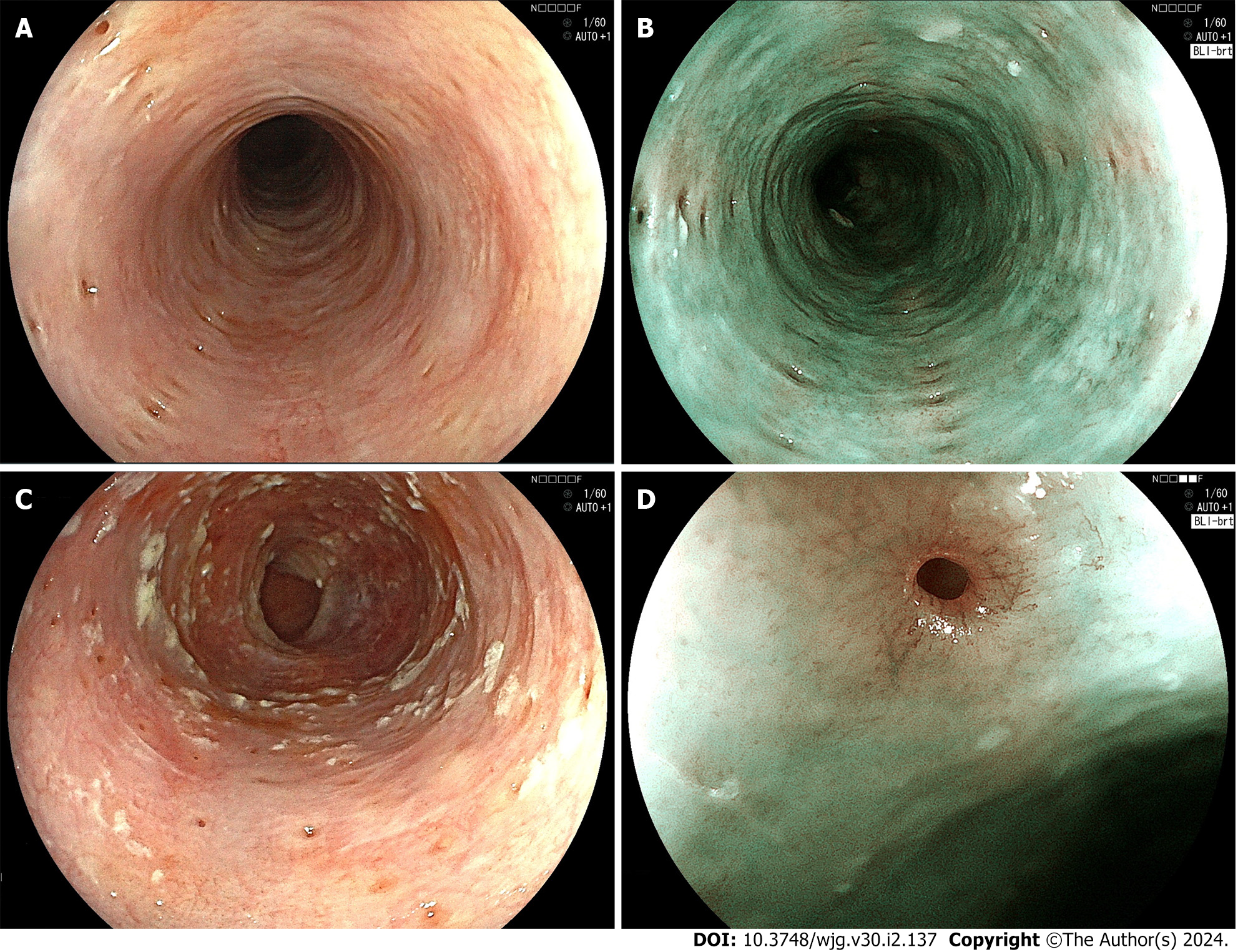
Figure 1 Endoscopic examination.
A: White-light endoscopic image. Multiple small diverticulum-like depressions of the mucosa with a diameter of 1-4 mm are seen. Fujifilm EG-L600ZW; B: Blue laser imaging (BLI)-bright image. The light-brown depressions are longitudinally aligned on the green-colored mucosa. Fujifilm EG-L600ZW; C: White-light endoscopic image. The orifices of pseudodiverticula are covered with whitish, plaque-like material. Fujifilm EG-L600ZW; D: BLI-bright, medium magnifying image. The orifice of the pseudodiverticulum is surrounded by blood vessels arranged in an eyelash-like pattern. Fujifilm EG-L600ZW.
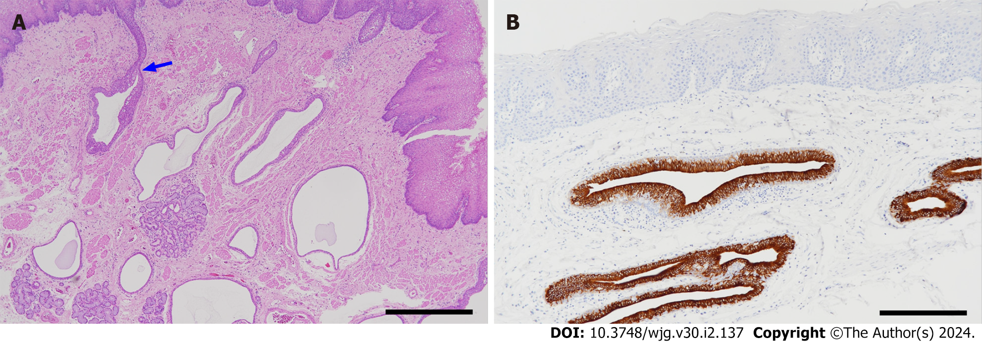
Figure 2 Pathological findings.
A: Histology of esophageal intramural pseudodiverticulosis (hematoxylin-eosin stain). Many cystically dilated ducts of the esophageal glands are seen in the lamina propria mucosae and submucosa. Some of them are continuous with the superficial epithelium (arrow) (scale bar: 1 mm); B: Cytokeratin 7 (CK7) immunostaining. Epithelial cells lining the ducts are positive for CK7. The epithelium of the duct shows hyperplasia and stratification. The esophageal mucosal epithelium (upper part of the figure) is negative for CK7 (scale bar: 200 µm).
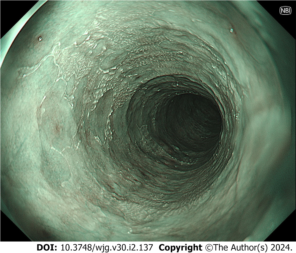
Figure 3 Narrow band imaging shows “epidermization”, which was widely spread in the middle esophagus 2.
5 years after the first endoscopic examination. The extent of epidermization has not markedly changed over a 3-year period. Olympus GIF-XZ1200.
- Citation: Shintaku M. Esophageal intramural pseudodiverticulosis. World J Gastroenterol 2024; 30(2): 137-145
- URL: https://www.wjgnet.com/1007-9327/full/v30/i2/137.htm
- DOI: https://dx.doi.org/10.3748/wjg.v30.i2.137


