Copyright
©The Author(s) 1997.
World J Gastroenterol. Dec 15, 1997; 3(4): 231-233
Published online Dec 15, 1997. doi: 10.3748/wjg.v3.i4.231
Published online Dec 15, 1997. doi: 10.3748/wjg.v3.i4.231
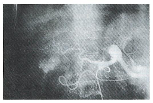
Figure 1 Primary liver cancer of the right lobe, with a hypervascular area diameter of 3 cm before treatment.
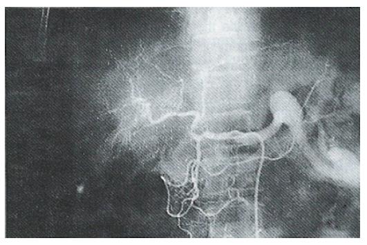
Figure 2 The same case as in Figure 1, showing the tumor stain having disappeared after the 6th treatment.
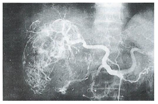
Figure 3 Celiac angiogram of gigantic nodular HCC with 12.
5 cm diameter in the right lobe before treatment.
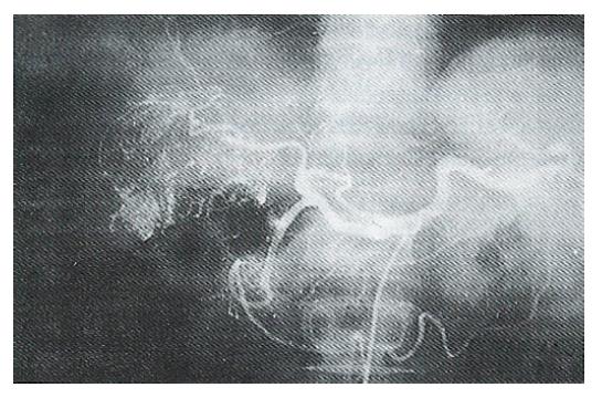
Figure 4 The same case as in Figure 3 after four treatments, showing necrosis in the center of the tumor and reduced presence of new vessels.
- Citation: Liu Q, Jia YC, Tian JM, Wang ZT, Ye H, Yang JJ, Sun F. Comparative study of different interventional therapies for primary liver cancer. World J Gastroenterol 1997; 3(4): 231-233
- URL: https://www.wjgnet.com/1007-9327/full/v3/i4/231.htm
- DOI: https://dx.doi.org/10.3748/wjg.v3.i4.231


