Copyright
©The Author(s) 1997.
World J Gastroenterol. Jun 15, 1997; 3(2): 64-66
Published online Jun 15, 1997. doi: 10.3748/wjg.v3.i2.64
Published online Jun 15, 1997. doi: 10.3748/wjg.v3.i2.64
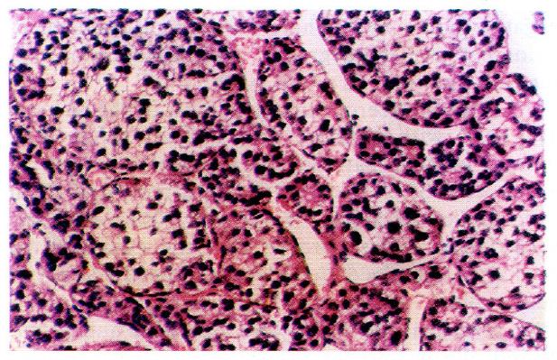
Figure 1 Clear cell hepatocellular carcinoma.
HE × 400.
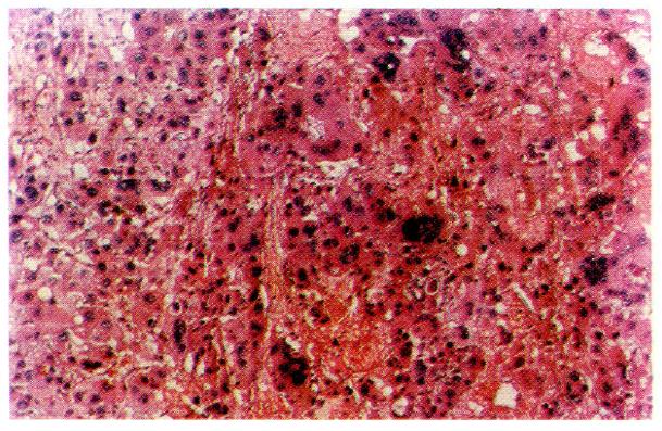
Figure 2 Giant cell hepatocellular carcinoma.
HE × 400.
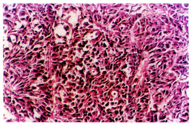
Figure 3 Spindle cell hepatocellular carcinoma.
HE × 400.
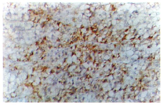
Figure 4 Clear cell hepatocellular carcinoma.
The AFP-positive portion in clear cells was expressed in the periphery of the cytoplasm. Immunohistochemical stains × 400. AFP: Alpha-fetoprotein.
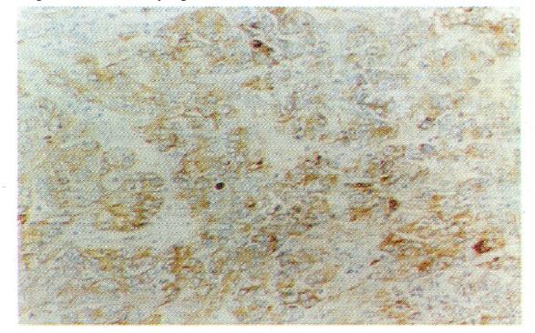
Figure 5 Clear cell hepatocellular carcinoma.
The AAT-positive portion was expressed in the cytoplasm. Immunohistochemical stains × 100. AAT: Alpha-1-antitrypsin.
Figure 6 Clear cell hepatocellular carcinoma.
The EMA-positive portion was expressed in the cancer cell membrane. Immunohistochemical stains × 100. EMA: Epithelial membrane antigen.
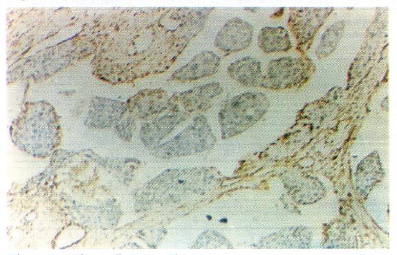
Figure 7 Clear cell hepatocellular carcinoma.
Tests for vimentin expression in cancer cells were negative, but those in the interstitial tissues appear to be positive. Immunohistochemical stains × 100
- Citation: Wu QM, Hu MH, Tan YS. Histopathology and immunohistochemistry of large hepatocellular carcinoma with undetectable or low serum levels of alpha-fetoprotein. World J Gastroenterol 1997; 3(2): 64-66
- URL: https://www.wjgnet.com/1007-9327/full/v3/i2/64.htm
- DOI: https://dx.doi.org/10.3748/wjg.v3.i2.64


