Copyright
©The Author(s) 2023.
World J Gastroenterol. Mar 7, 2023; 29(9): 1427-1445
Published online Mar 7, 2023. doi: 10.3748/wjg.v29.i9.1427
Published online Mar 7, 2023. doi: 10.3748/wjg.v29.i9.1427
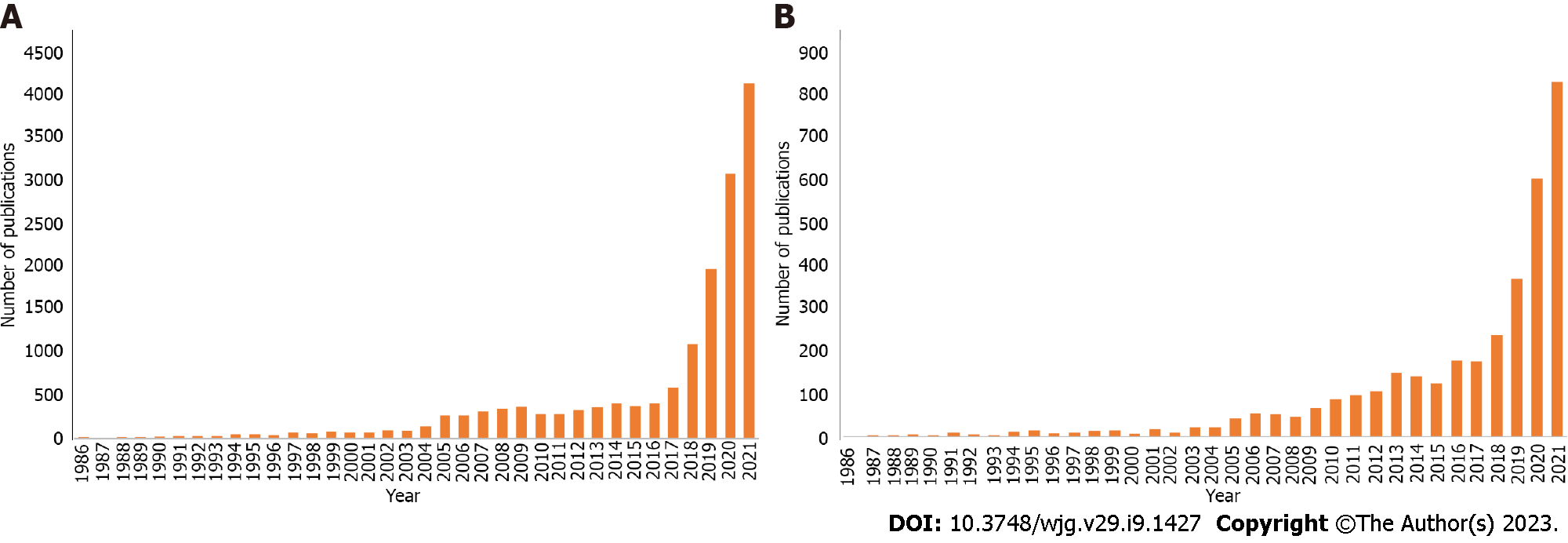
Figure 1 PubMed results by year using the search terms.
A and B: Artificial intelligence radiology (top) and artificial intelligence AND (liver OR pancreas) (bottom).
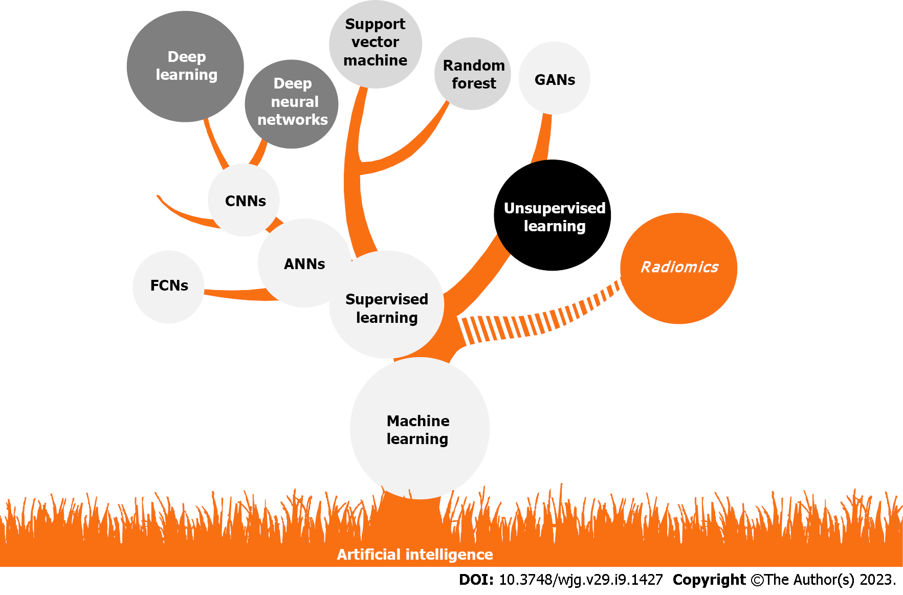
Figure 2 Relation between artificial intelligence and related subdisciplines, neural network architectures, and/or techniques.
ANN: Artificial neural network; FCN: Fully convolutional network; CNN: Convolutional neural network; GAN: Generation adversarial network.
Figure 3 Diagram of a convolutional neural network used for the classification of a focal liver lesion in a computerized tomography image.
HCC: Hepatocellular carcinoma; CT: Computed tomography.
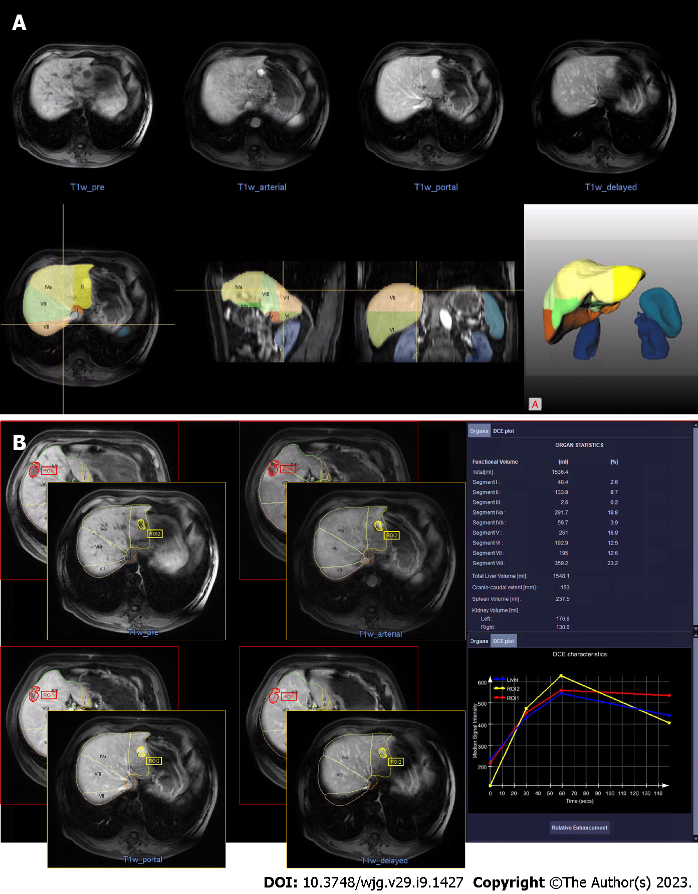
Figure 4 In-house experience on liver assessment with artificial intelligence.
Magnetic resonance studies of a patient with liver focal lesions (liver hemangiomas), processed with the Liver Analysis research application from Siemens Healthcare. A: Automatic segmentation of the whole liver, liver segments, and other abdominal organs; B: Automatic detection, segmentation, and measurement of the two liver hemangiomas.
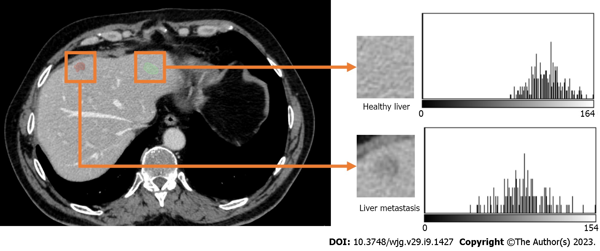
Figure 5 Computerized tomography scan of a 61-year-old male patient with colon carcinoma and liver metastases.
The intensity histograms of regions with and without metastases are different; hence, the first order radiomics features[109], which are based on the intensity histogram will potentially be different.
Figure 6 Sixty-seven-year-old patient with pancreatic carcinoma and liver metastases treated with chemotherapy.
The Digital Oncology Companion (Siemens Healthineers, Germany) artificial intelligence-based prototype automatically segments liver, portal and hepatic vessels, lesions, and surrounding anatomical structures. From left to right: screenshots of the segmented liver, vessels, and lesions; and generated 3D models.
- Citation: Berbís MA, Paulano Godino F, Royuela del Val J, Alcalá Mata L, Luna A. Clinical impact of artificial intelligence-based solutions on imaging of the pancreas and liver. World J Gastroenterol 2023; 29(9): 1427-1445
- URL: https://www.wjgnet.com/1007-9327/full/v29/i9/1427.htm
- DOI: https://dx.doi.org/10.3748/wjg.v29.i9.1427


