©The Author(s) 2023.
World J Gastroenterol. Dec 14, 2023; 29(46): 6049-6059
Published online Dec 14, 2023. doi: 10.3748/wjg.v29.i46.6049
Published online Dec 14, 2023. doi: 10.3748/wjg.v29.i46.6049
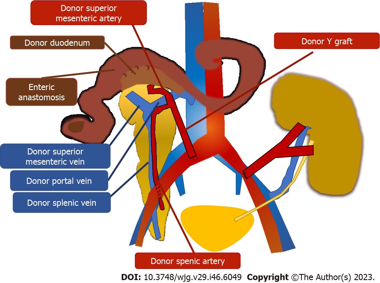
Figure 1 Schematic imaging representation of simultaneous pancreas and kidney transplantation.
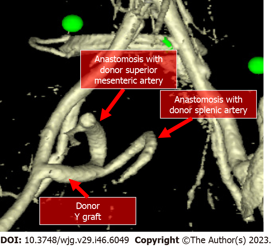
Figure 2 Volume rendering reformat of computed tomography images in the arterial phase demonstrates the Y conduit of donor iliac artery to the superior mesenteric and splenic arteries of the graft.
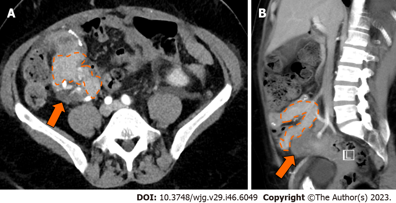
Figure 3 A 51-year-old woman, one month after simultaneous kidney-pancreatic transplantation is admitted to the emergency department for abdominal pain; final diagnosis at biopsy of the pancreatic graft was rejection.
A: Contrast enhanced computed tomography in the axial plane shows a minimally inhomogeneous pancreatic graft (dotted orange line) with peripancreatic fluid (arrows) posteriorly to the graft; B: Sagittal plane of the same contrast enhanced computes tomography.
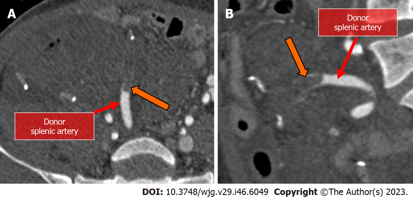
Figure 4 A 46-year-old woman, few days after surgery had low prothrombin time; final diagnosis was graft arterial thrombosis.
A: Contrast enhanced computed tomography in the angiographic phase demonstrated arterial thrombosis of the donor splenic artery. In this patient, the transplanted pancreas was explanted thereafter; B: Coronal plane of the same contrast enhanced computed tomography in the angiographic phase.
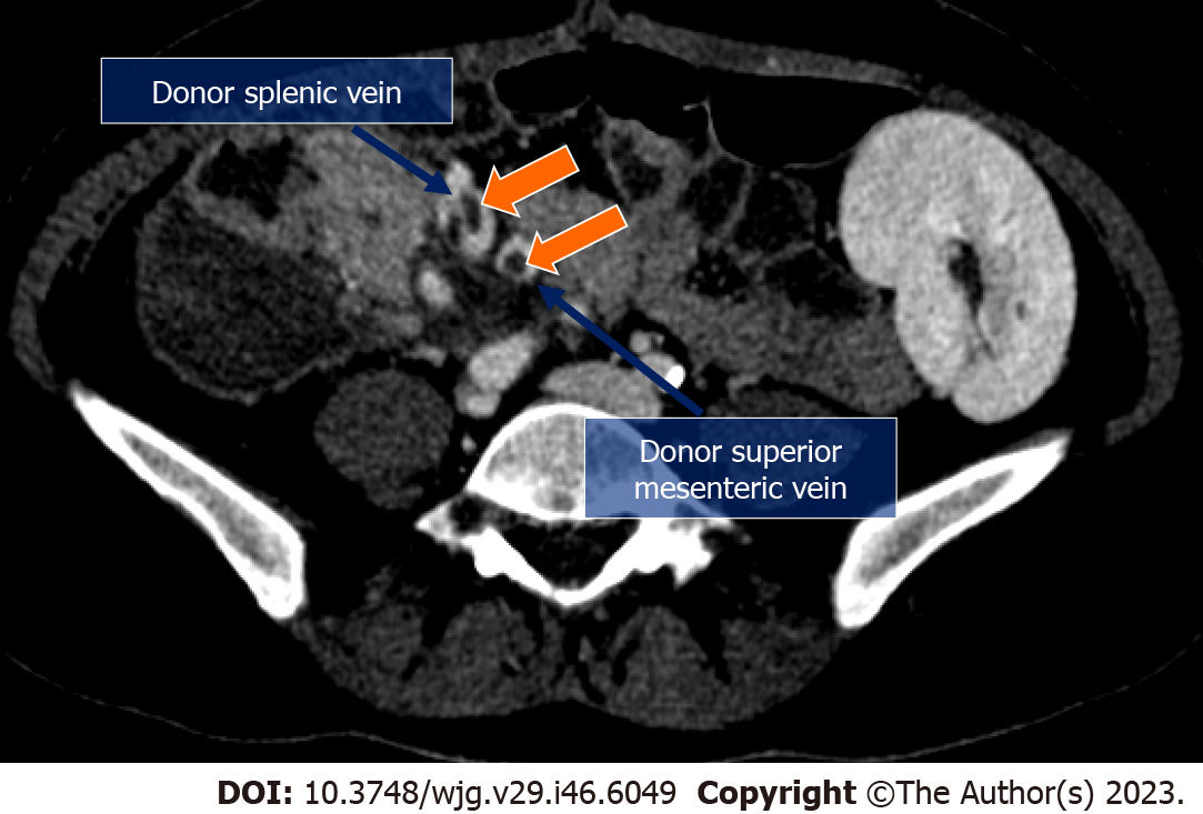
Figure 5 A 35-year-old woman is admitted to the emergency department for abdominal pain; final diagnosis was venous thrombosis.
Contrast enhanced computed tomography in the venous phase demonstrates venous thrombi (orange arrows) in the donor splenic vein and in the donor superior mesenteric vein.
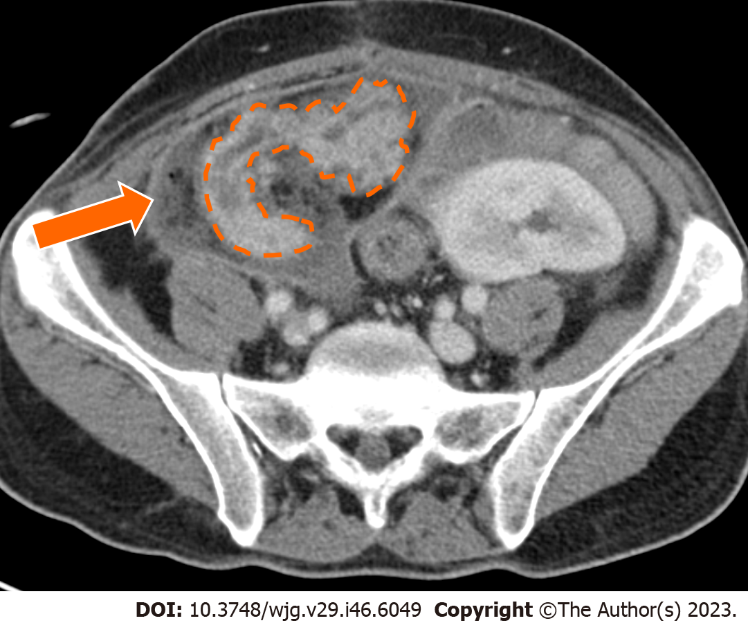
Figure 6 A 35-year-old man is admitted to the emergency department presenting hyperpyrexia and abdominal tenderness; final diagnosis was edematous pancreatitis with acute peripancreatic fluid collection.
Contrast enhanced computed tomography in the axial plane shows inhomogeneous transplanted pancreas (dotted orange lines) with slightly enlarged Wirsung duct and acute peripancreatic fluid collection (arrow) with thick wall and fat stranding surrounding the transplanted pancreas. A diagnosis of edematous pancreatitis was made.
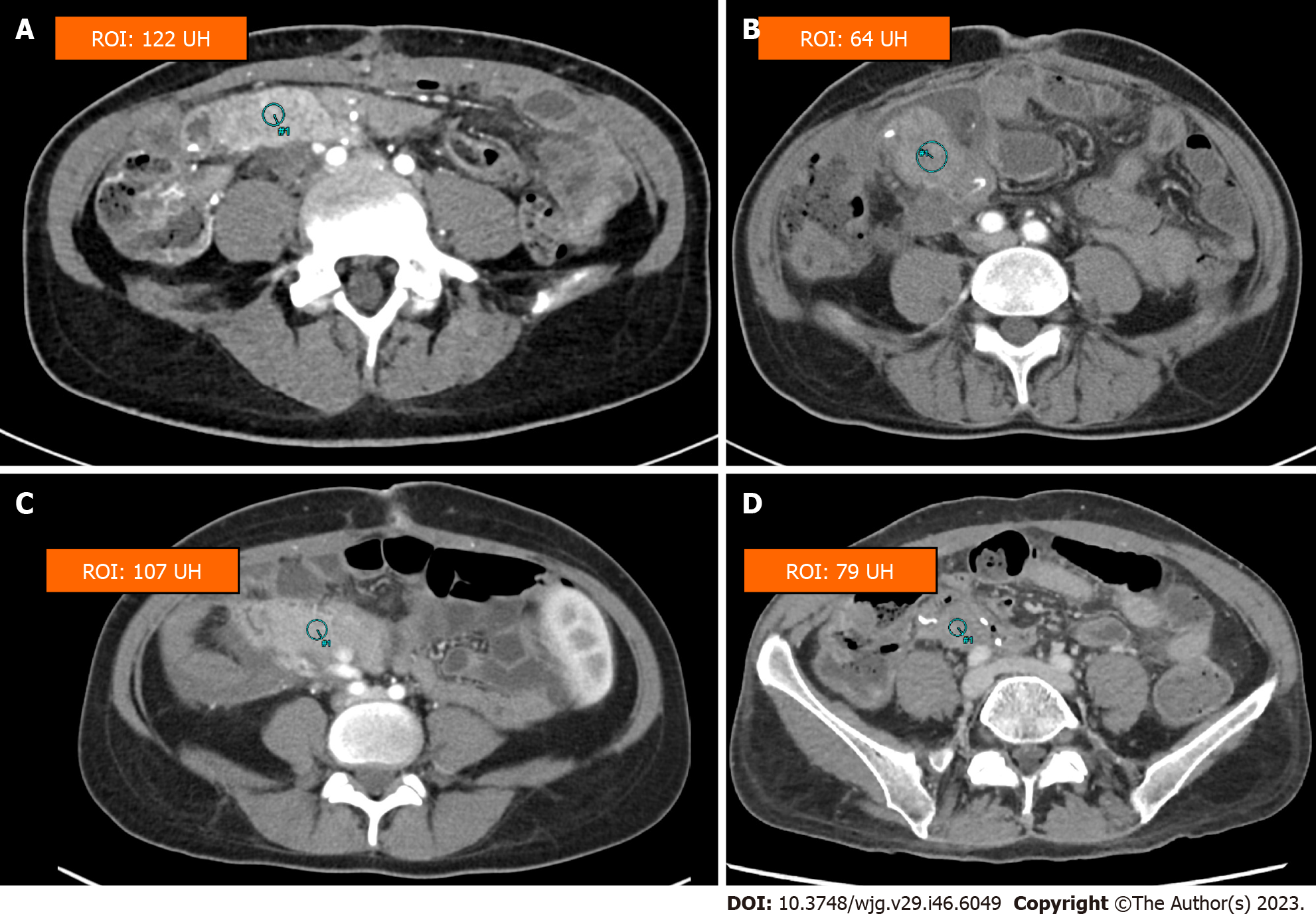
Figure 7 Contrast enhanced computed tomography in four different patients after pancreatic transplantation.
A region of interest is drawn in the pancreas in the four patients. A: Normal pancreatic parenchyma; B: Inflammatory pancreatitis; C: Decreased parenchymal enhancement from venous thrombosis; D: Decreased parenchymal enhancement from arterial thrombosis.
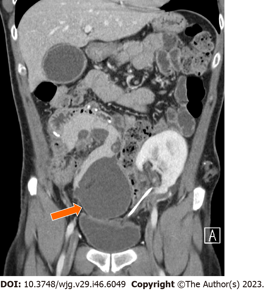
Figure 8 A 35-year-old man after four weeks from edematous pancreatitis.
Contrast enhanced computed tomography in the coronal plane shows an encapsulated fluid collection (arrow) of homogeneously low attenuation surrounded by a well-defined enhancing wall consistent with pancreatic pseudocyst.
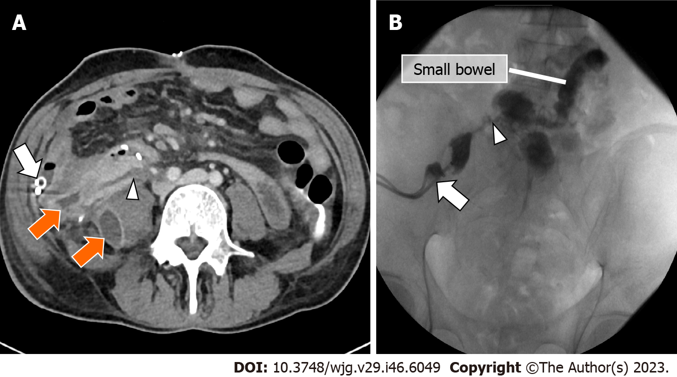
Figure 9 A 43-year-old man admitted to the emergency department for hyperpyrexia and abdominal tenderness; final diagnosis was enteric leakage.
A: Contrast enhanced CT demonstrated the presence of an enteric leakage as a fistulous tract (arrowhead) from the small bowel at the level of the duodenojejunostomy, which resulted into fluid collections (orange arrows). Abdominal drainage (white arrow) was inserted and the analysis of the fluid was consistent with the diagnosis of enteric leakage; B: Fluoroscopic image demonstrate the presence of an enteric leakage as a fistulous tract (arrowhead) from the small bowel at the level of the duodenojejunostomy. Abdominal drained is also evident (white arrow).
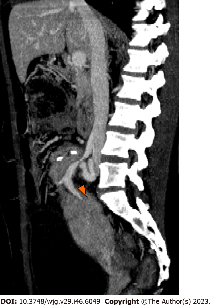
Figure 10 A 42-year-old woman admitted in the hospital for elevation of pancreatic amylase 6 mo after transplant; final diagnosis was venous stenosis.
Contrast enhanced computed tomography in the sagittal plane with Maximum Intensity Projection reconstruction demonstrates venous stenosis (arrowhead) at the anastomotic site. The pancreatic graft is enlarged with inhomogeneous enhancement which likely reflects graft dysfunction.
- Citation: D'Alessandro C, Todisco M, Di Bella C, Crimì F, Furian L, Quaia E, Vernuccio F. Surgical complications after pancreatic transplantation: A computed tomography imaging pictorial review. World J Gastroenterol 2023; 29(46): 6049-6059
- URL: https://www.wjgnet.com/1007-9327/full/v29/i46/6049.htm
- DOI: https://dx.doi.org/10.3748/wjg.v29.i46.6049


