©The Author(s) 2023.
World J Gastroenterol. Nov 28, 2023; 29(44): 5935-5944
Published online Nov 28, 2023. doi: 10.3748/wjg.v29.i44.5935
Published online Nov 28, 2023. doi: 10.3748/wjg.v29.i44.5935
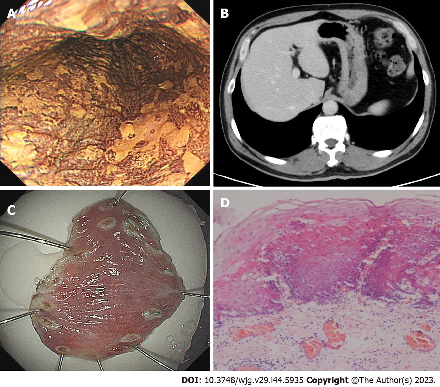
Figure 1 The first gastroendoscopy examination in August 2020.
A: After iodine staining, the esophageal mucosa was scattered in patchy light-stained areas, ranging from 0.2 cm to 0.4 cm, and a 2.0 cm × 1.0 cm, superficial, flat lesion (30 cm from the incisor teeth) did not stain, whereas the pink areas were positive; B: Computed tomography showed no abnormal thickening in the gastrointestinal wall; C: Endoscopic submucosal dissection was performed 1 wk after gastroendoscopy; D: Postoperative pathology revealed moderate to severe dysplasia of squamous epithelium.
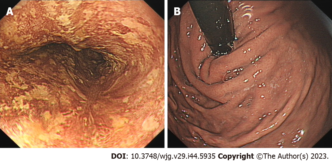
Figure 2 Re-examination of the patient 7 mo after endoscopic submucosal dissection in March 2021.
A: Gastroendoscopy revealed a 1.0 cm × 2.0 cm scar on the right posterior wall of the esophagus, 30 cm from the incisor teeth. Iodine staining revealed that the esophageal mucosa was scattered in the patchy, lightly stained area, indicated by the pink sign (-); B: The mucosa of gastric fundus and cardia was smooth without abnormality.
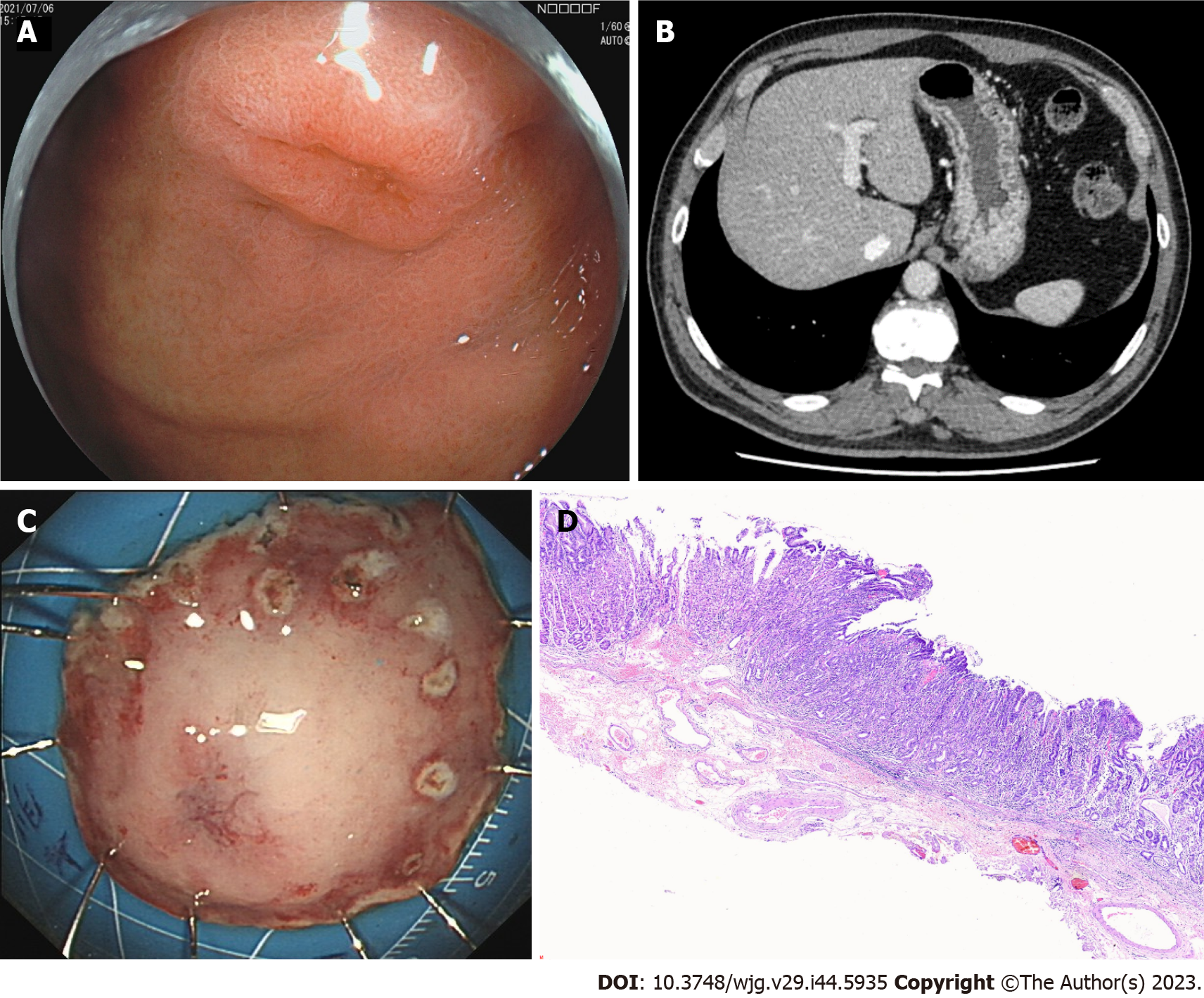
Figure 3 The patient was re-examined for the second time 1 year after endoscopic submucosal dissection in July 2021.
A: Gastroendoscopy revealed a 0.6 cm × 0.8 cm IIa + IIc lesion near the anterior wall of the gastric fundus near the cardia. B: Computed tomography showed no abnormal thickening in the gastrointestinal wall; C: The patient underwent Endoscopic submucosal dissection of the fundus lesion; D: Postoperative pathology revealed moderate to severe dysplasia of gastric mucous.
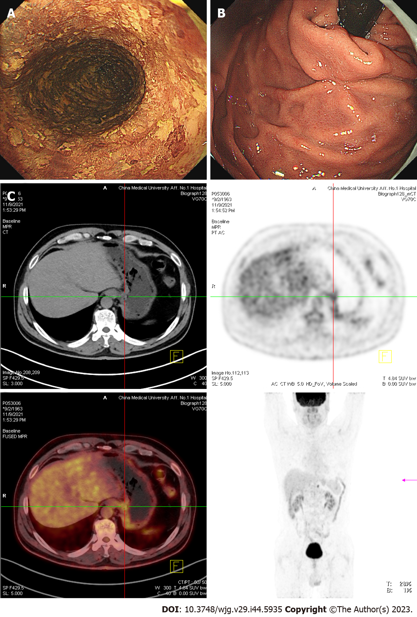
Figure 4 In November 2021, the patient underwent the third re-examination 15 mo after the first endoscopic submucosal dissection.
A: The entire esophagus showed patchy, lightly stained areas after iodine staining in gastroendoscopy. B: The mucosa of the gastric fundus and cardia was smooth except for the scar after Endoscopic submucosal dissection operation, and no abnormality was noted on gastroendoscopy. C: Computed tomography (CT) revealed no abnormal thickening in the gastrointestinal wall. However, positron emission tomography-CT revealed that the metabolism of the lower esophagus and stomach cardia wall slightly increased. A lymph node was noted in the retroperitoneal area with high-density shadow and increased metabolism.
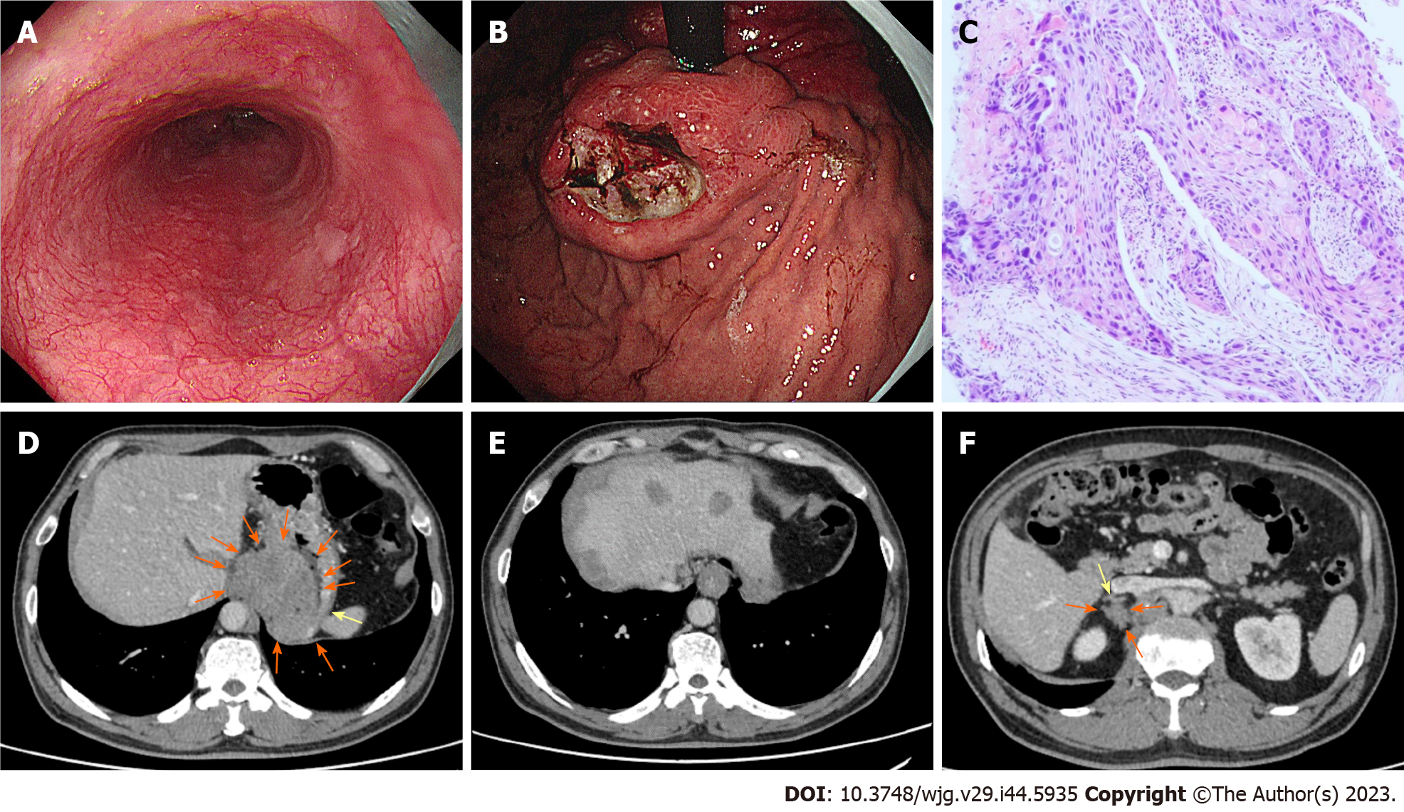
Figure 5 In November 2022, the patient underwent the fourth endoscopy re-examination.
A: Esophageal mucosa was smooth, and no advanced cancer was noted; B: Circumferential submucosal tumor-like swelling was observed in the cardia and stomach body, along with local ulceration and the formation of multiple ulcers; C: Biopsy of cardia revealed squamous-cell carcinoma; D: Computed tomography (CT) showed cardia tumor; E: CT revealed multiple metastatic tumors in the liver; F: CT showed metastatic tumors in the right adrenal gland.
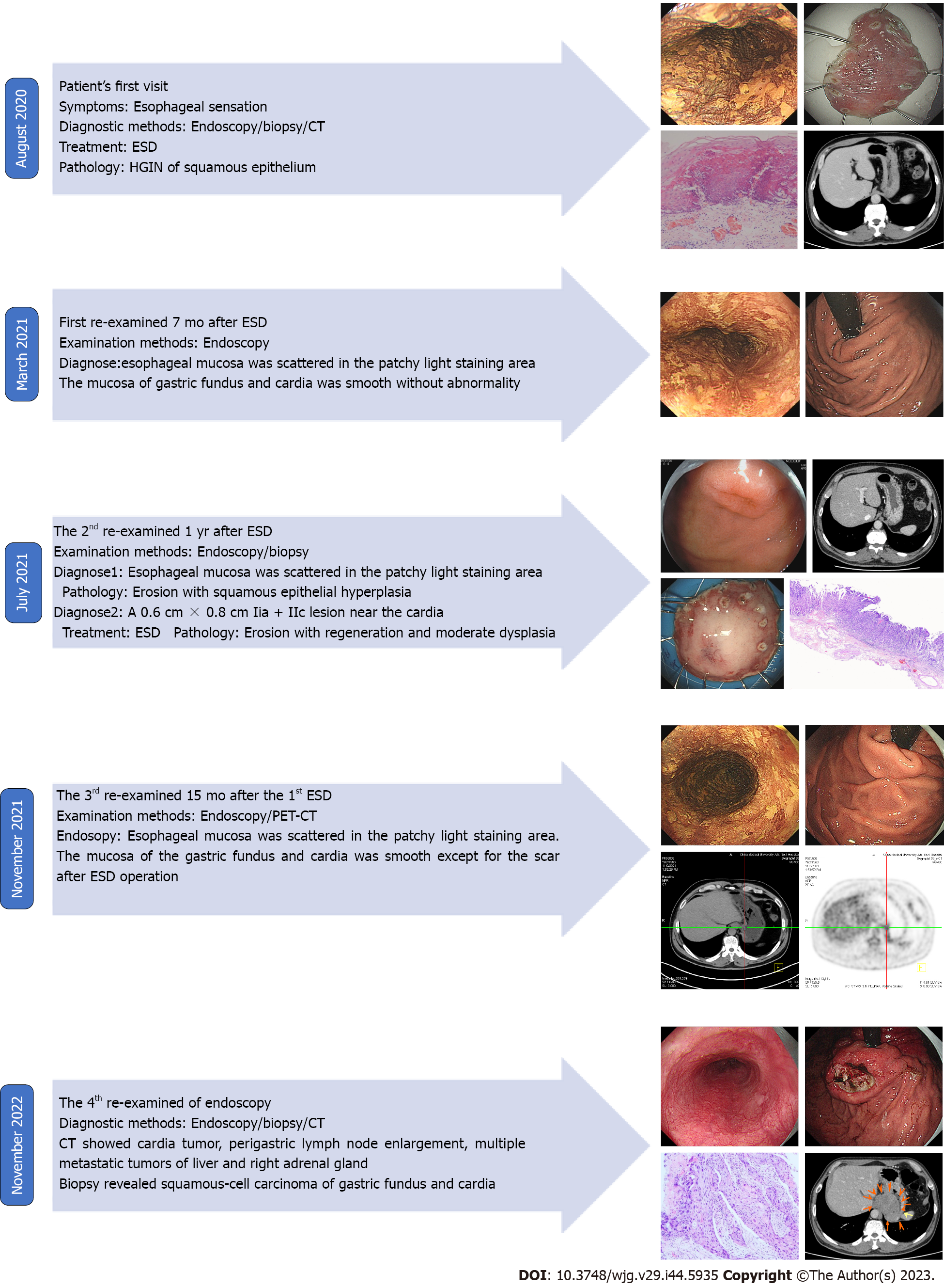
Figure 6 Summary of disease development and treatment of the present case.
- Citation: Yang MQ, Sun MJ, Zhang HJ. Mucosal esophageal carcinoma following endoscopic submucosal dissection with giant gastric metastasis: A case report and review of literature. World J Gastroenterol 2023; 29(44): 5935-5944
- URL: https://www.wjgnet.com/1007-9327/full/v29/i44/5935.htm
- DOI: https://dx.doi.org/10.3748/wjg.v29.i44.5935


