Copyright
©The Author(s) 2023.
World J Gastroenterol. Aug 21, 2023; 29(31): 4809-4814
Published online Aug 21, 2023. doi: 10.3748/wjg.v29.i31.4809
Published online Aug 21, 2023. doi: 10.3748/wjg.v29.i31.4809
Figure 1 Clinical features of the female patient with solitary rectal ulcer syndrome.
A: Colonoscopy image upon her admission; B: Chromoendoscopy result upon her admission using the Indicarmine dyeing; C: Ultrasonic endoscopy was used for solitary rectal ulcer observation; D: Pathological results of the solitary rectal ulcer; E: Imaging of the solitary rectal ulcer, using the intestine computed tomography enhancement; F: Colonoscopy image upon her reexamination after therapy.
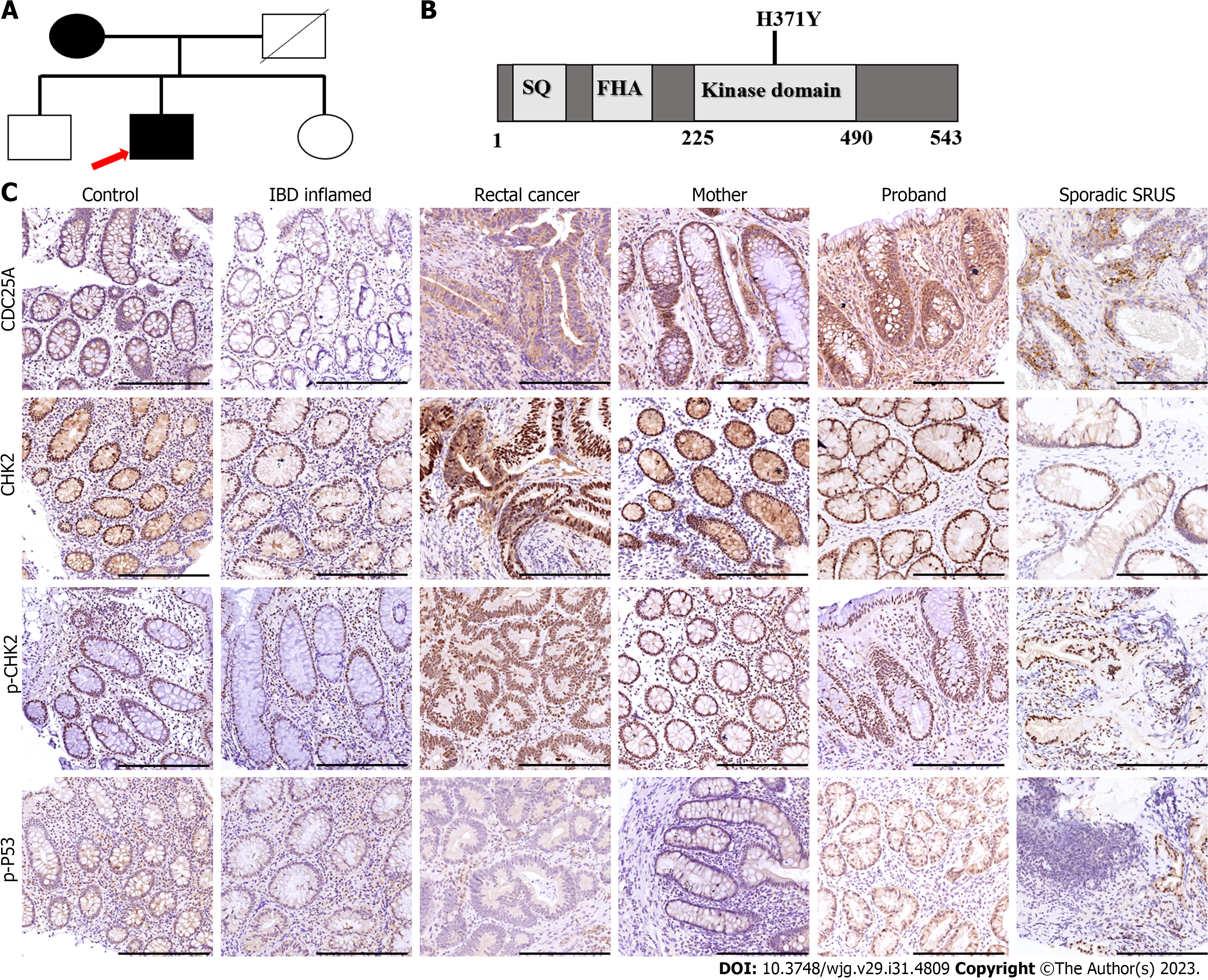
Figure 2 Genetic background of the patients with solitary rectal ulcer syndrome patients in a mother-son relationship.
A: Family tree of the familial patients with solitary rectal ulcer syndrome. Squares indicate male family members; circles indicate female family members; black indicate affected patients; white indicate healthy family members; slashes indicate deceased family members; arrow indicate the first diagnosed patient of the family; B: Schematic diagram of the CHEK2 protein; C: The differential expressions of CHEK2 and the downstream genes among the healthy control, and patients with inflammatory bowel diseases, rectal cancer, and solitary rectal ulcer syndrome, using the immunohistochemical staining; scale bars, 100 μm. IBD: Inflammatory bowel diseases; SRUS: Solitary rectal ulcer syndrome.
- Citation: He CC, Wang SP, Zhou PR, Li ZJ, Li N, Li MS. Inherited CHEK2 p.H371Y mutation in solitary rectal ulcer syndrome among familial patients: A case report. World J Gastroenterol 2023; 29(31): 4809-4814
- URL: https://www.wjgnet.com/1007-9327/full/v29/i31/4809.htm
- DOI: https://dx.doi.org/10.3748/wjg.v29.i31.4809


