©The Author(s) 2022.
World J Gastroenterol. Feb 14, 2022; 28(6): 675-682
Published online Feb 14, 2022. doi: 10.3748/wjg.v28.i6.675
Published online Feb 14, 2022. doi: 10.3748/wjg.v28.i6.675
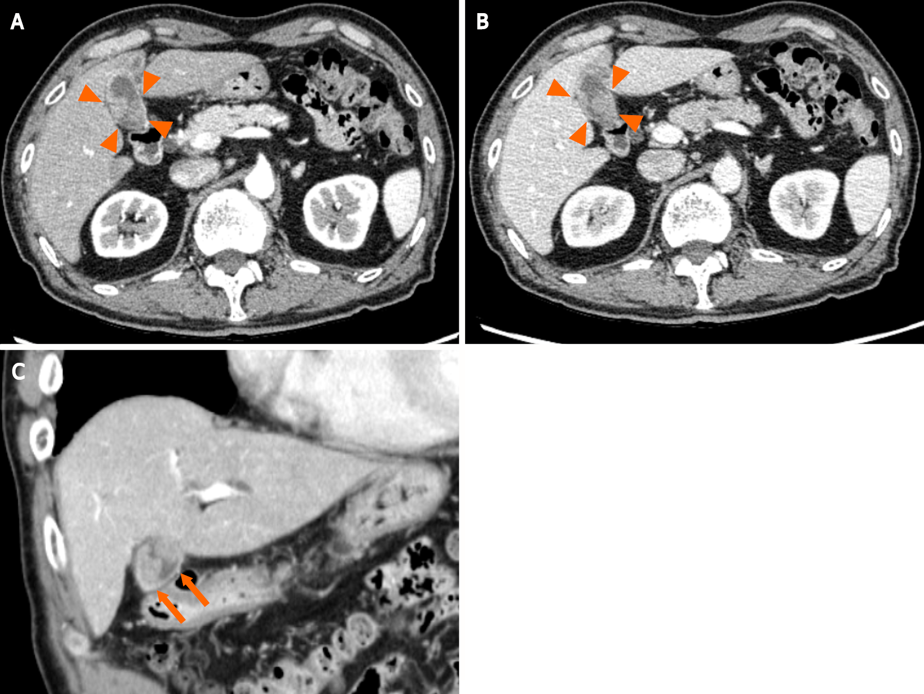
Figure 1 A contrast-enhanced abdominal computed tomography scan shows two irregular and highly contrast-enhanced masses (arrowheads and arrow) at the neck and body of the gallbladder as well as periportal lymph node enlargement, which is consistent with gallbladder cancer lymph node metastasis.
A: Axial section image in the early phase showing neck of the gallbladder; B: Axial section image in the delayed phase showing neck of the gallbladder in the delayed phase; C: Coronal sectional image showing body of the gallbladder.
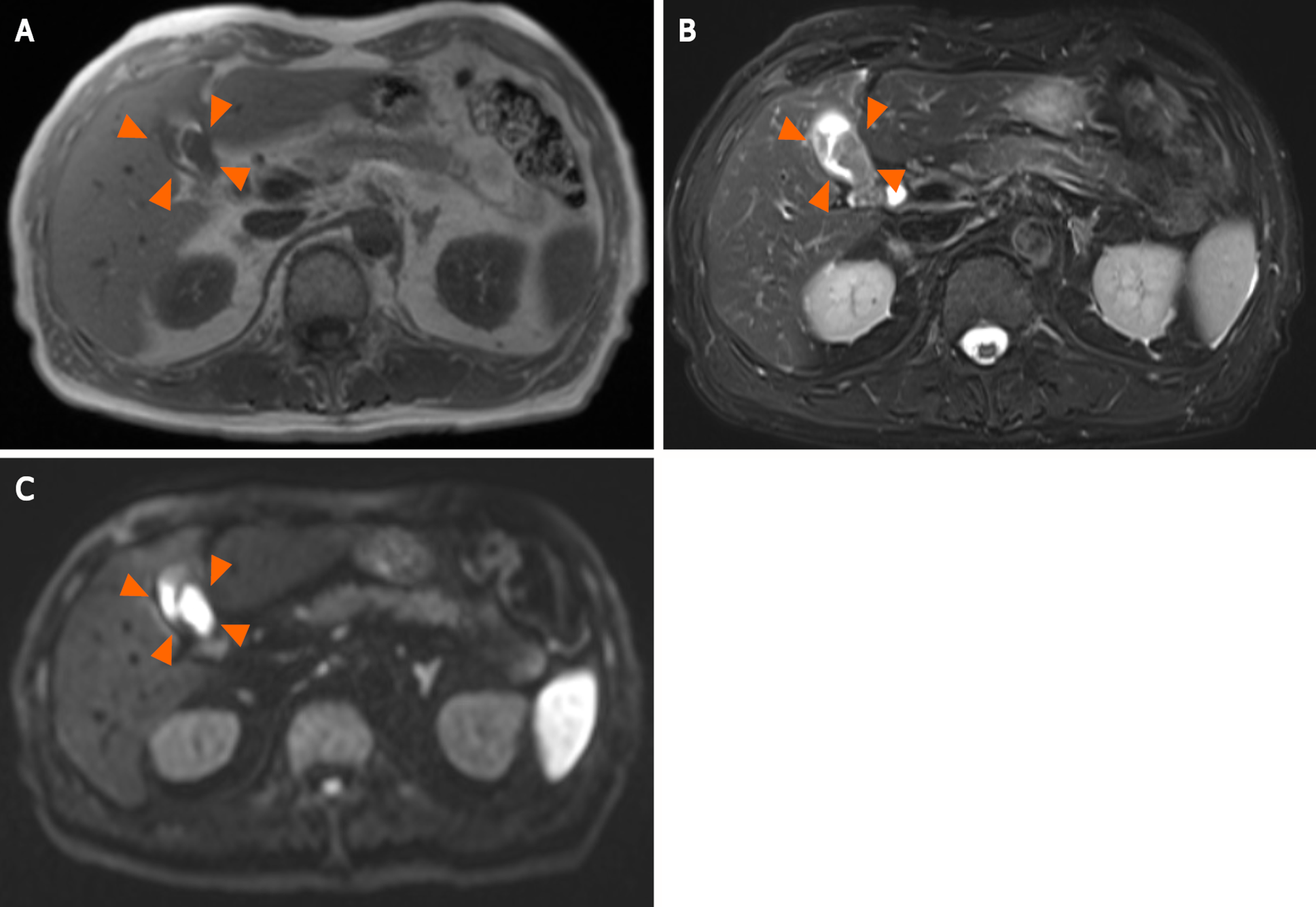
Figure 2 Magnetic resonance imaging reveals a hypointense tumor signal.
A: T1-weighted imaging (arrowheads); B: A hyperintense signal on T2-weighted imaging (arrowheads); C: Diffusion-weighted imaging (arrowheads).
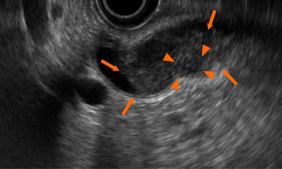
Figure 3 Endoscopic ultrasonography shows a heterogeneous echoic mass (arrows) with internal partially low echo (arrowheads).
The mass extends into the lumen but does not infiltrate the serosa.
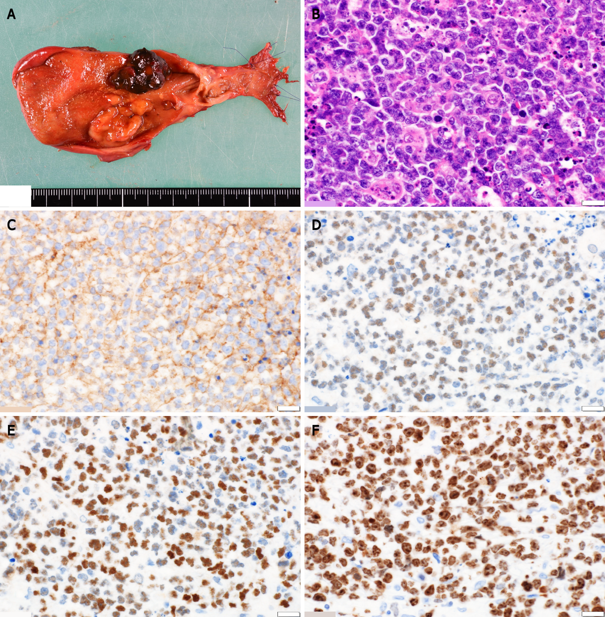
Figure 4 Histologic examination.
A: Periportal lymphadenopathy and two tumors at the neck and body of the gallbladder, measuring 27 mm × 20 mm and 20 mm × 18 mm in diameter, respectively; B: Histological findings reveal monotonous lymphoid cells with hemophagocytosis by macrophages; C-E: Immunohistochemical staining for markers shows the presence of CD10 (C), BCL6 (D), and c-Myc (E) and the absence of BCL2; F: The Ki-67 index is > 80%. The white scale bars represent 1 mm.
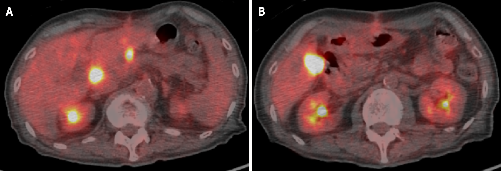
Figure 5 Positron emission tomography reveals increased 18F-fluorodeoxyglucose uptake at the superior pancreaticoduodenal lymph nodes and hepatic radical margin.
A: Superior pancreaticoduodenal lymph nodes; B: Hepatic radical margin.
- Citation: Hosoda K, Shimizu A, Kubota K, Notake T, Hayashi H, Yasukawa K, Umemura K, Kamachi A, Goto T, Tomida H, Yamazaki S, Narusawa Y, Asano N, Uehara T, Soejima Y. Gallbladder Burkitt’s lymphoma mimicking gallbladder cancer: A case report. World J Gastroenterol 2022; 28(6): 675-682
- URL: https://www.wjgnet.com/1007-9327/full/v28/i6/675.htm
- DOI: https://dx.doi.org/10.3748/wjg.v28.i6.675


