©The Author(s) 2022.
World J Gastroenterol. Mar 28, 2022; 28(12): 1284-1287
Published online Mar 28, 2022. doi: 10.3748/wjg.v28.i12.1284
Published online Mar 28, 2022. doi: 10.3748/wjg.v28.i12.1284
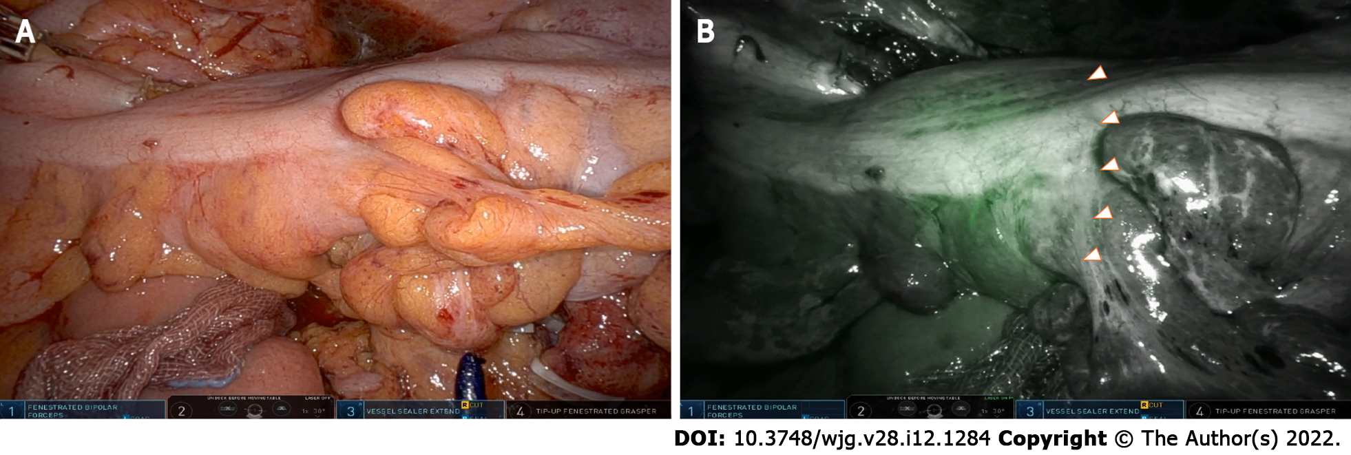
Figure 1 Near infrared fluorescence in conjunction with indocyanine green allowing visualization of the microcirculation before development of the colorectal anastomosis.
A: A white light image before visualizing the ischemic zone of the sigmoid colon using excited fluorescence; B: An intraoperative near infrared fluorescence image after visualizing the ischemic zone of the sigmoid colon using excited fluorescence.
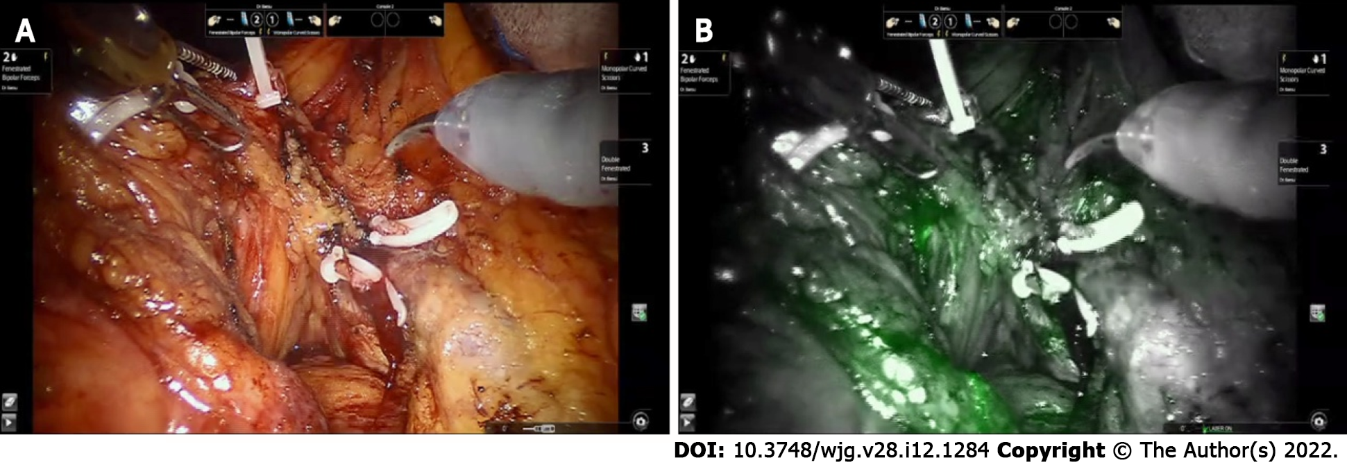
Figure 2 Mapping of additional lymph nodes outside the proposed resection margins to achieve curative radical lymphadenectomy in robot-assisted right hemicolectomy.
A: A white light image after D3 Lymphadenectomy around superior mesenteric vessels; B: A near infrared fluorescence image after visualizing the remained lymph nodes after lymphadenectomy using excited fluorescence.
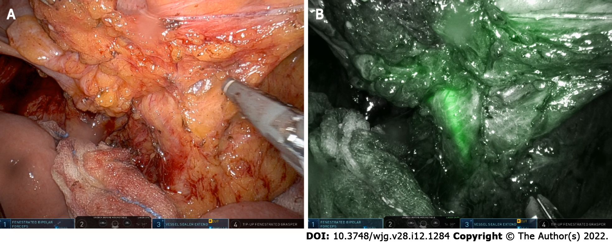
Figure 3 Robot-assisted lymph node dissection around the inferior mesenteric artery with preservation of the left colic artery using near infrared fluorescence imaging.
A: Dissection around the root of the inferior mesenteric artery (white light image); B: A near infrared fluorescence image visualizing the left colic artery using excited fluorescence.
- Citation: Bae SU. Near-infrared fluorescence imaging guided surgery in colorectal surgery. World J Gastroenterol 2022; 28(12): 1284-1287
- URL: https://www.wjgnet.com/1007-9327/full/v28/i12/1284.htm
- DOI: https://dx.doi.org/10.3748/wjg.v28.i12.1284


