©The Author(s) 2021.
World J Gastroenterol. Aug 28, 2021; 27(32): 5351-5361
Published online Aug 28, 2021. doi: 10.3748/wjg.v27.i32.5351
Published online Aug 28, 2021. doi: 10.3748/wjg.v27.i32.5351
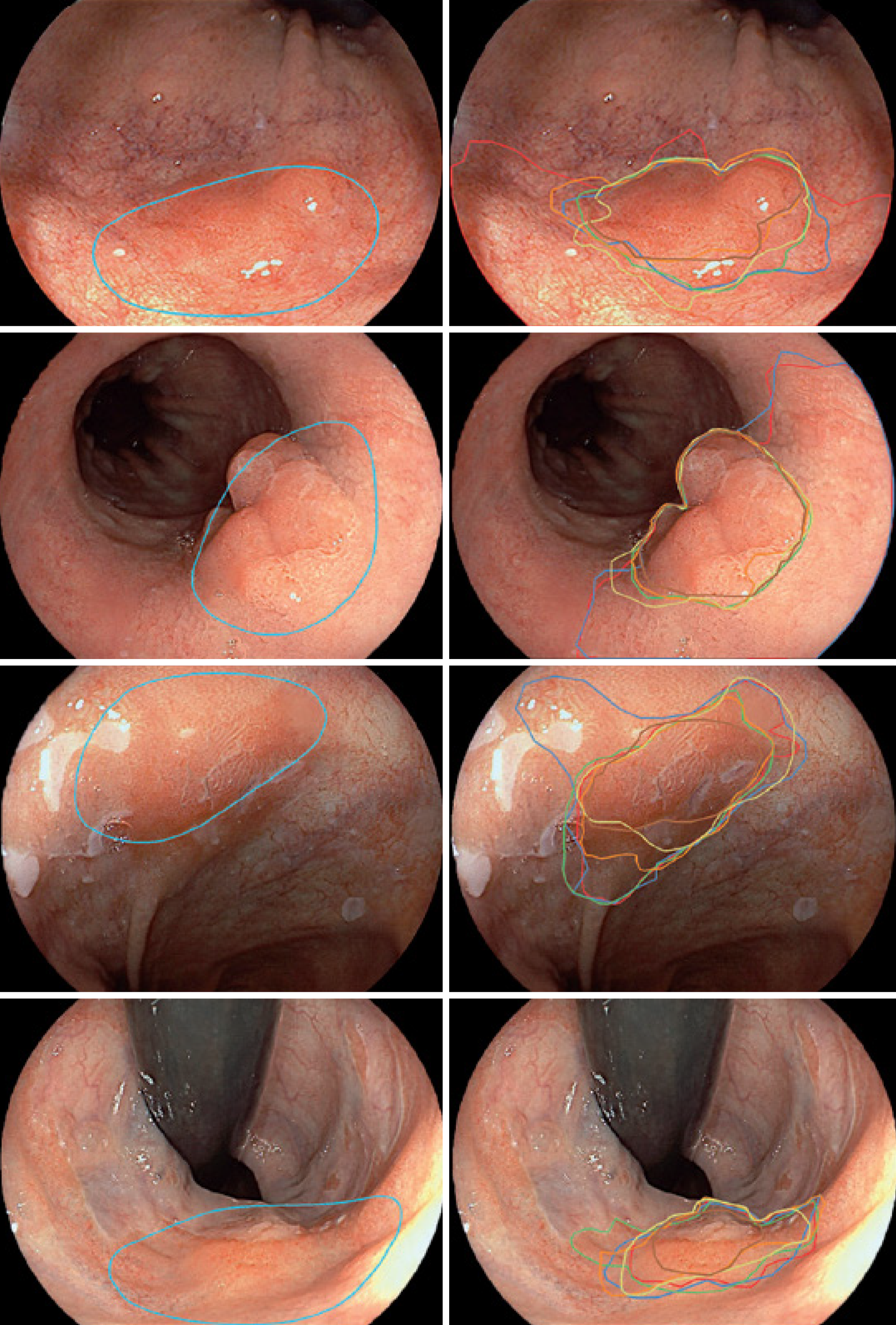
Figure 1 Delineation of Barrett's esophagus by computer-aided detection system and expert endoscopists.
Images on the left are from a computer-aided detection system to identify and delineate dysplastic lesions in Barrett’s esophagus with white-light imaging. Images on the right show the outlines of the same lesions made by expert endoscopists[13]. Citation: de Groof J, van der Sommen F, van der Putten J, Struyvenberg MR, Zinger S, Curvers WL, Pech O, Meining A, Neuhaus H, Bisschops R, Schoon E, With PH, Bergman JJ. The Argos project: The development of a computer-aided detection system to improve detection of Barrett's neoplasia on white-light endoscopy. United European Gastroenterol J 2019; 7: 538-47. Copyright © The Author(s) 2019. Published by SAGE Publications.
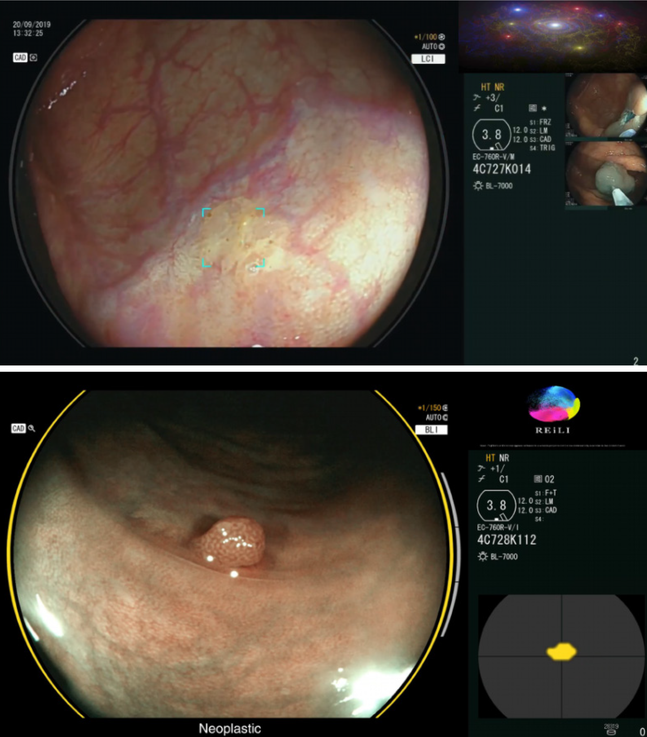
Figure 2 Computer-aided detection system for detecting and classifying colorectal polyps.
The top image is a polypoid lesion (delimited by blue marks) identified by a CADe system. The bottom image is a lesion classified as neoplastic by the CADx system with a high degree of confidence (three gray bars on the right side of the image)[33]. Citation: Mori Y, Neumann H, Misawa M, Kudo SE, Bretthauer M. Artificial intelligence in colonoscopy - Now on the market. What's next? J Gastroenterol Hepatol 2021; 36(1): 7-11. Copyright © The Author(s) 2020. Published by Journal of Gastroenterology and Hepatology Foundation and John Wiley & Sons Australia, Ltd.
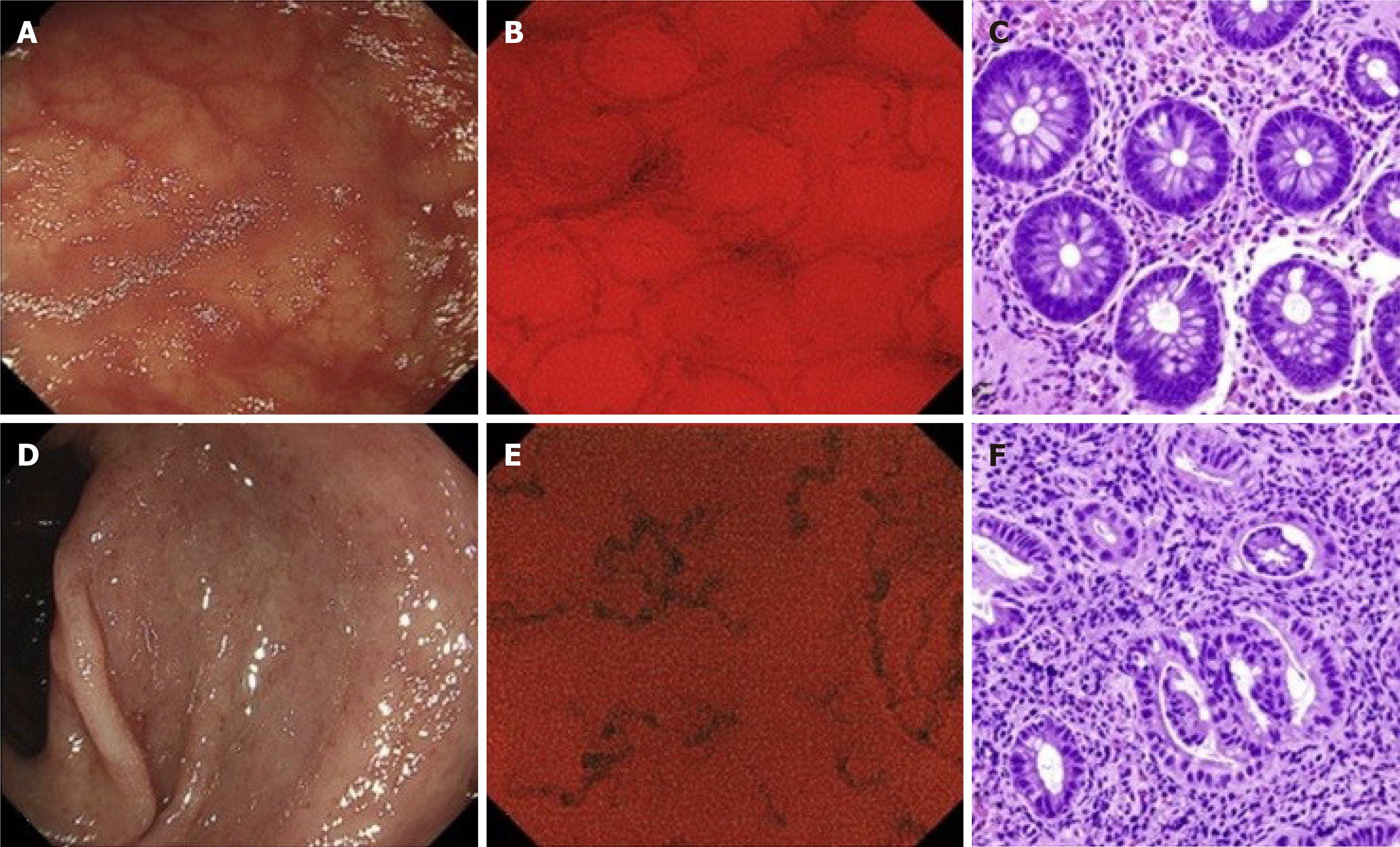
Figure 3 Application of computer-aided detection systems to endocytoscopy.
A, D: Conventional endoscopic images in WLI; B, E: Endocytoscopic images; C, F: Histological images. A CAD system evaluated the endocytoscopy image B: “healing” and E: active inflammation. The classifications were later confirmed by the pathologist[48]. Citation: Maeda Y, Kudo SE, Mori Y, Misawa M, Ogata N, Sasanuma S, Wakamura K, Oda M, Mori K, Ohtsuka K. Fully automated diagnostic system with artificial intelligence using endocytoscopy to identify the presence of histologic inflammation associated with ulcerative colitis (with video). Gastrointest Endosc 2019; 89(2): 408-415. Copyright © The Author(s) 2019 by the American Society for Gastrointestinal Endoscopy. Published by Elsevier, Inc.
- Citation: Correia FP, Lourenço LC. Artificial intelligence application in diagnostic gastrointestinal endoscopy - Deus ex machina? World J Gastroenterol 2021; 27(32): 5351-5361
- URL: https://www.wjgnet.com/1007-9327/full/v27/i32/5351.htm
- DOI: https://dx.doi.org/10.3748/wjg.v27.i32.5351


