Copyright
©The Author(s) 2021.
World J Gastroenterol. Jul 28, 2021; 27(28): 4536-4554
Published online Jul 28, 2021. doi: 10.3748/wjg.v27.i28.4536
Published online Jul 28, 2021. doi: 10.3748/wjg.v27.i28.4536
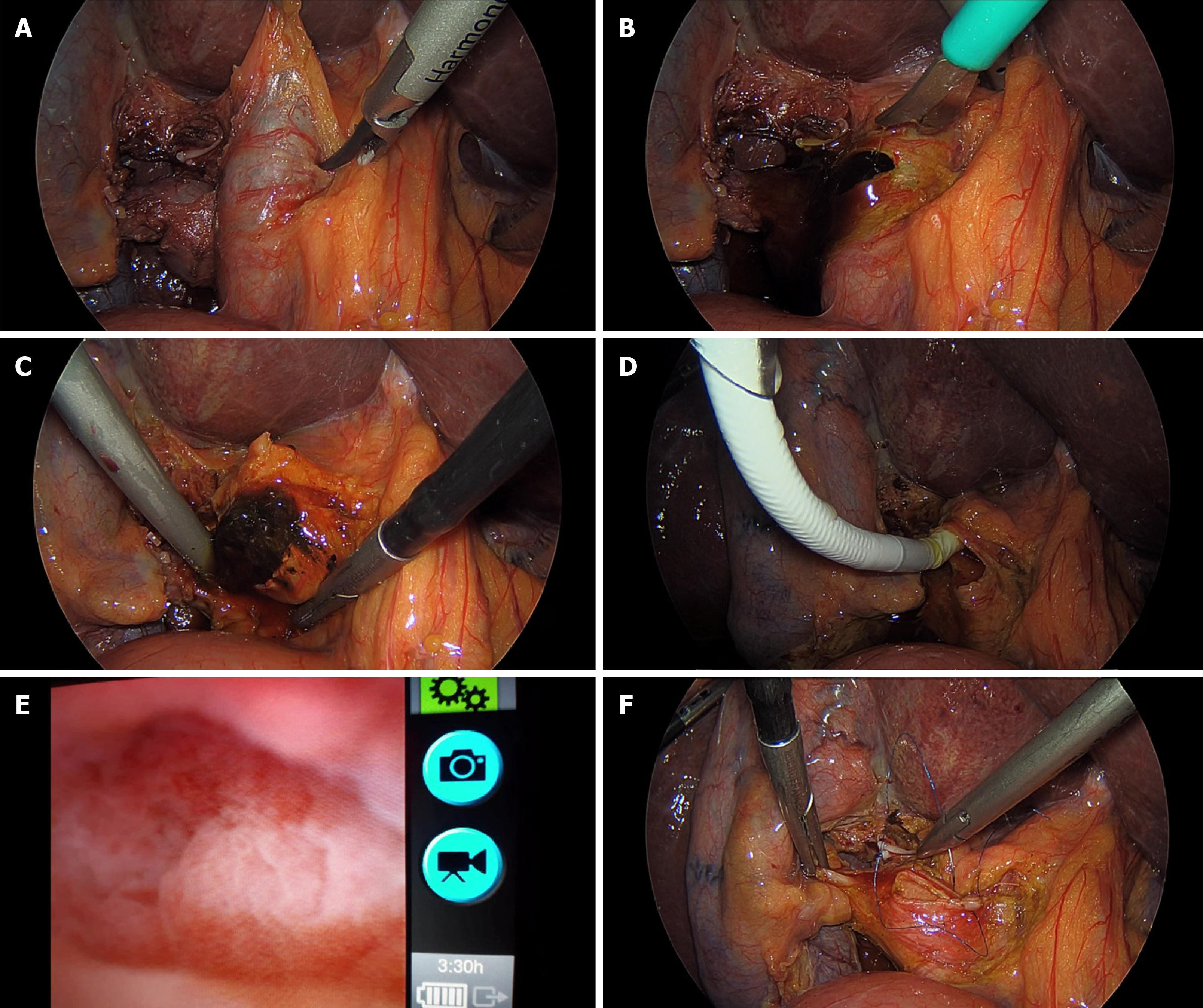
Figure 1 Intraoperative images: Laparoscopic exploration of the common bile duct.
A: Common bile duct (CBD) dilation; B: CBD section; C: Stone extraction; D: Insertion of the choledochoscope into the CBD; E: Choledochoscopic image of the CBD; F: Suture of CBD.
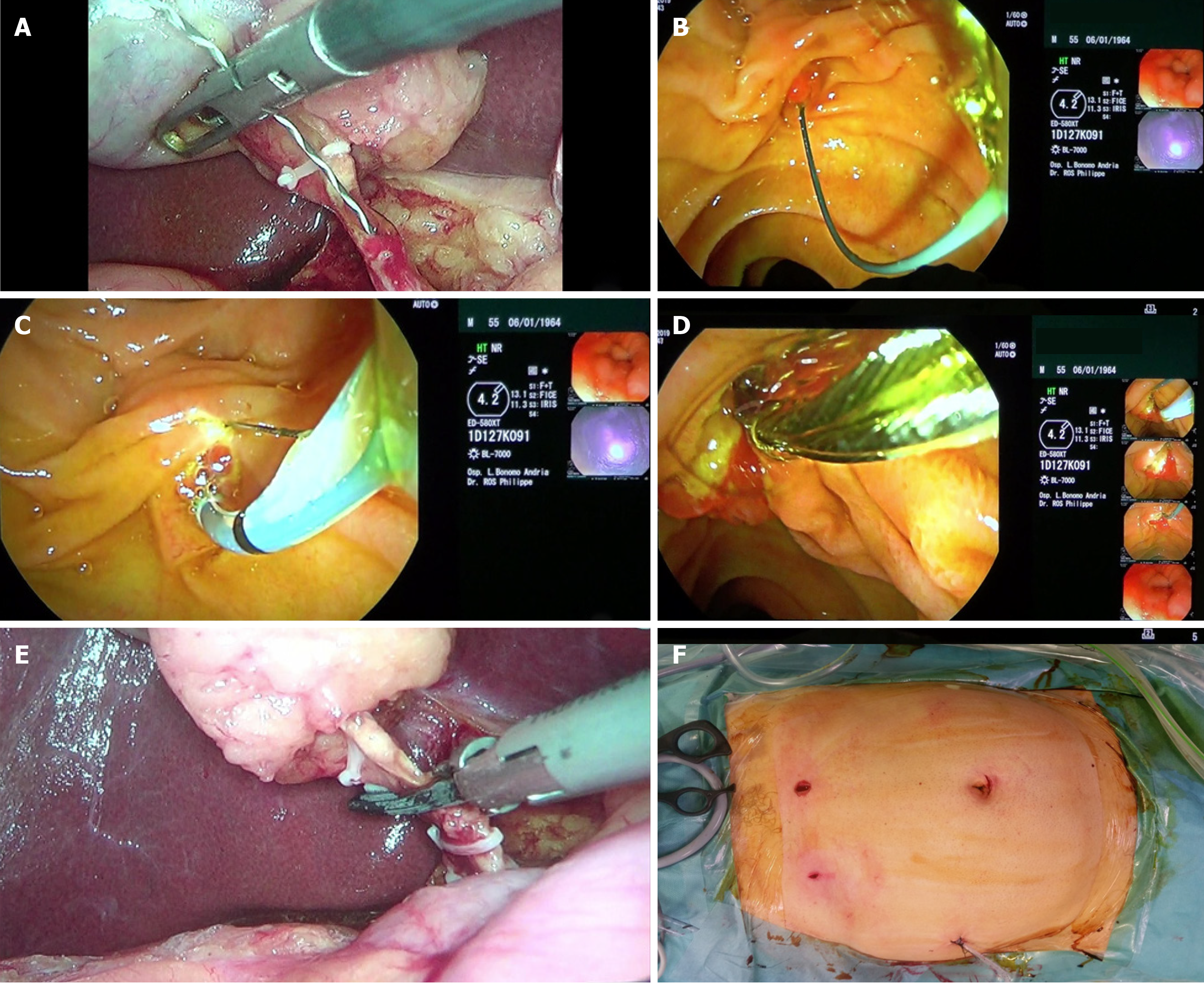
Figure 2 Intraoperative images: Endoscopic retrograde cholangio-pancreatography during laparoscopic cholecystectomy (“rendezvous technique”).
A: Insertion of the guide wire into the cystic duct; B: Guide wire exits through the papilla into the duodenum; C: Endoscopic sphincterotomy on guide wire; D: Extraction of stones from the common bile duct with a dormia basket; E: Section between clips of the cystic duct and subsequent retrograde cholecystectomy; F: Postoperative final scars.
- Citation: Cianci P, Restini E. Management of cholelithiasis with choledocholithiasis: Endoscopic and surgical approaches. World J Gastroenterol 2021; 27(28): 4536-4554
- URL: https://www.wjgnet.com/1007-9327/full/v27/i28/4536.htm
- DOI: https://dx.doi.org/10.3748/wjg.v27.i28.4536


