©The Author(s) 2021.
World J Gastroenterol. Jun 14, 2021; 27(22): 2963-2978
Published online Jun 14, 2021. doi: 10.3748/wjg.v27.i22.2963
Published online Jun 14, 2021. doi: 10.3748/wjg.v27.i22.2963
Figure 1 Common upper gastrointestinal endoscopic and microscopic presentations in ulcerative colitis.
UGI: Upper gastrointestinal; UC: Ulcerative colitis.
Figure 2 Endoscopic and microscopic presentations of esophageal ulcers associated with ulcerative colitis.
A: Endoscopic view showing multiple ulcerative lesions of the esophagus; B: Microscopic view showing squamous epithelial hyperplasia, interstitial fibrous tissue proliferation, inflammatory cell infiltration, and visible neutrophil aggregation.
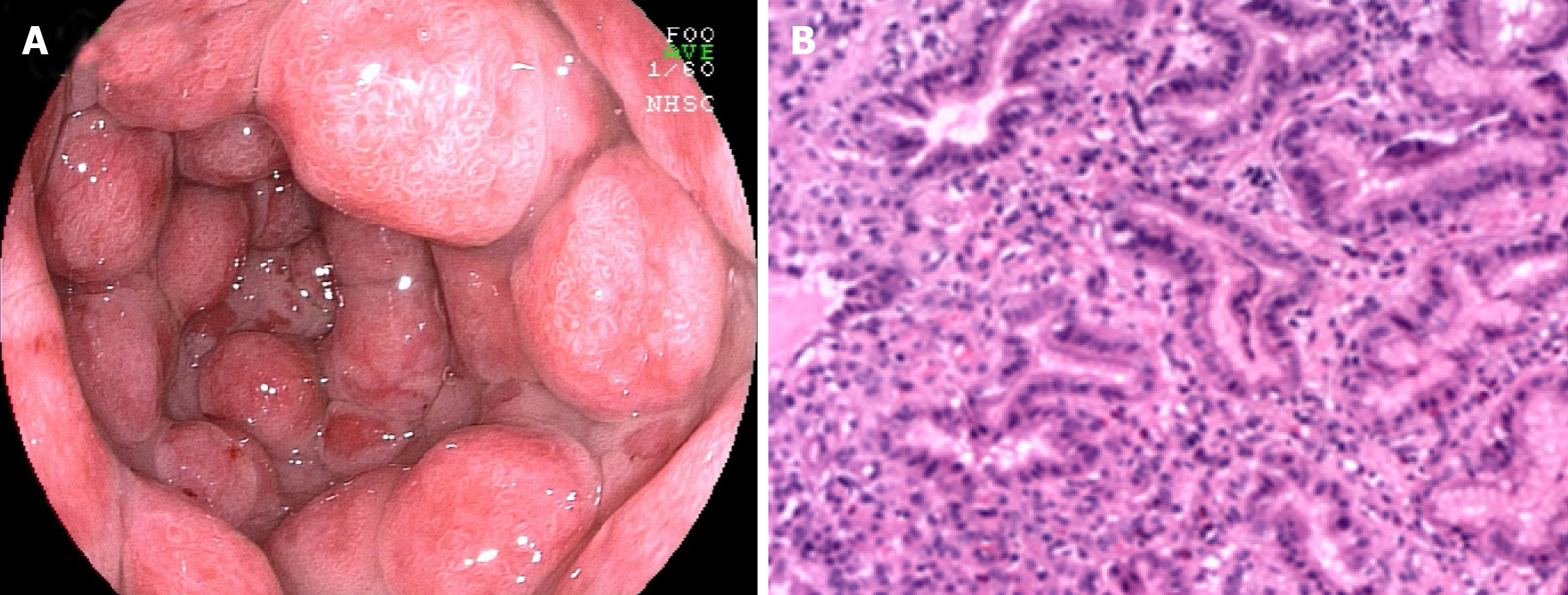
Figure 3 Endoscopic and microscopic manifestations of gastritis-associated ulcerative colitis.
A: Endoscopic image displaying multiple protrusion lesions of the gastric mucosa; B: Microscopic image showing mucosal inflammation with hyperplastic polypoid changes.
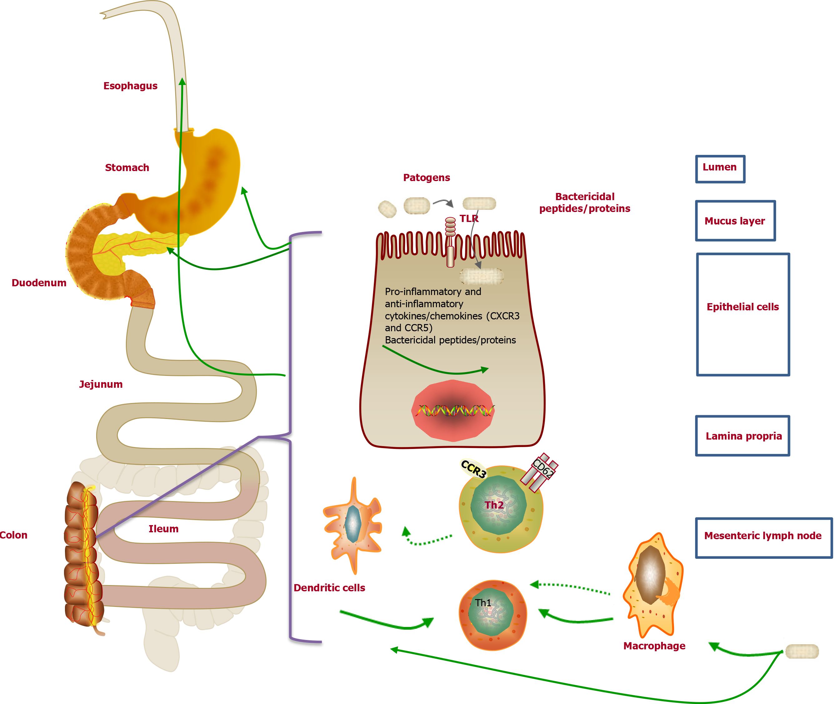
Figure 4 Pathogenesis of upper gastrointestinal mucosal lesions.
Figure 5 Endoscopic pictures of esophageal ulcer- and gastritis-associated ulcerative colitis after treatment.
A: Endoscopic picture revealing a healing scar of multiple esophageal ulcers after administration of remicade (infliximab for injection); B: Endoscopic picture demonstrating multiple protrusion lesions of the gastric mucosa after treatment with remicade and methylprednisolone.
- Citation: Sun Y, Zhang Z, Zheng CQ, Sang LX. Mucosal lesions of the upper gastrointestinal tract in patients with ulcerative colitis: A review. World J Gastroenterol 2021; 27(22): 2963-2978
- URL: https://www.wjgnet.com/1007-9327/full/v27/i22/2963.htm
- DOI: https://dx.doi.org/10.3748/wjg.v27.i22.2963


