©The Author(s) 2020.
World J Gastroenterol. Dec 28, 2020; 26(48): 7619-7632
Published online Dec 28, 2020. doi: 10.3748/wjg.v26.i48.7619
Published online Dec 28, 2020. doi: 10.3748/wjg.v26.i48.7619
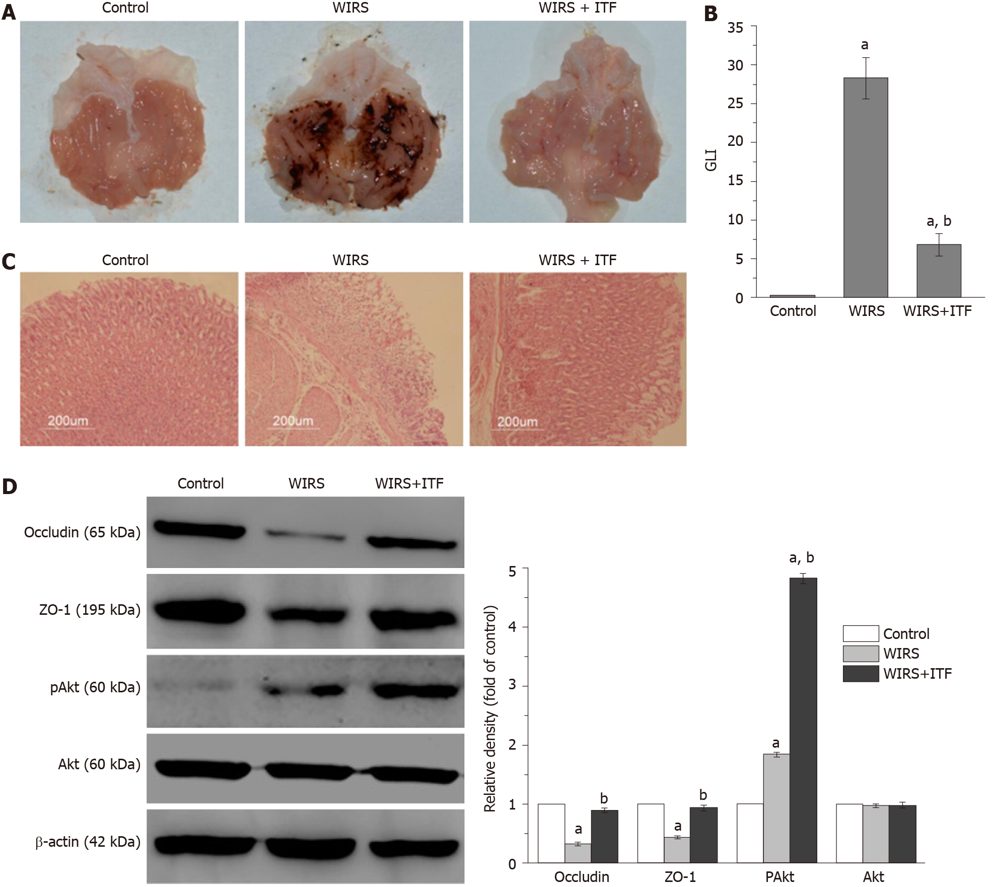
Figure 1 Intestinal trefoil factor alleviated gastric mucosal damage induced by acute stress.
Gastric mucosal changes and protein expressions were presented. A: Macroscopic appearance; B: Gastric lesion index; C: Histopathological analysis (Hematoxylin and eosin staining, magnification: 100 ×); D: Western blotting analysis of occludin, zonula occludens-1 (ZO-1), Akt, and pAkt. aP < 0.05 vs control group; bP < 0.05 vs water immersion restraint stress (WIRS) group. ITF: Intestinal trefoil factor.
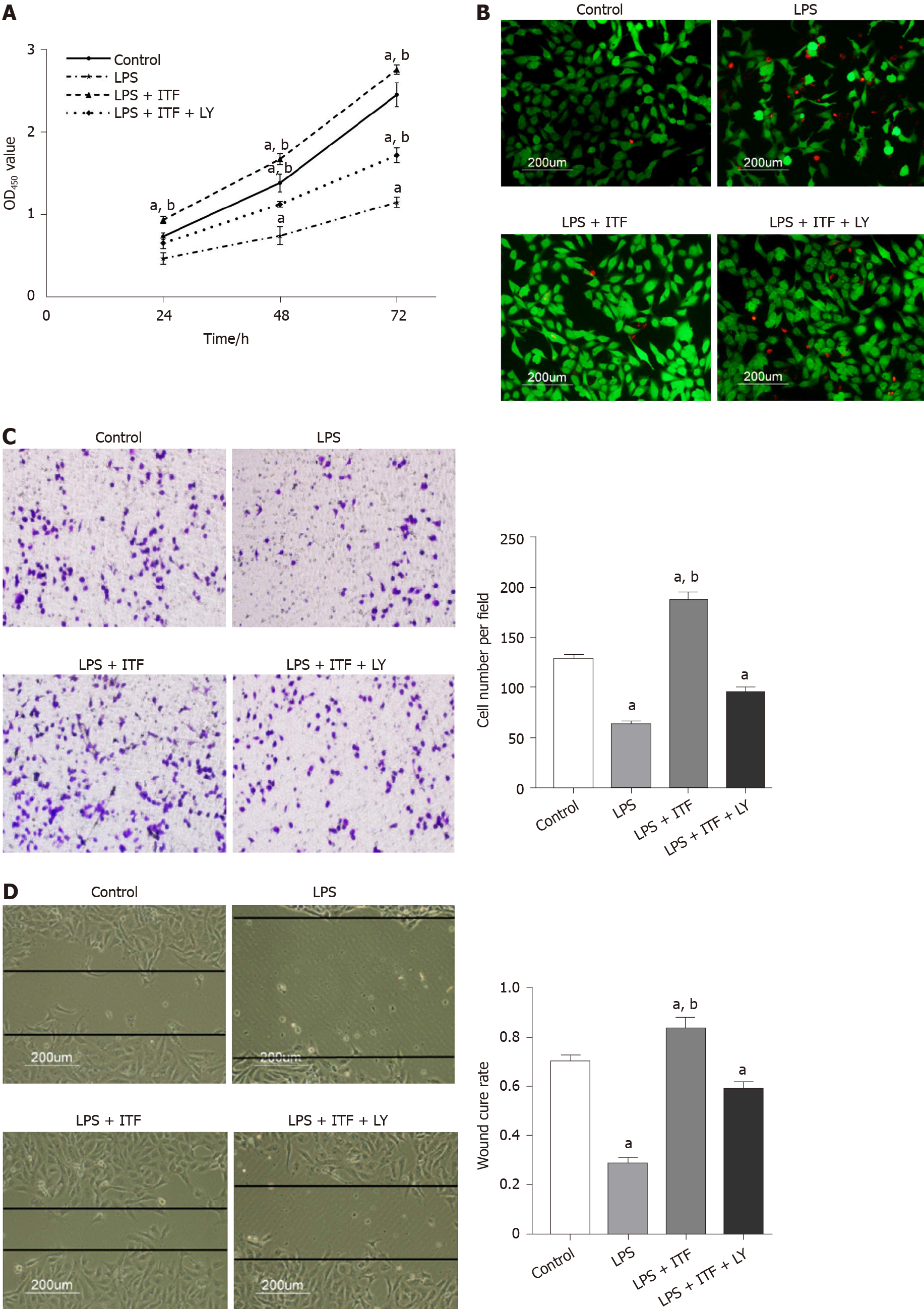
Figure 2 Intestinal trefoil factor promoted the proliferation and migration and inhibited necrosis of GES-1 cells.
A: Intestinal trefoil factor (ITF) promoted proliferation of GES-1 cells, and LY294002 decreased the cell viability; B: Fluorescent images of treated cells following fluorescein diacetate (FDA)/propidium iodide (PI) staining. Viable cells were stained with FDA (green), and necrotic cells were stained with propidium iodide (red), the scale bar = 200 μm; C: Transwell migration; and D: Wound healing assay analyzed the migration of GES-1 cells treated with lipopolysaccharides (LPS), ITF, or LY294002. The images are representative of three independent experiments; aP < 0.05 vs control cells; bP < 0.05 vs LPS-treated cells.
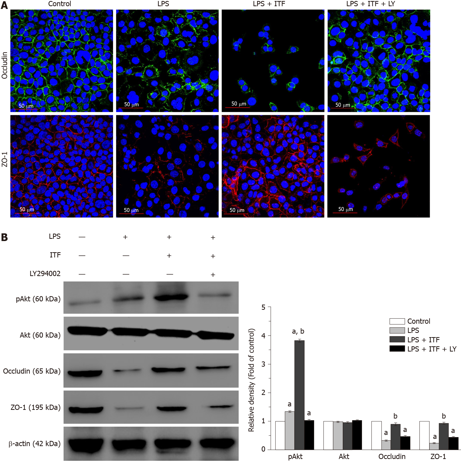
Figure 3 Intestinal trefoil factor preserved integrity of the gastric mucosa by activating the Akt signaling pathway.
A: Immunofluorescence staining of tight junction markers [occludin and zonula occludens-1 (ZO-1)] demonstrated that intestinal trefoil factor (ITF) maintained the integrity of GES-1 cells after treated with lipopolysaccharides (LPS). LY294002 undermined the protective effects of ITF via inhibiting the Akt signaling pathway in vitro; and B: Expression of occludin, ZO-1, Akt and pAkt were detected by Western blotting, suggesting that activation of the Akt signaling was essential for ITF to protect epithelium from damage induced by LPS. aP < 0.05 vs control cells; bP < 0.05 vs LPS-treated cells.
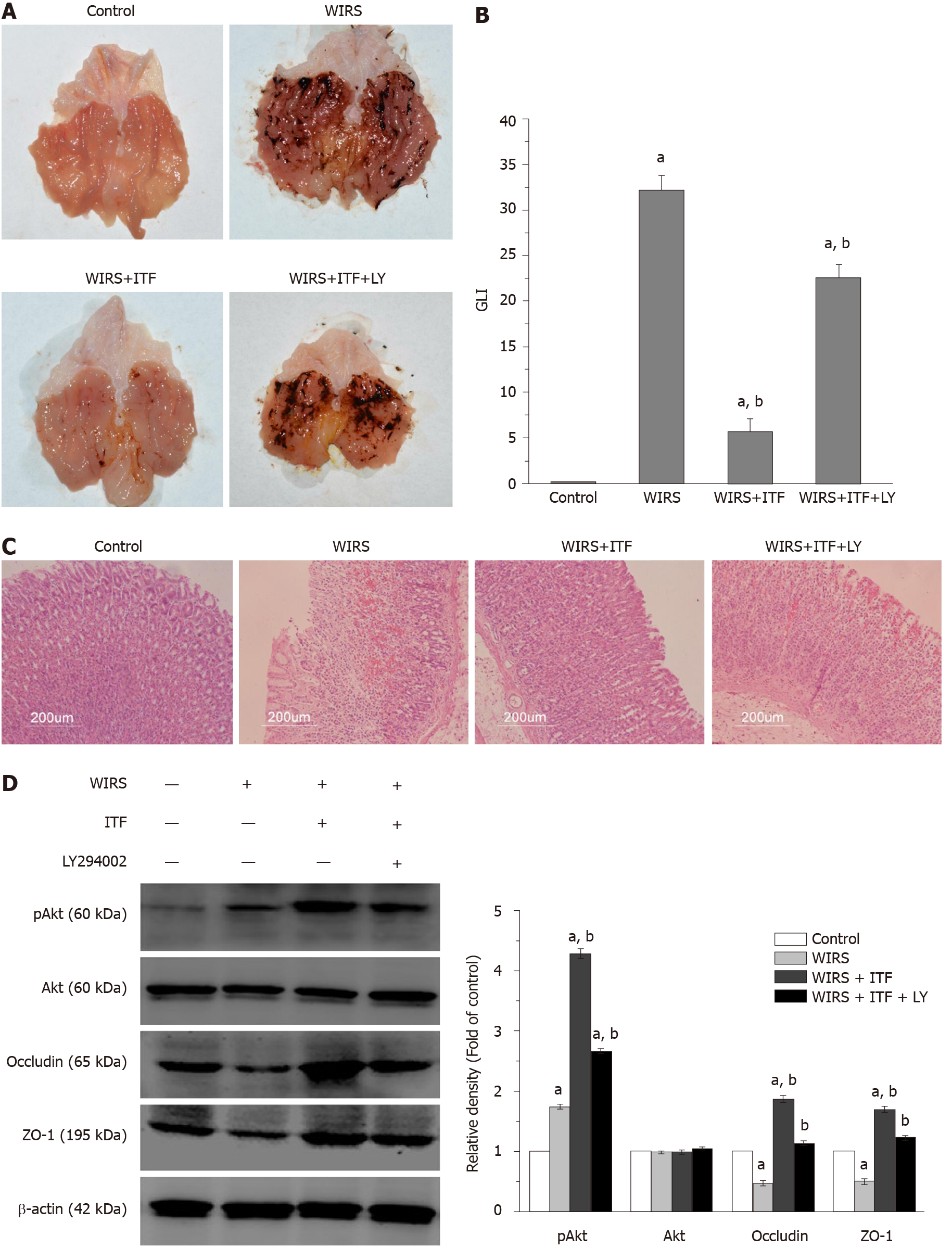
Figure 4 Inhibition of the Akt signaling pathway attenuated the protective effects of intestinal trefoil factor in vivo.
Gastric mucosal changes were presented in each group. A: Gross morphological appearance; B: Gastric lesion index (GLI); C: Histopathological analysis (Hematoxylin and eosin staining, magnification: 100 ×); and D: The expression of occludin, zonula occludens-1 (ZO-1), Akt, and pAkt were determined. Inhibition of the Akt signaling pathway by LY294002 significantly attenuated the protective effects of intestinal trefoil factor (ITF) in vivo. aP < 0.05 vs control group; bP < 0.05 vs water immersion restraint stress (WIRS) group.
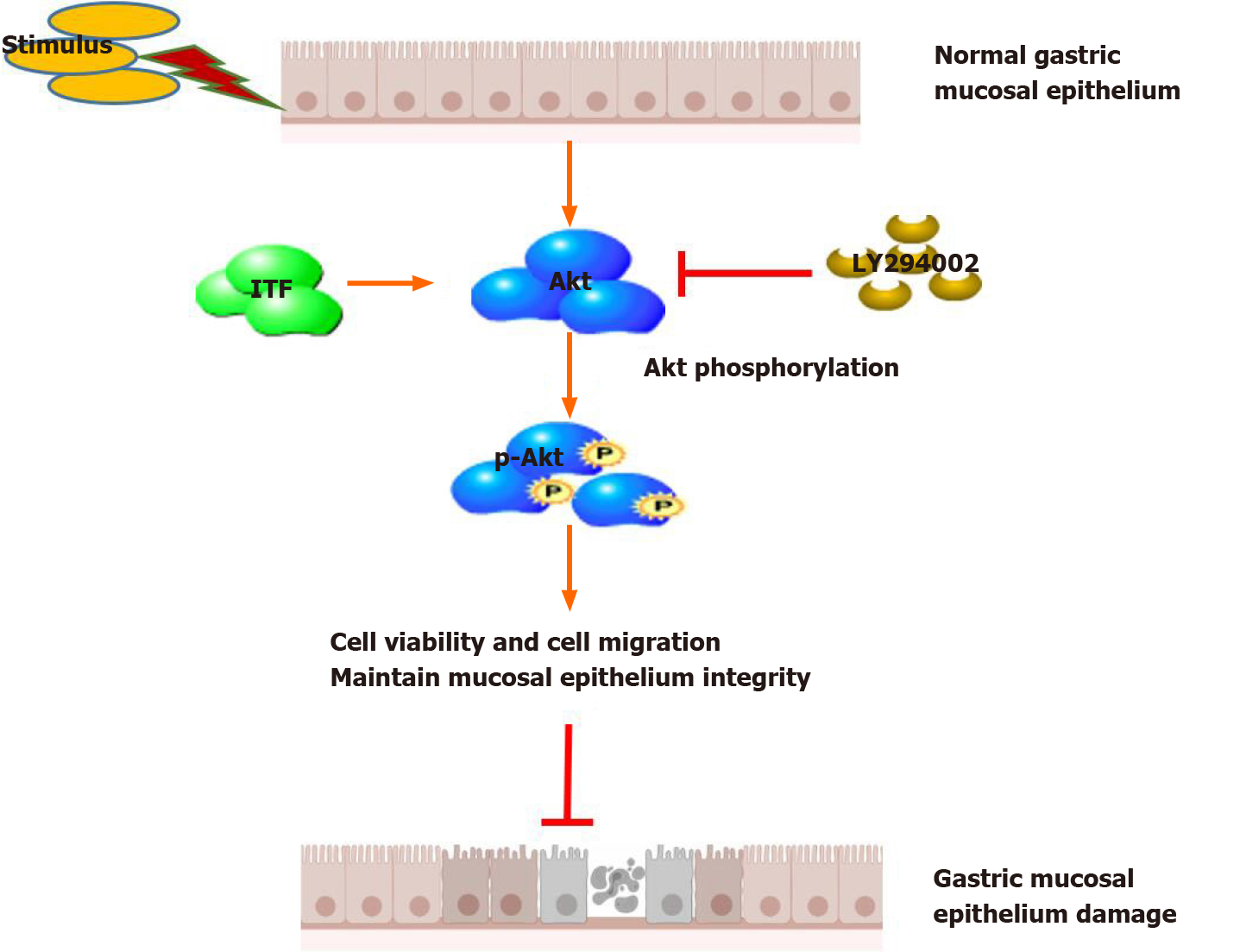
Figure 5 Schematic diagram of the protective effects of intestinal trefoil factor on gastric mucosal epithelium.
Intestinal trefoil factor (ITF) promoted the proliferation and migration of gastric epithelial cells and preserved integrity of the mucosal epithelium via activating the Akt signaling pathway.
- Citation: Huang Y, Wang MM, Yang ZZ, Ren Y, Zhang W, Sun ZR, Nie SN. Pretreatment with intestinal trefoil factor alleviates stress-induced gastric mucosal damage via Akt signaling. World J Gastroenterol 2020; 26(48): 7619-7632
- URL: https://www.wjgnet.com/1007-9327/full/v26/i48/7619.htm
- DOI: https://dx.doi.org/10.3748/wjg.v26.i48.7619


