©The Author(s) 2020.
World J Gastroenterol. Nov 21, 2020; 26(43): 6822-6836
Published online Nov 21, 2020. doi: 10.3748/wjg.v26.i43.6822
Published online Nov 21, 2020. doi: 10.3748/wjg.v26.i43.6822
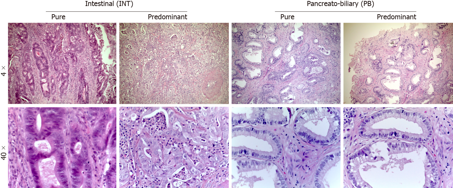
Figure 1 Representation of predominant histological sub-types of ampullary adenocarcinomas.
Right side: Pancreatobiliary types; Left side: Intestinal types (Up magnification 4 ×, down magnification 40 ×). PB: Pancreato-Biliary; INT: Intestinal.
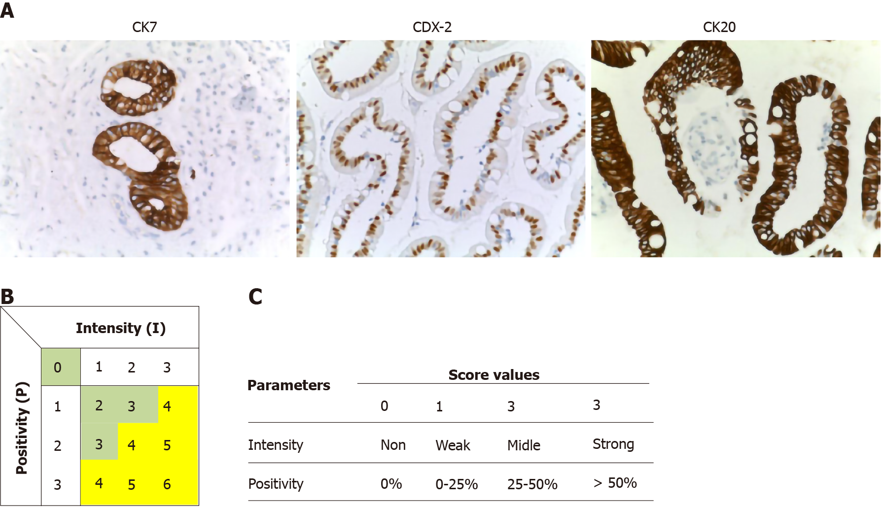
Figure 2 The analyses and quantification of three markers was based on the expression in normal controls.
A: Normal control for CK7, CDX-2 and CK20; B: The total score was established before the analyses; and C: Intervals of values associated with each parameter of the immunohistochemical score.
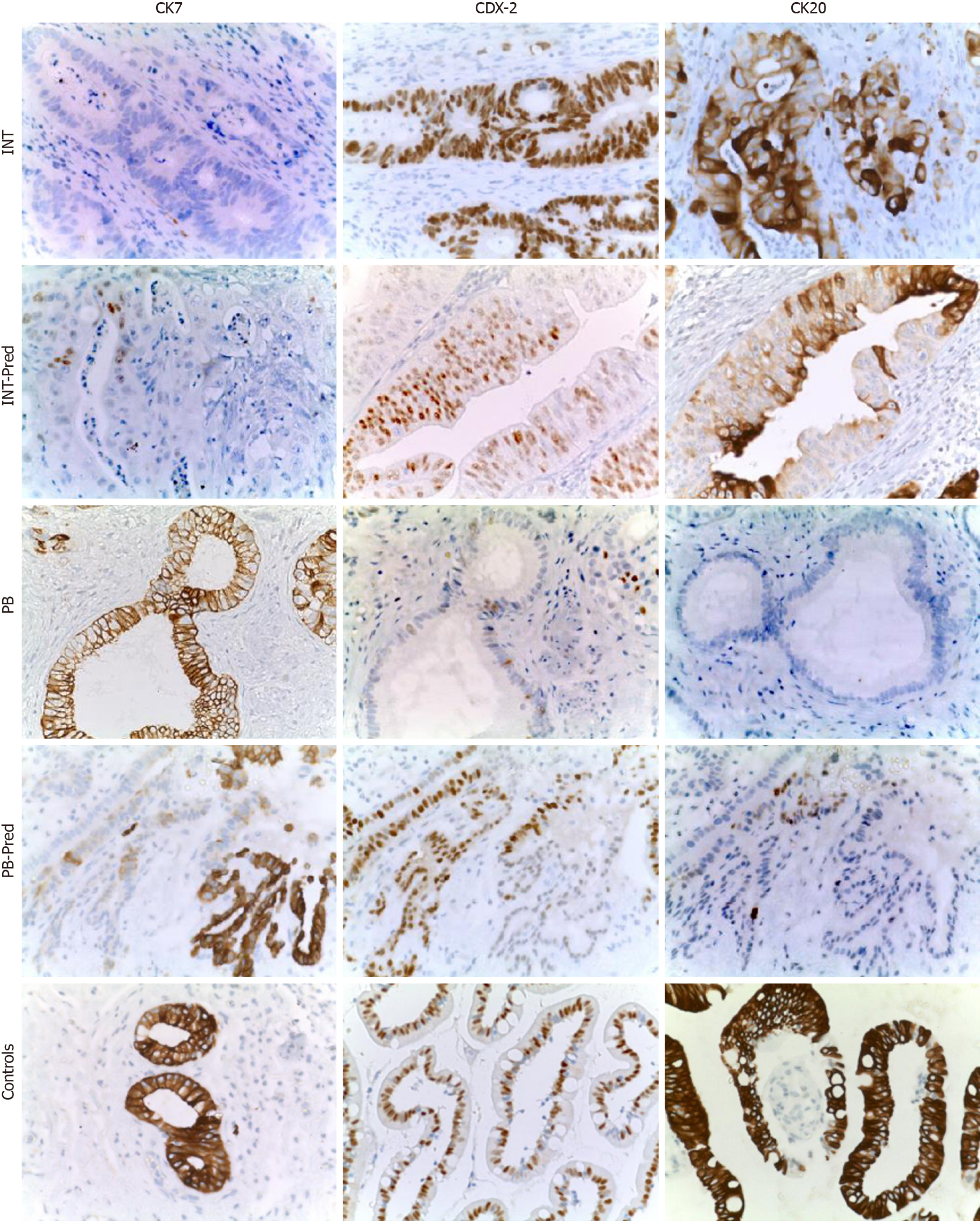
Figure 3 Representation of immunohistochemical staining in all sub-types observed in ampullary adenocarcinoma samples.
All images were acquired using a magnification 40 ×. IHC: Immunohistochemical; PB: Pancreato-Biliary; INT: Intestinal.
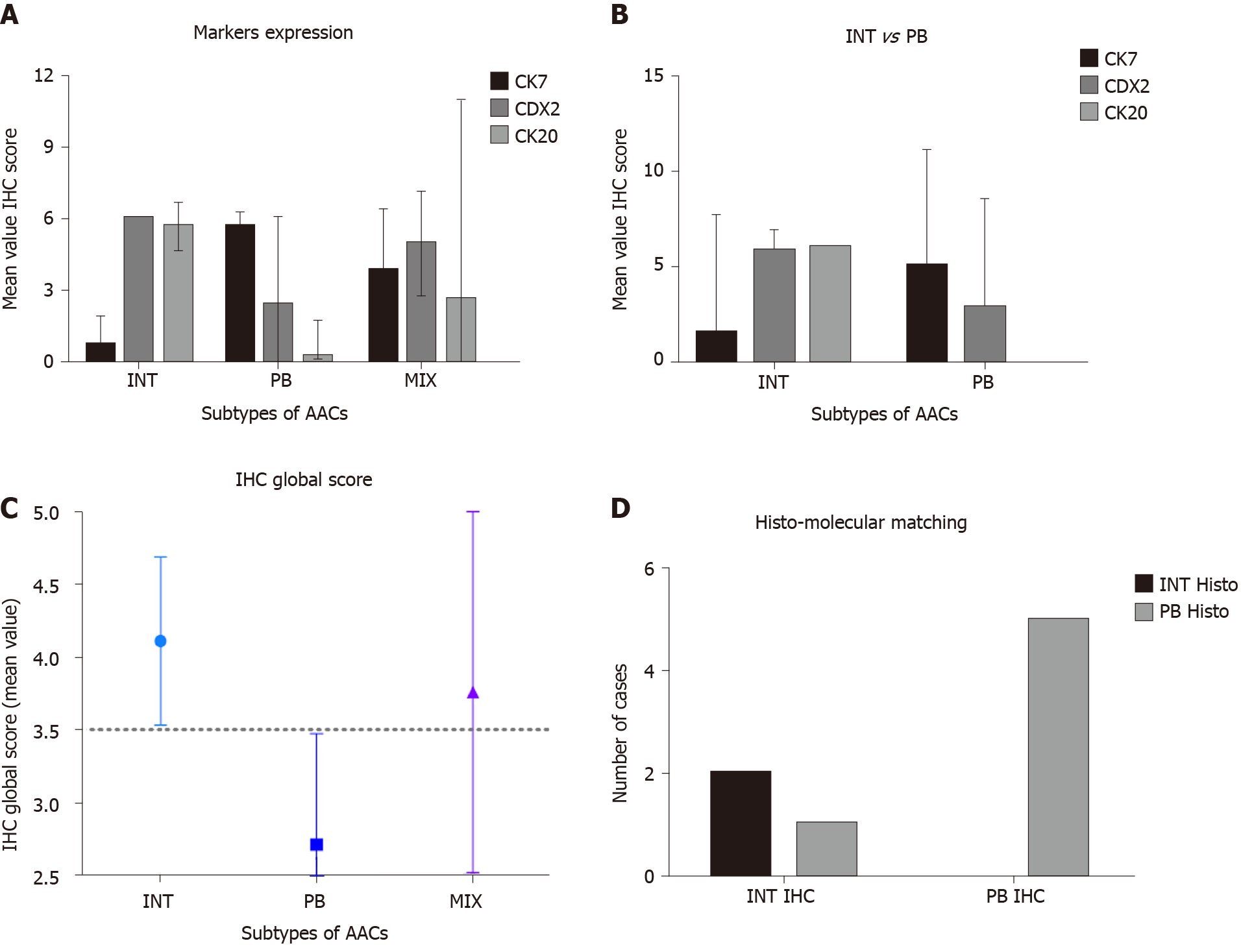
Figure 4 Statistical analyses of immunohistochemical markers and their values.
A: Mean immunohistochemical value for each marker in INTestinal, Pancreato-Biliary and MIX types of ampullary adenocarcinomas, according to histological analyses (hematoxylin-eosin staining); B: Mean immunohistochemical value to compare intestinal vs Pancreato-Biliary; C: Global Score mean value in all sub-types of AACs; and D: Chi-square test to compare the histological vs molecular sub-types. AACs: Ampullary adenocarcinomas; IHC: Immunohistochemical; INT: Intestinal; PB: Pancreato-Biliary.
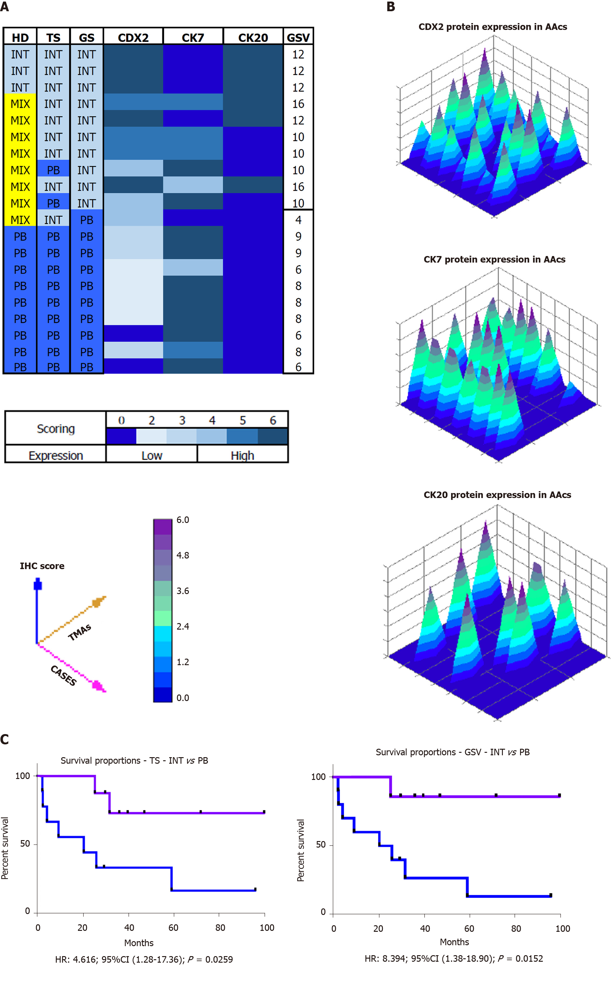
Figure 5 Results elaborating the computerized analyses.
A: Classification according to the three different data sets obtained: Histology, Total score and Global score; B: 3D representation of immunohistochemical analyses of all samples; C: Kaplan–Meier curves of INTestinal vs Pancreato-Biliary according the molecular partition by Total score (above) or Global Score (below). IHC: Immunohistochemical; TS: Total score; Intestinal; PB: Pancreato-Biliary; AACs: Ampullary adenocarcinomas; TMA: Three tissue microarrays; INT: Intestinal.
- Citation: Palmeri M, Funel N, Di Franco G, Furbetta N, Gianardi D, Guadagni S, Bianchini M, Pollina LE, Ricci C, Del Chiaro M, Di Candio G, Morelli L. Tissue microarray-chip featuring computerized immunophenotypical characterization more accurately subtypes ampullary adenocarcinoma than routine histology. World J Gastroenterol 2020; 26(43): 6822-6836
- URL: https://www.wjgnet.com/1007-9327/full/v26/i43/6822.htm
- DOI: https://dx.doi.org/10.3748/wjg.v26.i43.6822


