©The Author(s) 2020.
World J Gastroenterol. Sep 21, 2020; 26(35): 5328-5342
Published online Sep 21, 2020. doi: 10.3748/wjg.v26.i35.5328
Published online Sep 21, 2020. doi: 10.3748/wjg.v26.i35.5328
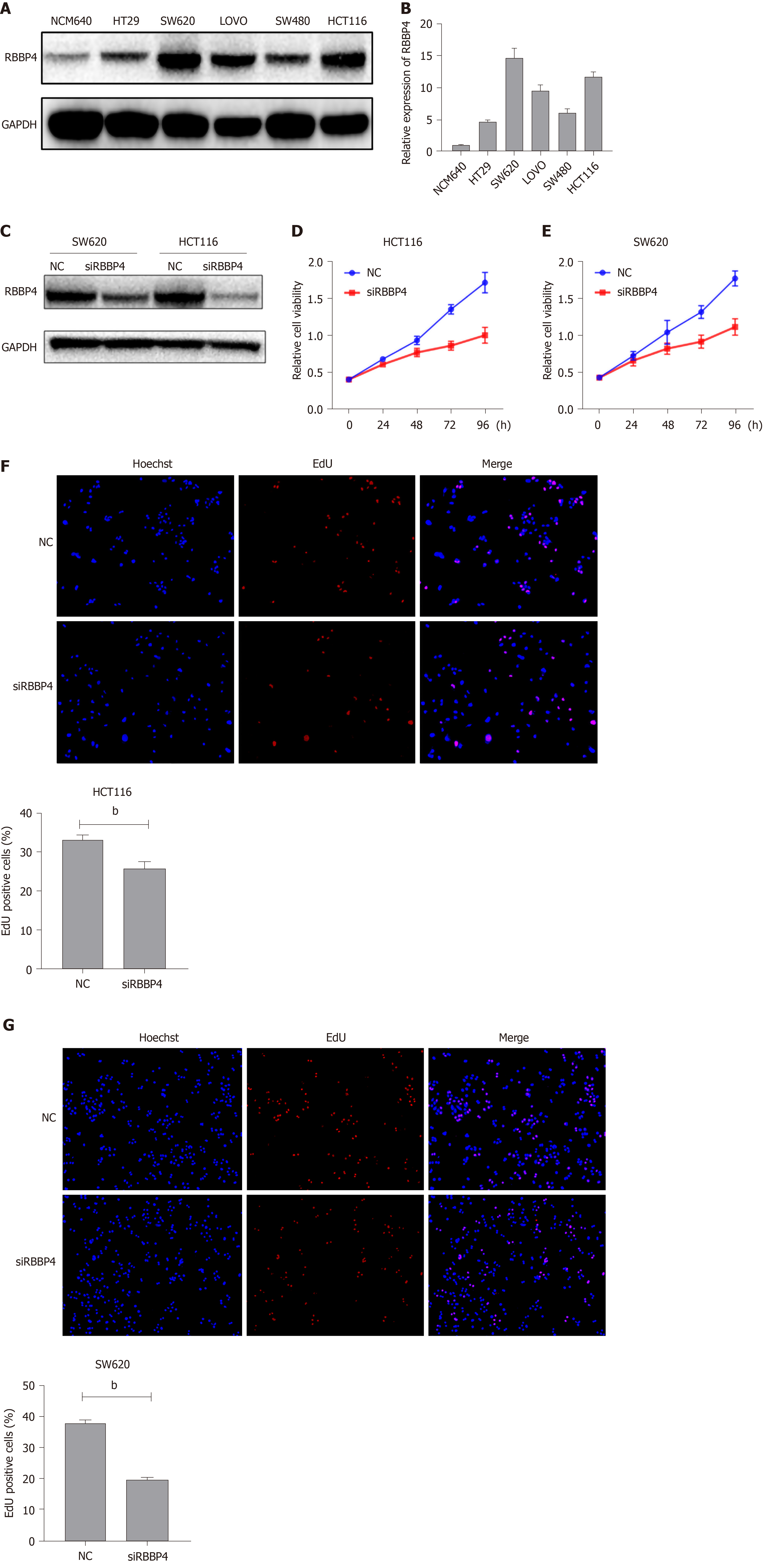
Figure 1 Expression of RBBP4 in colon cancer cell lines and its effect on cell proliferation.
A: Protein level of RBBP4 in the colon cancer cell lines quantified by western blotting; B: mRNA level of RBBP4 in the colon cancer cell lines quantified by polymerase chain reaction; C: RBBP4 siRNA efficiency verified by western blotting in SW620 and HCT116 cells; D: Cell viability was detected by the Cell Counting Kit-8 assay in HCT116 cells; E: Cell viability was detected by the Cell Counting Kit-8 assay in SW620 cells; F and G: Cell proliferation was detected by 5-ethynyl-2’-deoxyuridine assay in HCT116 cells and SW620 cells. bP < 0.01 vs controls. EdU: 5-Ethynyl-2’-deoxyuridine.
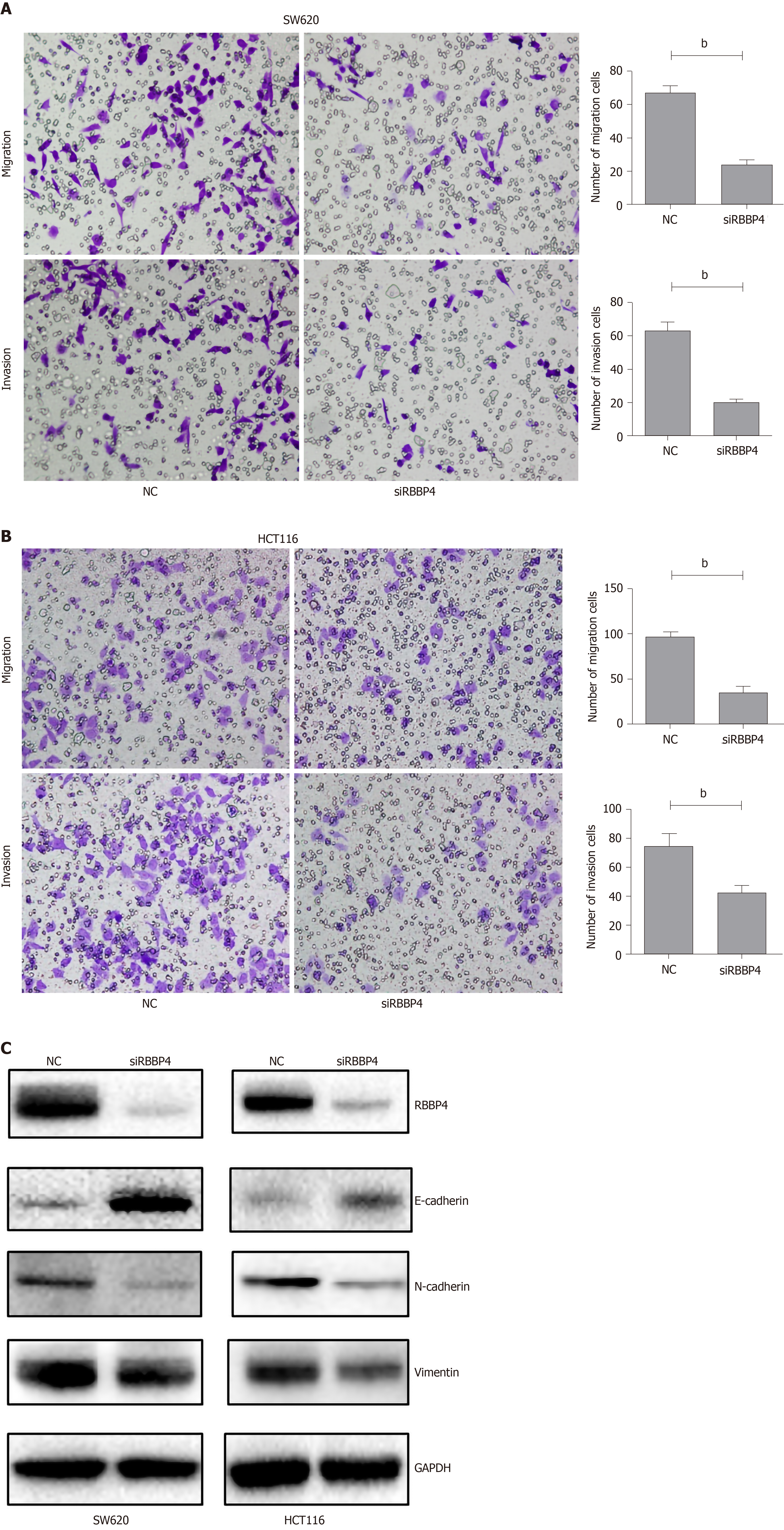
Figure 2 RBBP4 knockdown inhibits migration and invasion of colon cancer cell lines.
A: Migration and invasion of RBBP4 knockdown SW620 cells were measured by the transwell assay. Results were quantitated by counting migrating and invasive cells in five randomly chosen high-power fields for each replicate; B: Migration and invasion of RBBP4 knockdown HCT116 cells were measured by the transwell assay; C: Western blotting examination for epithelial-mesenchymal transition related proteins. bP < 0.01 vs controls.
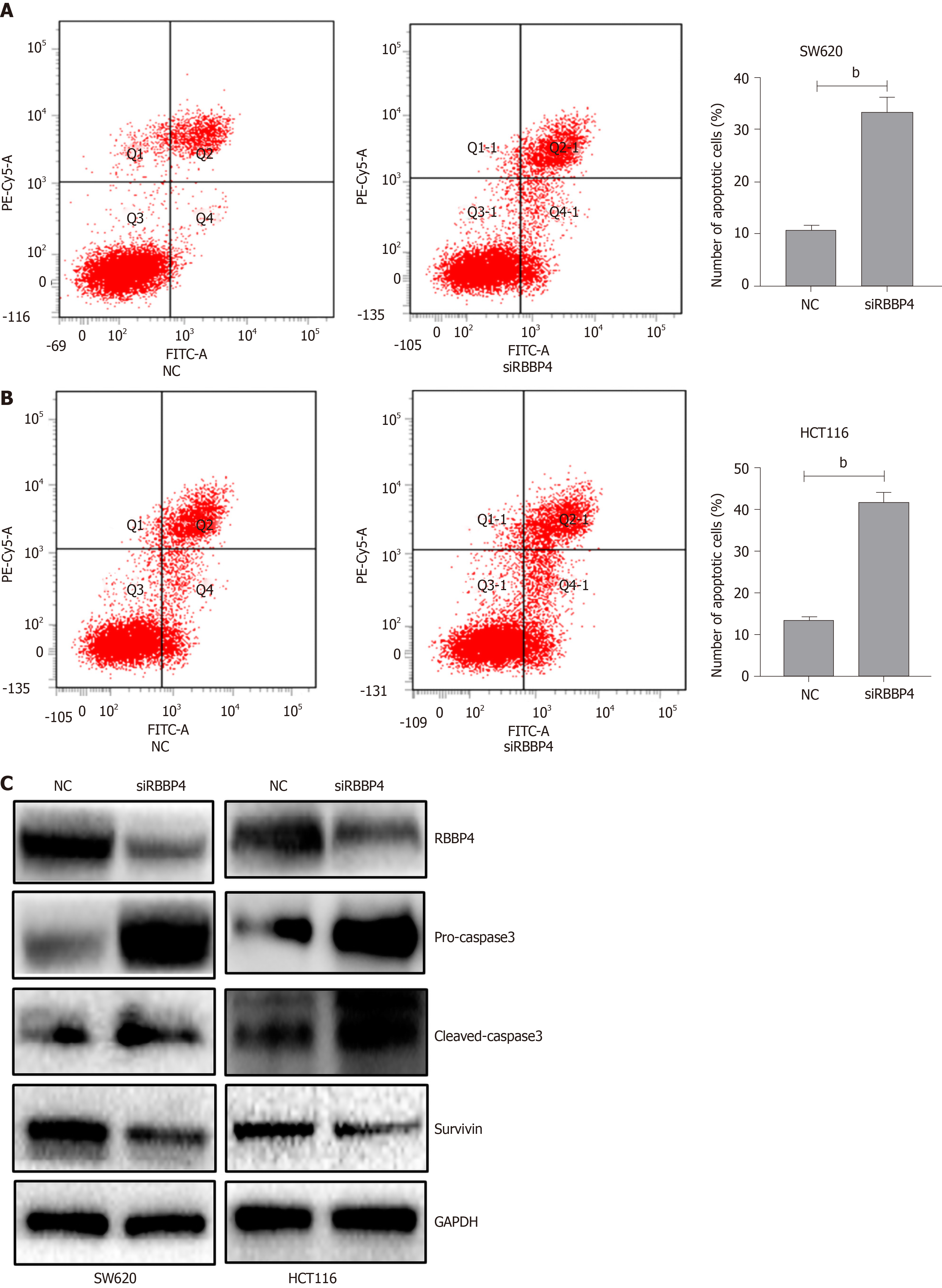
Figure 3 RBBP4 knockdown inhibits apoptosis of colon cancer cell lines.
A: Apoptosis of SW620 cells examined by flow cytometry with the Annexin V-FITC/propidium iodide kit. The cells in the Q4 quadrant were defined as the apoptotic cells; B: Apoptosis of HCT116 cells examined by flow cytometry with the Annexin V-FITC/propidium iodide kit; C: Western blotting examination for apoptotic proteins. bP < 0.01 vs controls.
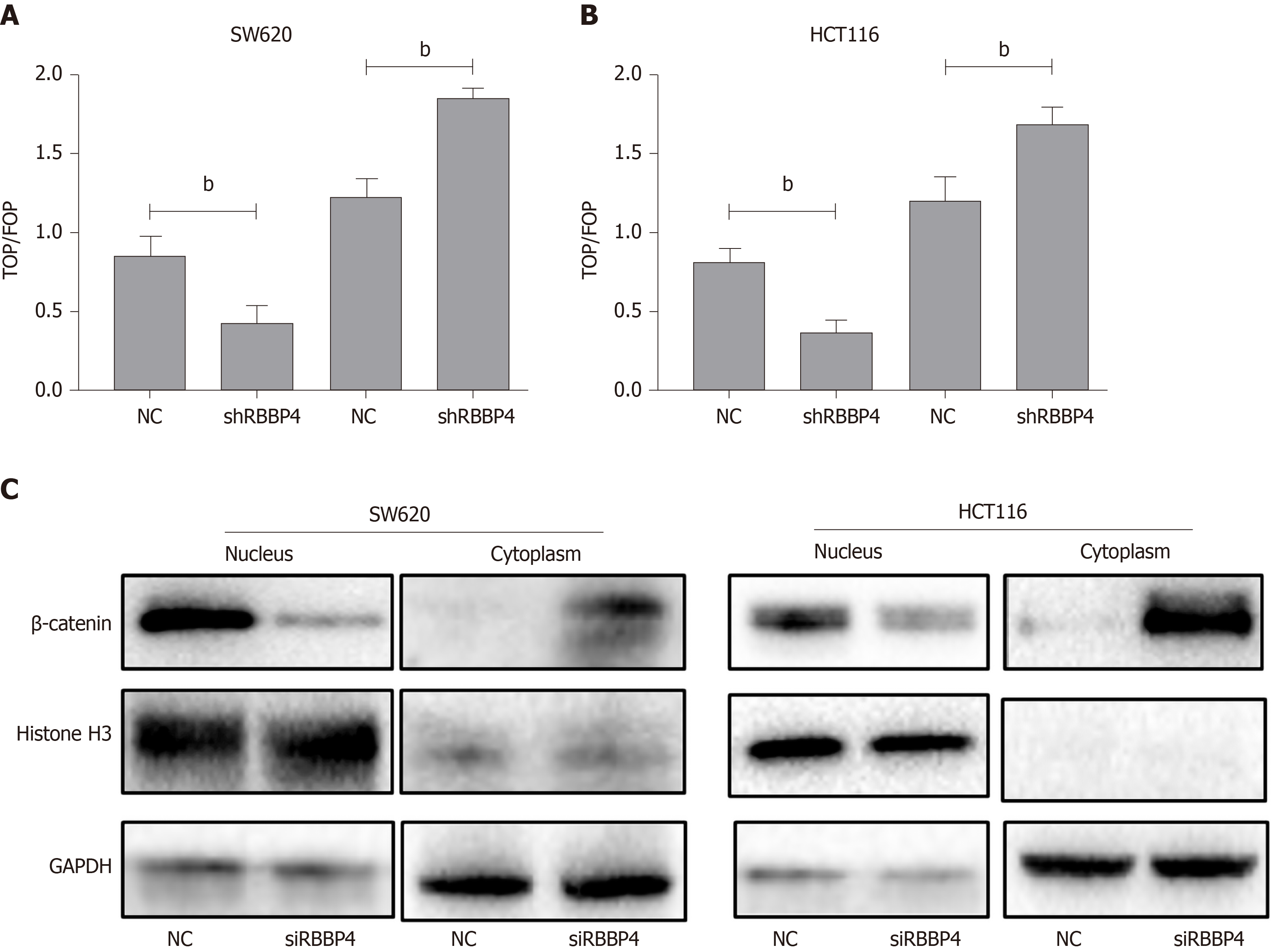
Figure 4 Effect of RBBP4 on activation of Wnt/β-catenin pathway.
A: The TOPFlash experiment in SW620 cells with RBBP4 knockdown or RBBP4 overexpression; B: The TOPFlash experiment in HCT116 cells with RBBP4 knockdown or RBBP4 overexpression; C: Western blotting of the level of β-catenin in cell nucleus and cytoplasm. bP < 0.01 vs controls.
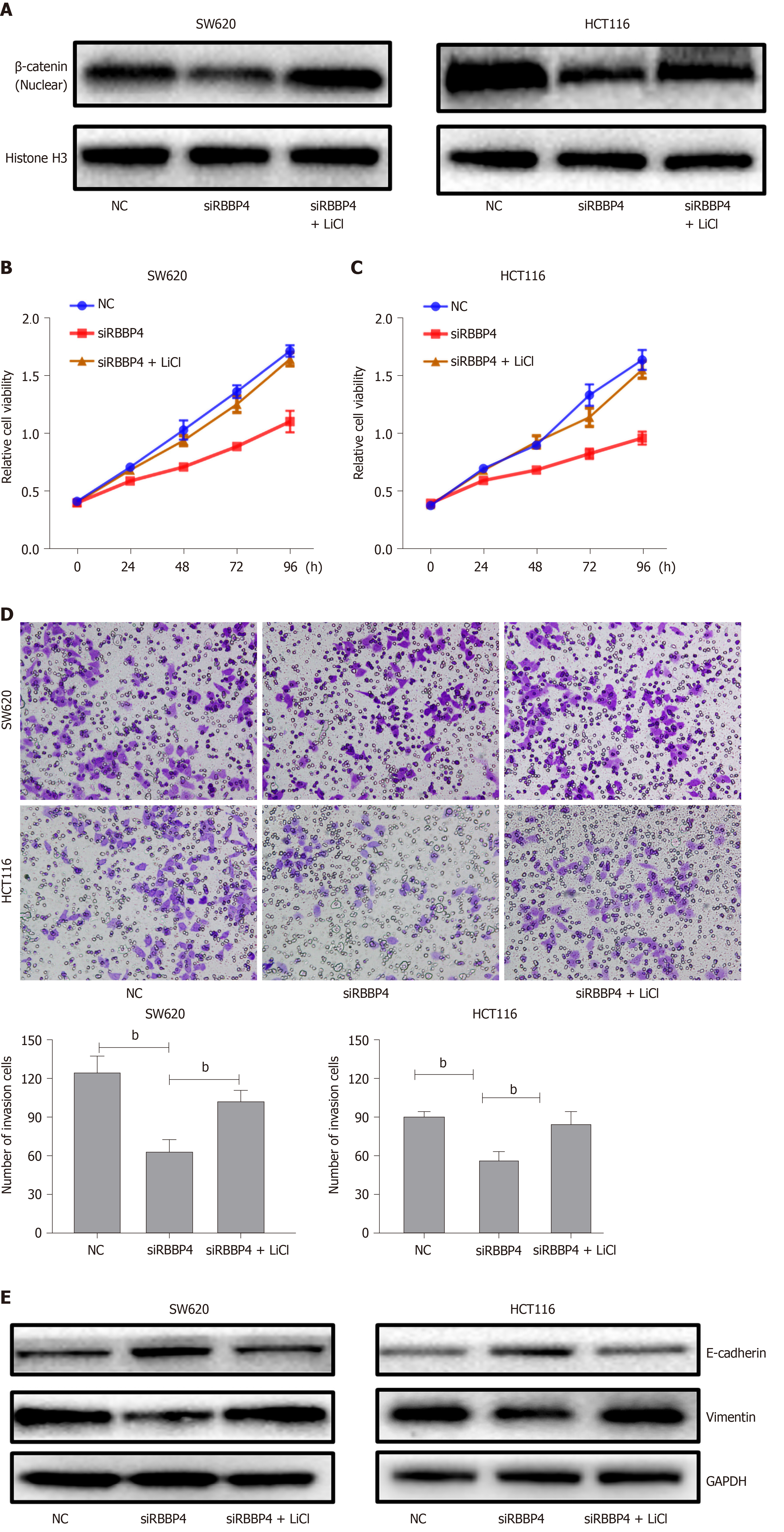
Figure 5 Rescue experiments of RBBP4.
A: SW620 and HCT116 cells with RBBP4 siRNA or negative control transfection were incubated with or without 20 mmol/L LiCl, then the level of β-catenin in the nucleus was detected by western blotting; B: The viability of SW620 cells with RBBP4 siRNA or negative control transfection cultured in medium with or without 20 mmol/L LiCl; C: The viability of HCT116 cells with RBBP4 siRNA or negative control transfection cultured in medium with or without 20 mmol/L LiCl; D: Invasion of SW620 and HCT116 cells with RBBP4 siRNA or negative control transfection cultured in medium with or without 20 mmol/L LiCl; E: The protein expression of E-cadherin and vimentin in SW620 and HCT116 cells with RBBP4 siRNA or negative control transfection cultured in medium with or without 20 mmol/L LiCl. bP < 0.01 vs controls.
- Citation: Li YD, Lv Z, Zhu WF. RBBP4 promotes colon cancer malignant progression via regulating Wnt/β-catenin pathway. World J Gastroenterol 2020; 26(35): 5328-5342
- URL: https://www.wjgnet.com/1007-9327/full/v26/i35/5328.htm
- DOI: https://dx.doi.org/10.3748/wjg.v26.i35.5328


