©The Author(s) 2020.
World J Gastroenterol. Jul 21, 2020; 26(27): 3889-3898
Published online Jul 21, 2020. doi: 10.3748/wjg.v26.i27.3889
Published online Jul 21, 2020. doi: 10.3748/wjg.v26.i27.3889
Figure 1 Necrosis of the hilar region due to ischemia of the proximal bile duct.
A: Model diagram; B: General pathology; C: H&E staining.
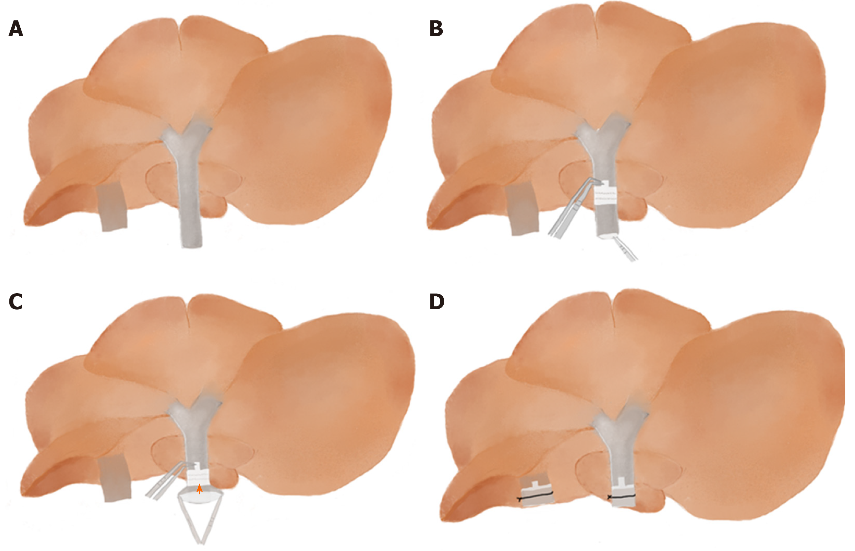
Figure 2 A simplified method of cuff technique.
A: Make sure the whole donor liver is immersed in water and rotate the donor liver floating in the dish so that the inferior surface faces upward; B: Pass the forceps through the lumen of the cuff to grasp the vessel so that the cuff is slipped over the vessel; C: Use forceps with a 45-degree angle to hold the extension of the cuff and use another curved round handle forceps to fold the end of the vessel over the cuff body to expose the inner endothelial surface and from one side to the other (orange arrow); D: Secure the PV(IHIVC) to the cuff by ligation. (CRITICAL STEP: Immerse donor liver under water so that the vessel wall naturally opens to facilitate vessel eversion).
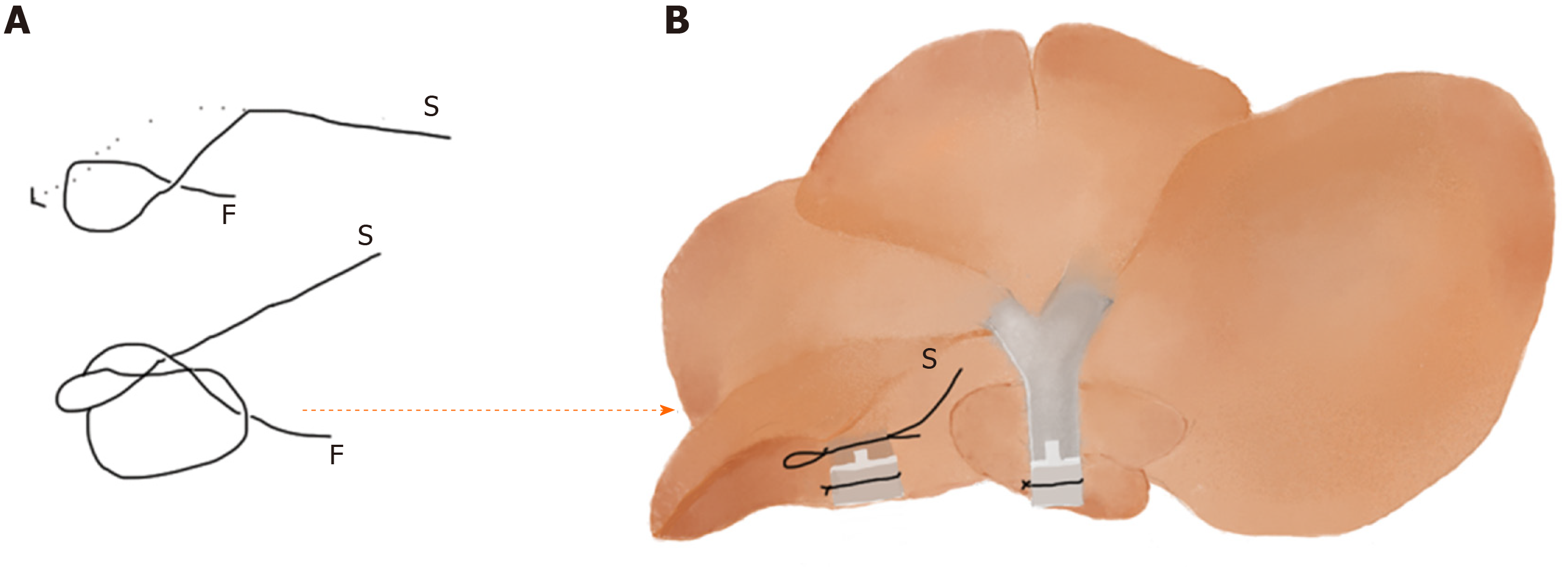
Figure 3 Use a slipknot to temporarily occlude donor infrahepatic inferior vena cava.
A: Demonstration of a slipknot: cross the fixed end (F) and the slip end (S), then keep F immobile and fold S to pass through the circle; B: Put the slipknot around the donor infrahepatic inferior vena cava (IHIVC) and fasten the slipknot. (Tip: After IHIVC anastomosis, pull S to loosen the slipknot).
- Citation: Li T, Hu Z, Wang L, Lv GY. Details determining the success in establishing a mouse orthotopic liver transplantation model. World J Gastroenterol 2020; 26(27): 3889-3898
- URL: https://www.wjgnet.com/1007-9327/full/v26/i27/3889.htm
- DOI: https://dx.doi.org/10.3748/wjg.v26.i27.3889


