Copyright
©The Author(s) 2020.
World J Gastroenterol. Jul 7, 2020; 26(25): 3603-3610
Published online Jul 7, 2020. doi: 10.3748/wjg.v26.i25.3603
Published online Jul 7, 2020. doi: 10.3748/wjg.v26.i25.3603
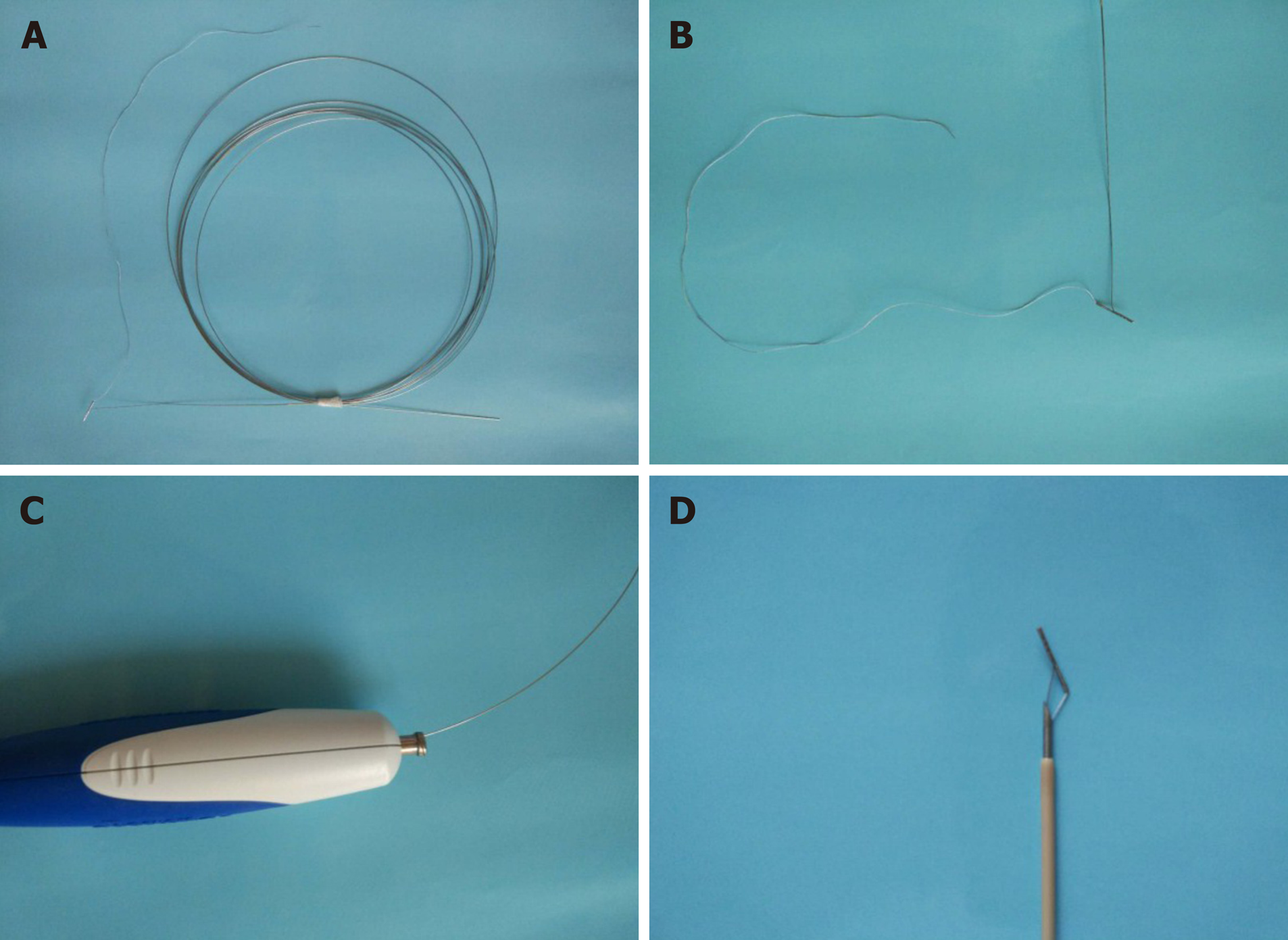
Figure 1 The retrievable puncture anchor.
A: The retrievable puncture anchor (Vedkang Inc., Changzhou, Jiangsu, China) in its entirety; B: The head of the anchor; C and D: A view of the passage of the anchor along the shaft of the puncture needle.
Figure 2 The retrievable puncture anchor traction method.
A: Schematic diagram: The bowel is punctured under endoscopic ultrasound (EUS) guidance; B: Endoscopic ultrasound image: The bowel is punctured under EUS guidance; C: Schematic diagram: The retrievable puncture anchor is passed through the needle; D: Endoscopic ultrasound image: The anchor is inserted into the bowel; E: Schematic diagram: After pulling out the needle, the anchor attaches to the bowel; F: Endoscopic ultrasound image: After pulling out the needle, the anchor attaches to the bowel; G: Schematic diagram: The identified small-bowel loop is punctured again under EUS guidance. A guidewire is inserted through the needle. The small bowel is shown being punctured and drained using electrocautery-enhanced delivery of lumen-apposing metal stents over the guidewire; H: Endoscopic ultrasound image: The anchor-grasped bowel is shown under traction for stent implantation; I: Schematic diagram: The retrieval cord is pulled using a pair of forceps; J: Endoscopy image: The retrieval cord is pulled using a pair of forceps; K: Schematic diagram: The retrievable puncture anchor is easily removed; L: Endoscopy image: The retrievable puncture anchor is easily removed.
Figure 3 Endoscopic imaging of stent placement and anastomoses.
A and B: Endoscopic imaging shows the well-reflected proximal flange of the stent immediately after deployment in the initial session; C: The stents were still in the same position 4 wk later and no traumatic changes were observed; D: A standard gastroscope was easily advanced through the anastomosis to observe the afferent and efferent bowel loops.
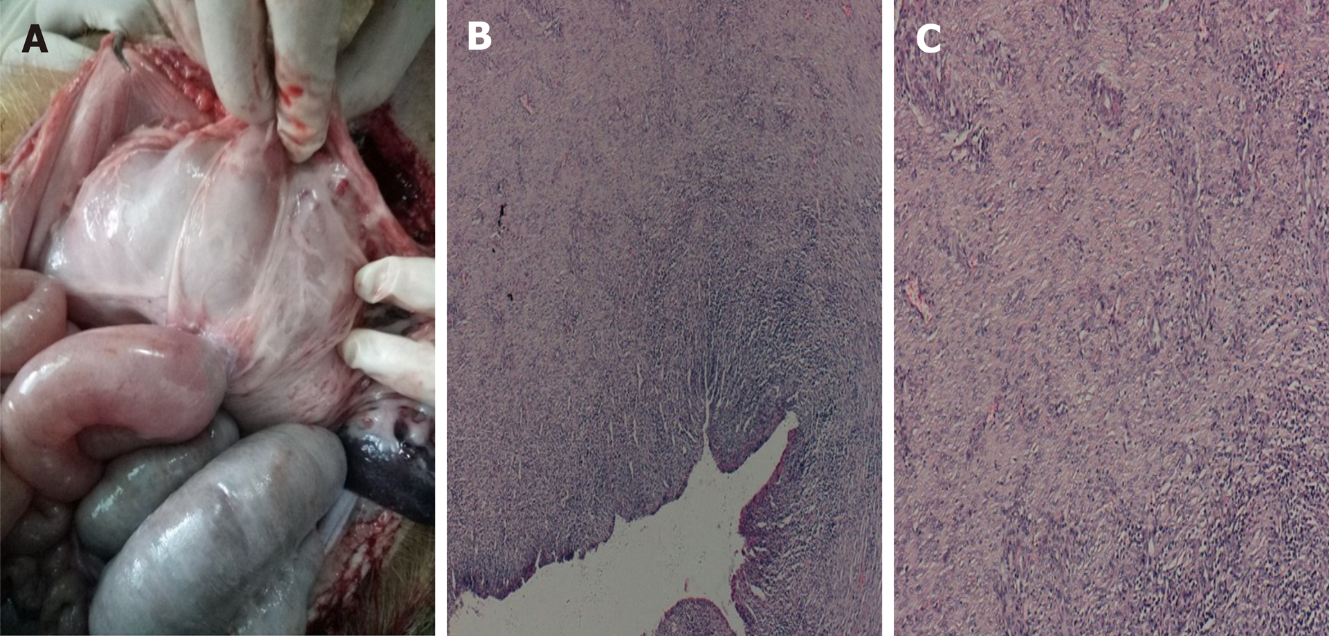
Figure 4 Autopsy and histological findings.
A-C: Necropsy and histological evidence shows complete adhesion between the bowel and the stomach wall (20 ×).
- Citation: Wang GX, Zhang K, Sun SY. Retrievable puncture anchor traction method for endoscopic ultrasound-guided gastroenterostomy: A porcine study. World J Gastroenterol 2020; 26(25): 3603-3610
- URL: https://www.wjgnet.com/1007-9327/full/v26/i25/3603.htm
- DOI: https://dx.doi.org/10.3748/wjg.v26.i25.3603


