Copyright
©The Author(s) 2020.
World J Gastroenterol. May 28, 2020; 26(20): 2618-2631
Published online May 28, 2020. doi: 10.3748/wjg.v26.i20.2618
Published online May 28, 2020. doi: 10.3748/wjg.v26.i20.2618
Figure 1 Fundic gland type adenocarcinoma.
A: Cancer cells with enlarged nuclei formed irregular glands. The surface of the tumor was covered with non-cancerous epithelium, hematoxylin and eosin, original magnification 4 ×; B: The tumor invaded into the submucosal layer in a state that kept the muscularis mucosa. The distance from muscularis mucosa was 75 μm, hematoxylin and eosin, original magnification 40 ×; C: PG-I stains showed focal positive for cancer cells; D: The submucosal tumor like a small protrusion on the greater curvature of the upper gastric body, white light endoscopy; E: The surface of the tumor was covered with normal epithelium, magnifying endoscopy with narrow-band imaging.
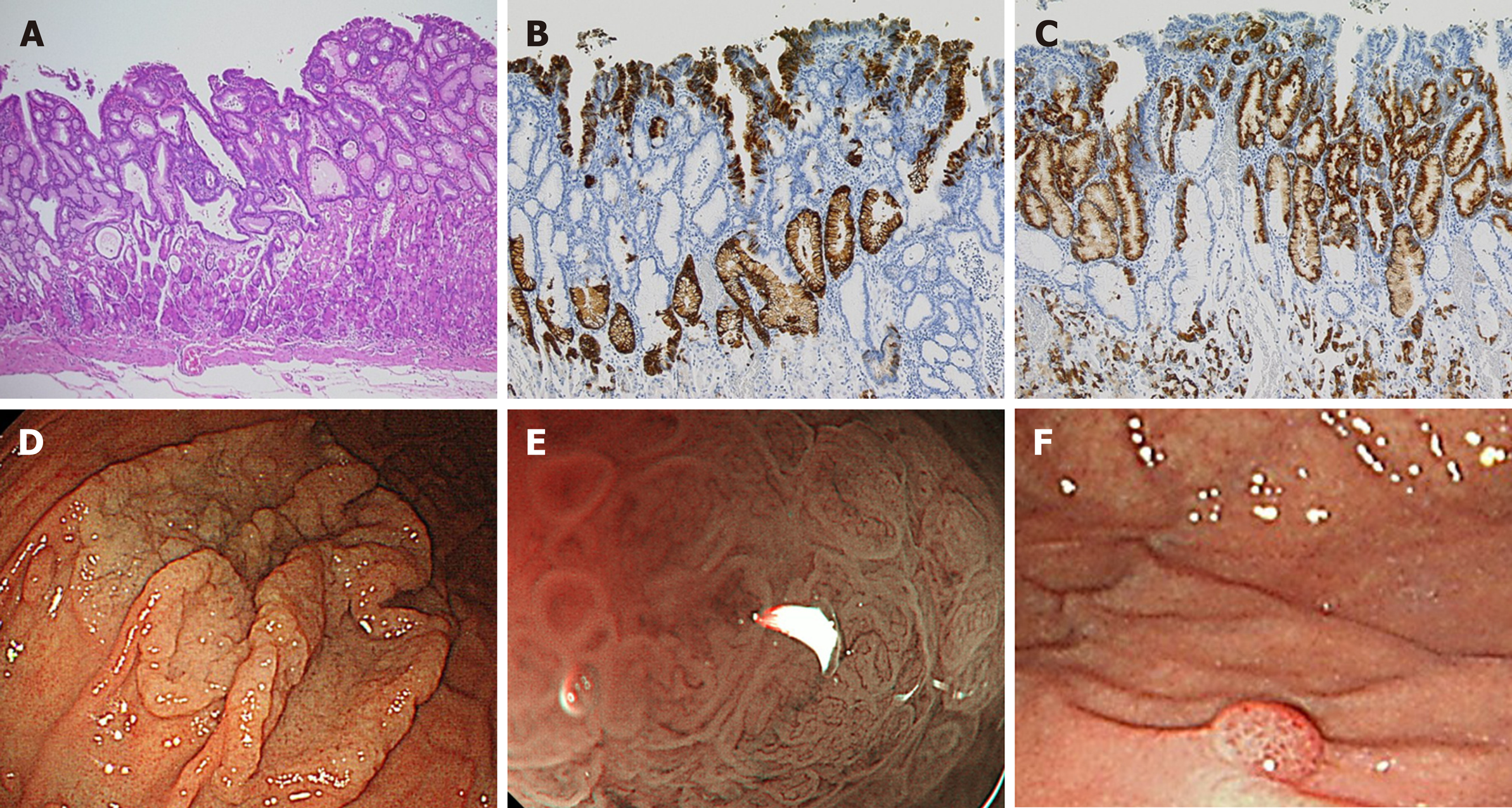
Figure 2 Foveolar type adenocarcinoma.
A: The villous structure composed of dysplastic columnar cells with clear cytoplasm. Hematoxylin and eosin, original magnification 4 ×; B: MUC5AC was positive for cancer; C: MUC6 was negative for cancer but positive for expanded noncancerous glands in the middle layer of mucosa; D: Whitish laterally spreading elevated lesion was seen at the greater curvature of the upper gastric body, white light endoscopy; E: A papillary or villous like fine mucosal pattern with intra-structural irregular vessels in all lesions, one lesion showed raspberry-like appearance magnifying endoscopy with narrow-band imaging; F: One lesion showed raspberry-like appearance.
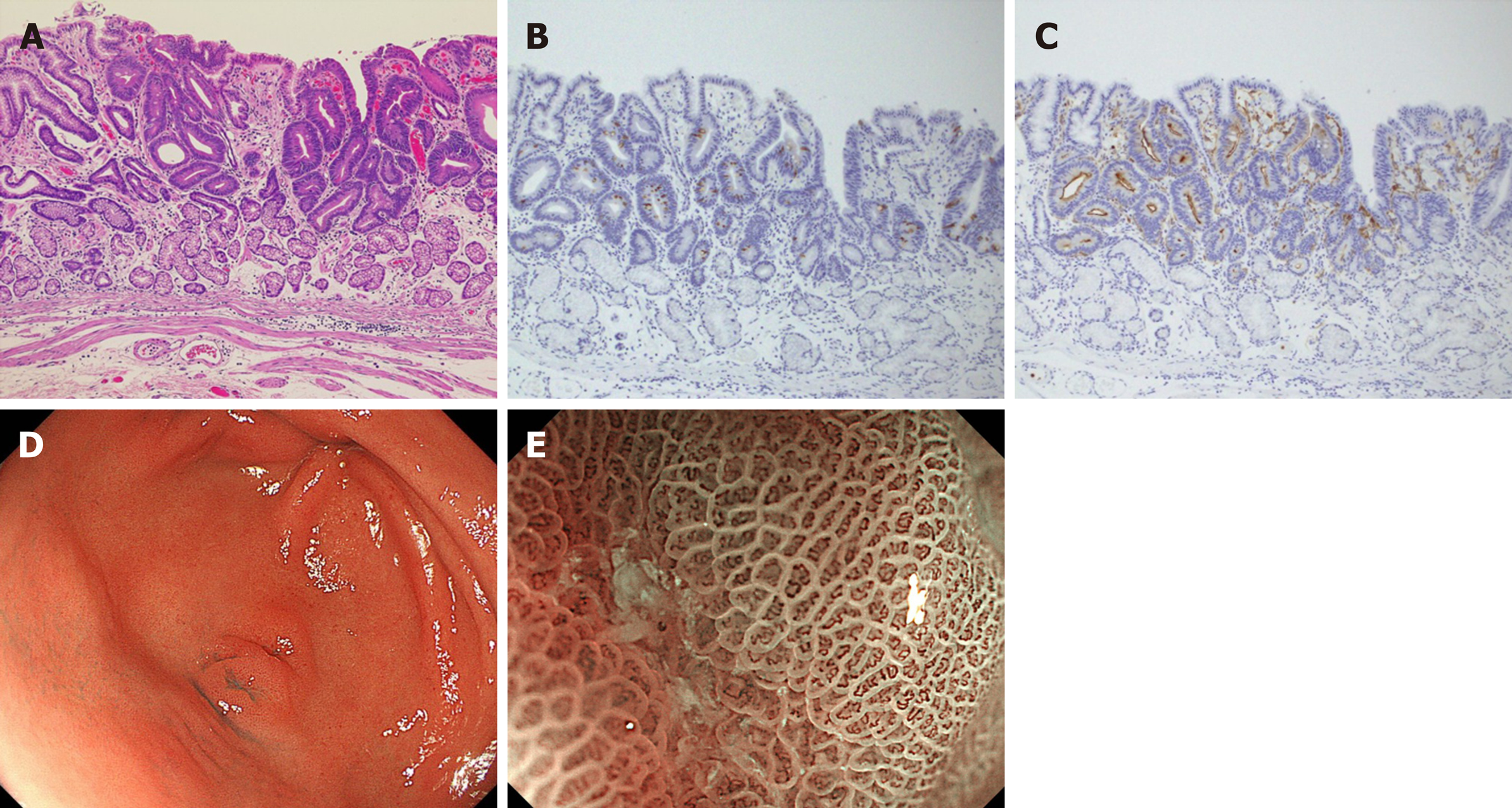
Figure 3 Intestinal-type adenocarcinoma.
A: The tubular structures lined by tall columnar cells with hyperchromatic, pencillate, and pseudostratified nuclei, hematoxylin and eosin, original magnification 4 ×; B: MUC2 was weakly positive for cancer cells; C: CD10 was positive for cancer cells; D: 0-IIa+IIc type tumor mimicking verrucosa on the gastric antrum, white light endoscopy; E: An irregular microvascular pattern and irregular microsurface pattern with a demarcation line were observed just in the recessed area, magnifying endoscopy with narrow-band imaging.
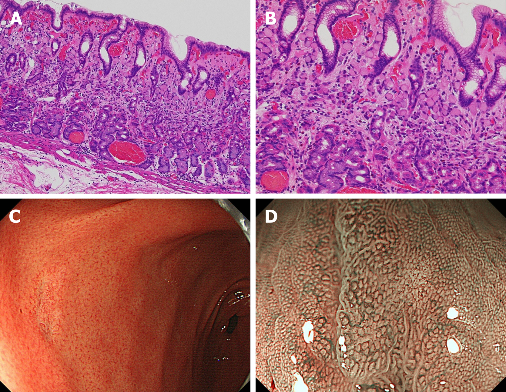
Figure 4 Pure signet-ring cell carcinoma.
A, B: Pure signet ring cell carcinoma were observed proliferative zone to the surface layer of mucosa, hematoxylin and eosin, A: Original magnification 4 ×; B: High magnification 40 ×; C: Discolored slightly depressed lesion on the gastric antrum, white light endoscopy; D: The surface of the tumor was covered with normal mucosa, magnifying endoscopy with narrow-band imaging
- Citation: Sato C, Hirasawa K, Tateishi Y, Ozeki Y, Sawada A, Ikeda R, Fukuchi T, Nishio M, Kobayashi R, Makazu M, Kaneko H, Inayama Y, Maeda S. Clinicopathological features of early gastric cancers arising in Helicobacter pylori uninfected patients. World J Gastroenterol 2020; 26(20): 2618-2631
- URL: https://www.wjgnet.com/1007-9327/full/v26/i20/2618.htm
- DOI: https://dx.doi.org/10.3748/wjg.v26.i20.2618


