©The Author(s) 2020.
World J Gastroenterol. May 7, 2020; 26(17): 2012-2029
Published online May 7, 2020. doi: 10.3748/wjg.v26.i17.2012
Published online May 7, 2020. doi: 10.3748/wjg.v26.i17.2012
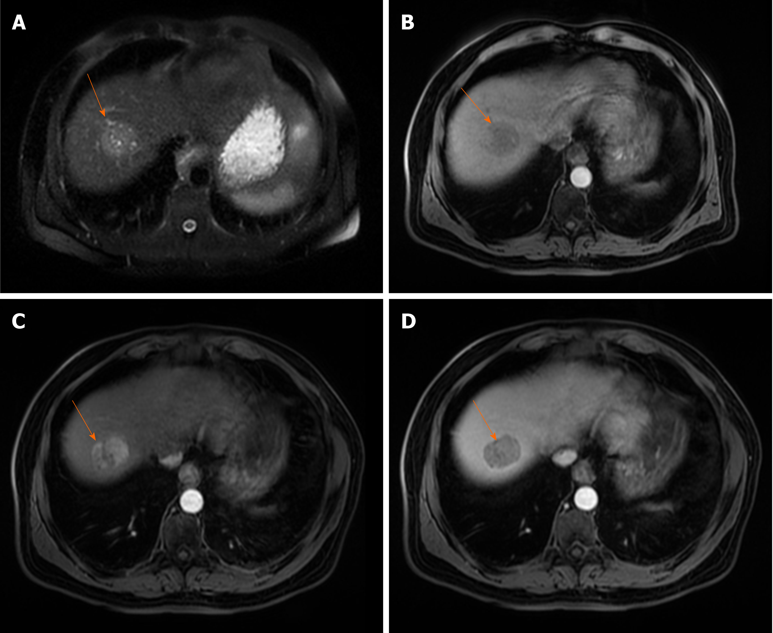
Figure 1 Typical hepatocellular carcinoma in 68-year old man with cirrhosis.
A: On axial T2-weighted image a nodular heterogeneously hyperintense tumor (arrow) is seen; B-D: The lesion (arrow) is hypointense on T1-weighted image (B), hypervascular on arterial phase (C) with washout in portal venous phase (D).
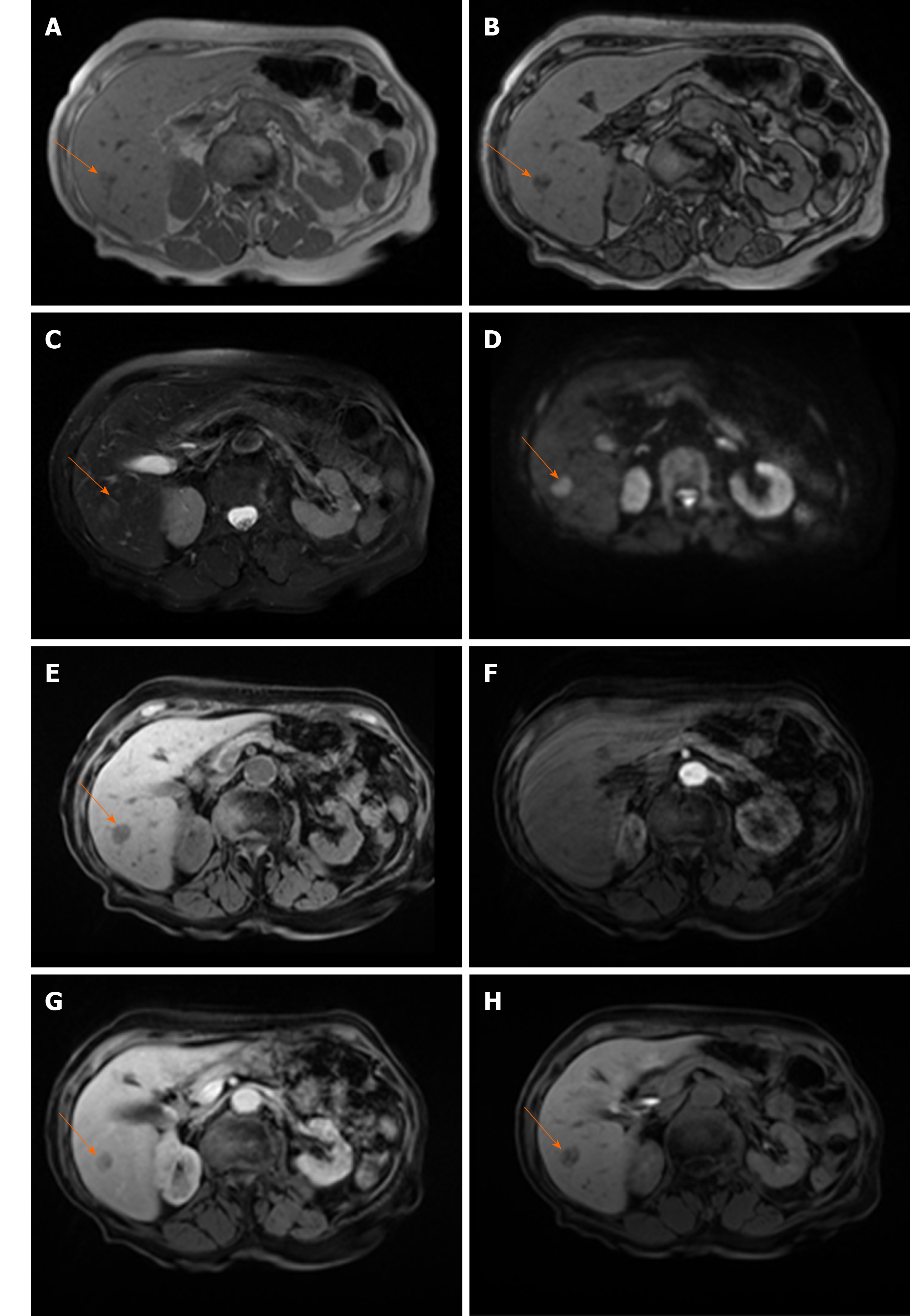
Figure 2 Hypovascular hepatocellular carcinoma in 58-year old woman with cirrhosis.
A: On axial in-phase image tumor (arrow) is isointense with surrounding liver parenchyma; B: On opposed-phase image there is a partial drop of signal intensity in the lesion corresponding to the fatty component; C-E: The lesion (arrow) is slightly hyperintense on T2-weighted FS image (C), hyperintense on diffusion weighted imaging (D), and hypointense on T1-weighted FS image (E); F and G: On arterial phase (F) the lesion is isointense with washout in portal venous phase (G); H: On hepatobiliary phase after administration of gadoxetic acid the tumor (arrow) is hypointense.
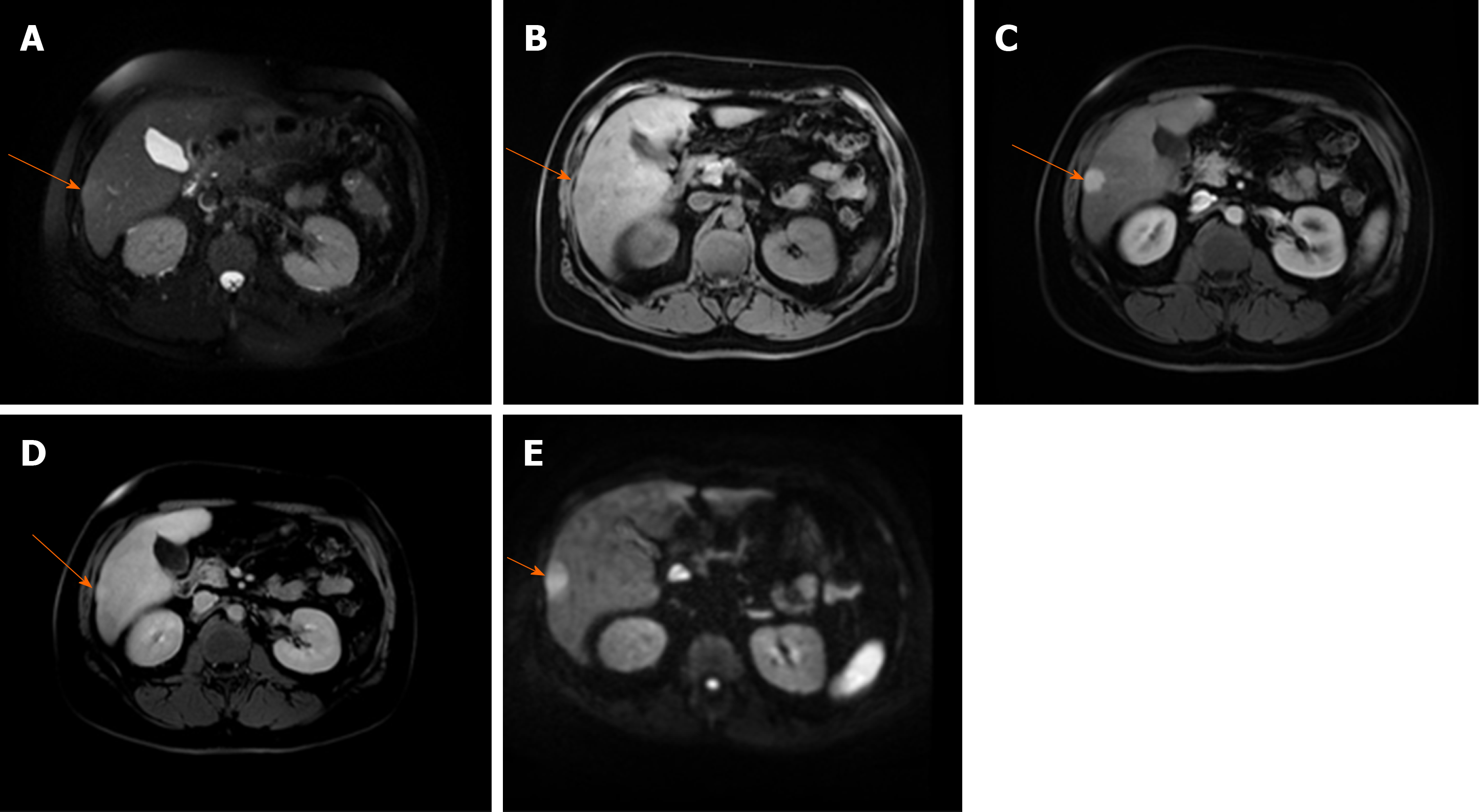
Figure 3 Hepatocellular carcinoma in 64-year old man with cirrhosis.
A: Axial T2-weighted FS image shows slightly hyperintense nodular lesion (arrow) located in segment VI; B-D: The lesion is hypointense on T1-weighted fat-saturated image (B), hypervascular on arterial phase (C) without washout on portal-venous phase (D); E: The tumor (arrow) is hyperintense on diffusion weighted imaging.
Figure 4 Hepatocellular carcinoma in 73-year old man with alcoholic cirrhosis.
A: Axial T2-weighted fat-saturated image shows slightly hyperintense well-defined nodular lesion (arrow) in segment Iva; B-D: The lesion (arrow) is hypointense on T1-weighted FS image (B), hypevascular on arterial phase (C) without washout on portal-venous phase (D); E and F: On hepatobiliary phase the tumor is strongly hyperintense (E) with diffusion restriction on diffusion-weighted image (F).
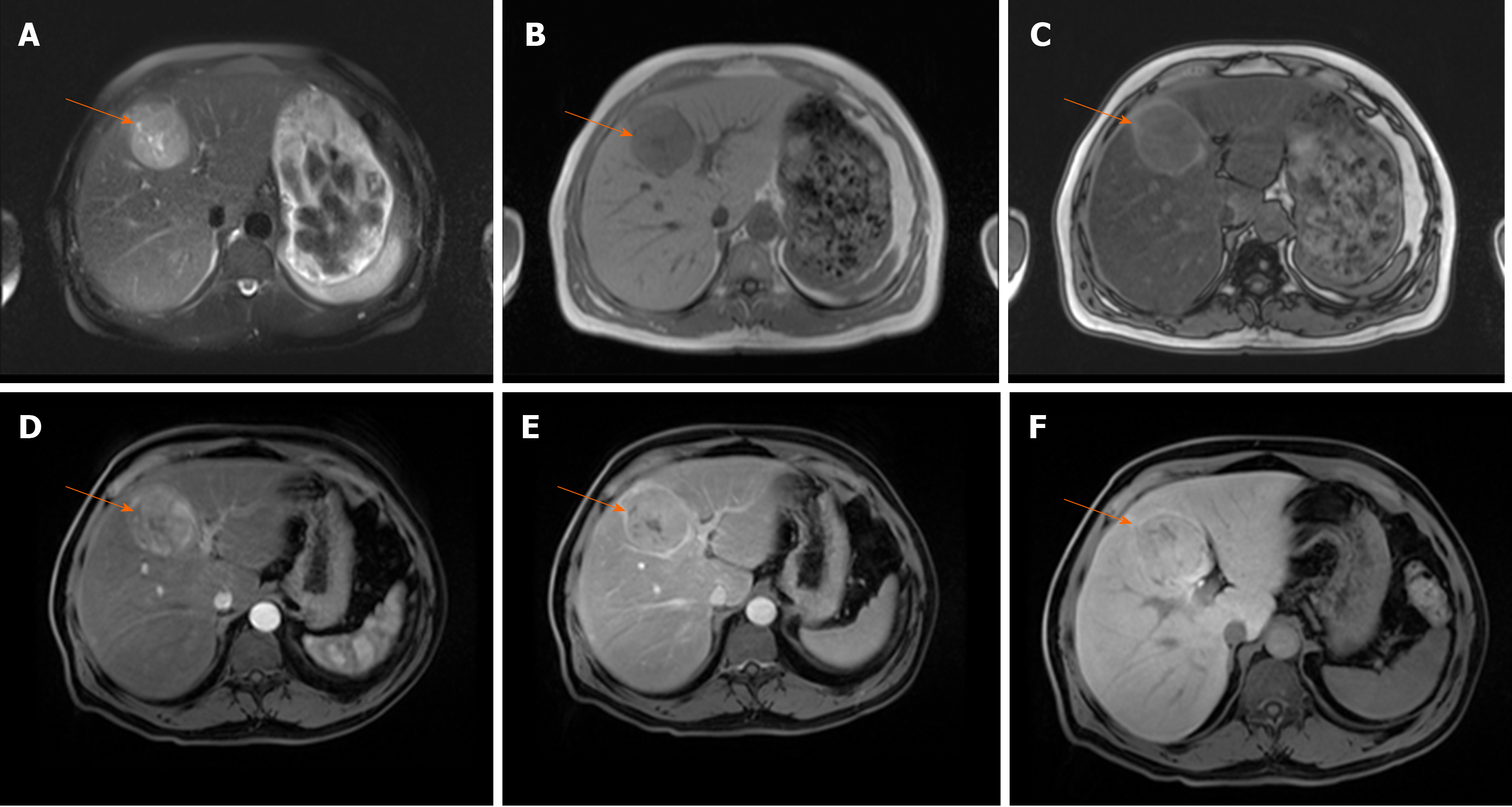
Figure 5 Hepatocellular carcinoma in 54-year old man with non-alcoholic fatty liver disease.
A: Axial T2-weighted FS image shows hyperintense lesion (arrow) in segment Ivb; B and C: Dual-echo images show that tumor (arrow) is hypointense on in-phase image (B) without signal drop on opposed-phase image, while background liver parenchyma shows diffuse signal drop as a consequenence of fatty liver disease (C); D and E: The lesion (arrow) is hypervascular on arterial phase (D) without washout on portal-venous phase (E); F: On hepatobiliary phase the nodule (arrow) is isointense with surrounding liver parenchyma.
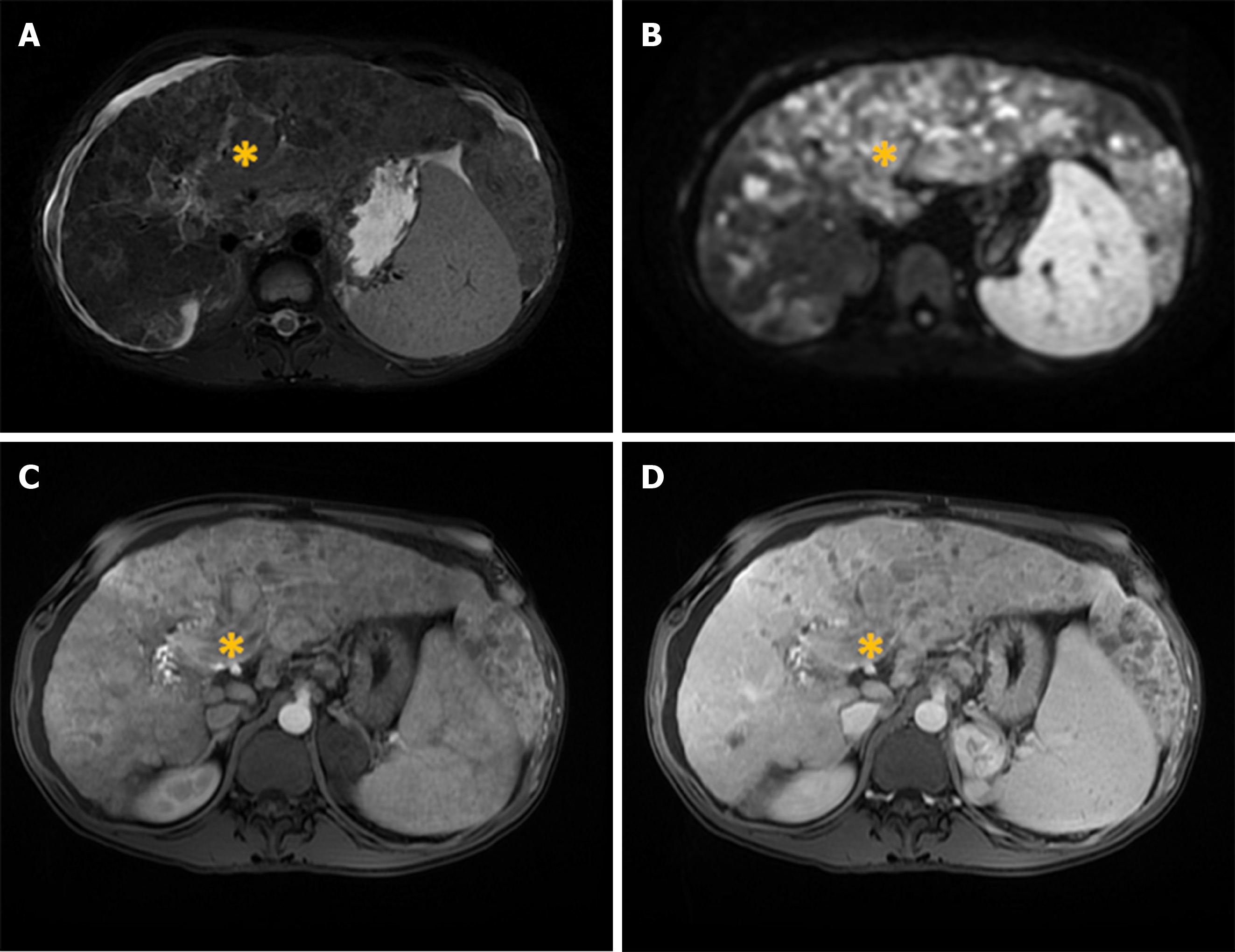
Figure 6 Diffuse hepatocellular carcinoma in 38-year-old man with long-standing Wilson disease.
Axial T2-weighted FS image shows multiple moderately hyperintense nodules scattered throughout liver parenchyma. A: Note also portal vein thrombosis with signal intensity of the thrombus similar to tumor nodules in the liver; B: On diffusion weighted image diffuse hyperintensity is seen corresponding to multiple tumor nodules; C and D: On arterial phase patchy enhancement can be seen including portal vein thrombus (C), with heterogeneous washout in portal vein phase (D).
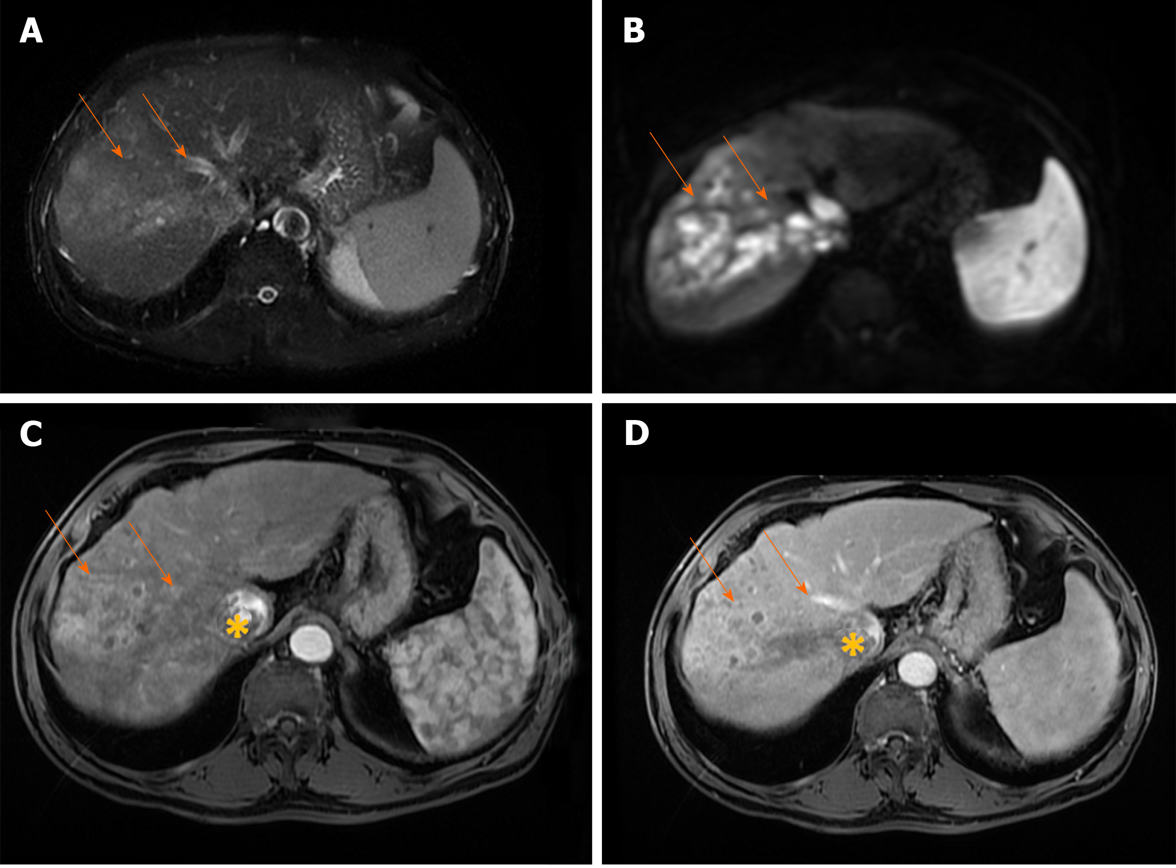
Figure 7 Infiltrative hepatocellular carcinoma in 71-year old man with cirrhosis.
A: Axial T2-weighted FS image shows ill-defined mass (arrows) in segments VII and VIII; B: On diffusion weighted image the lesion is hyperintense; C and D: The tumor is heterogeneously hyperintense on arterial phase (C), with washout in portal vein phase (D). Note also right hepatic vein thrombosis with propagation of the tumor thrombus in vena cava inferior (asterix).
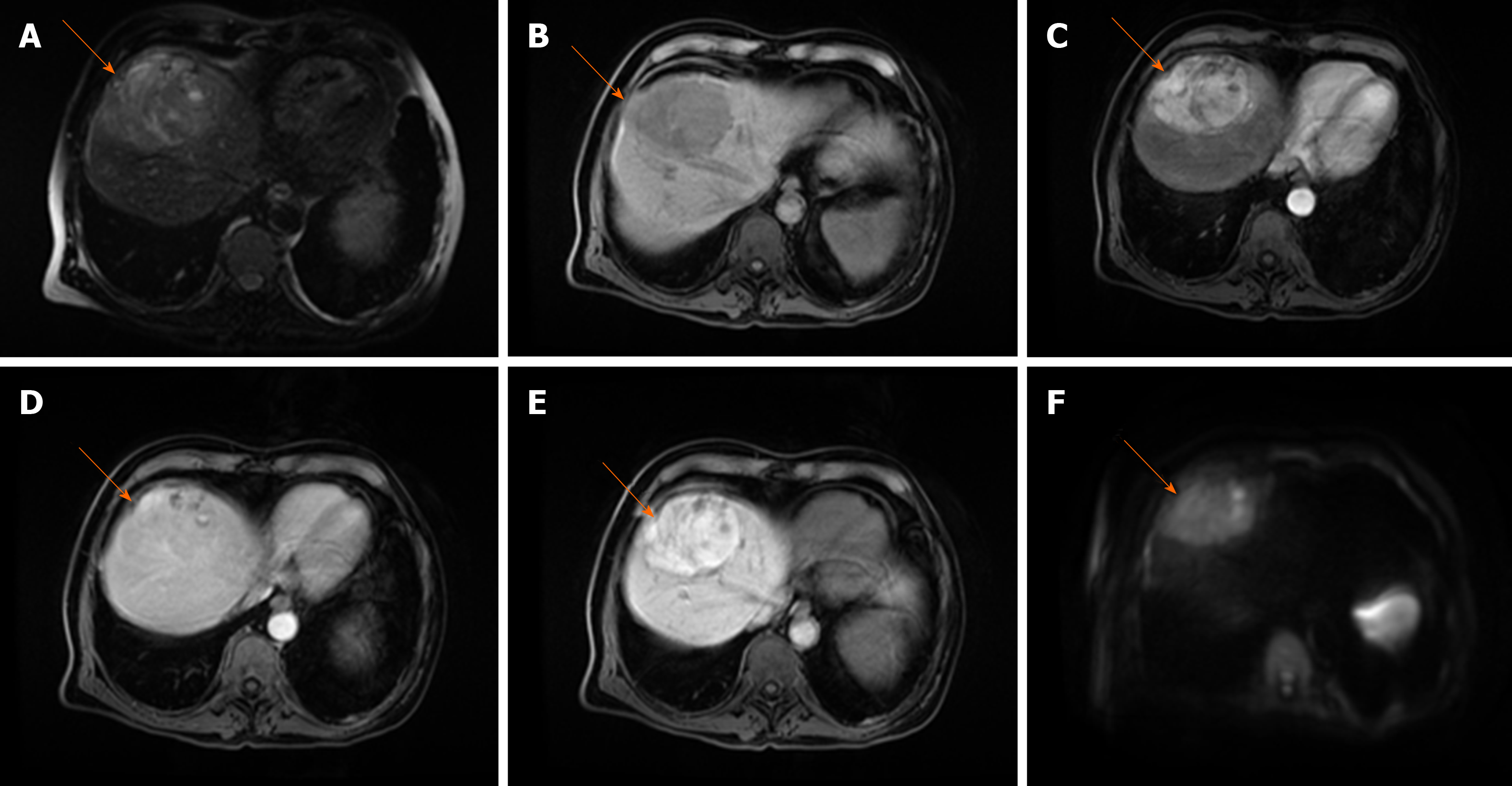
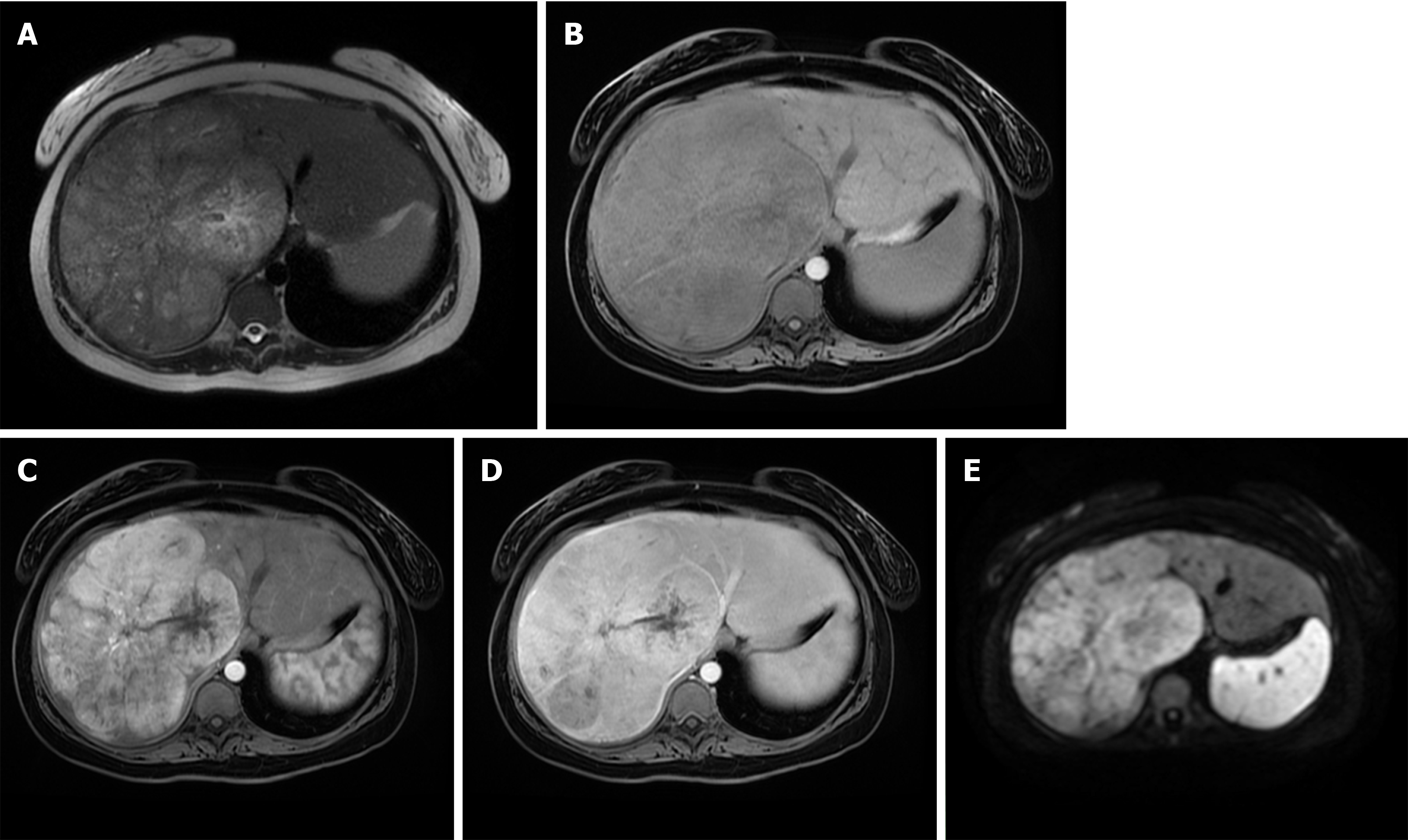
Figure 8 Fibrolamellar hepatocellular carcinoma in 23-year old woman without chronic liver disease.
A: Axial T2-weighted image shows large heterogeneous mass occupying almost whole right liver lobe; B-E: The tumor is hypointense on T1-weghted FS image (B), hypervascular on arterial phase (C) with washout in parts of the lesion on portal venous phase (D) and diffusion restriction (E).
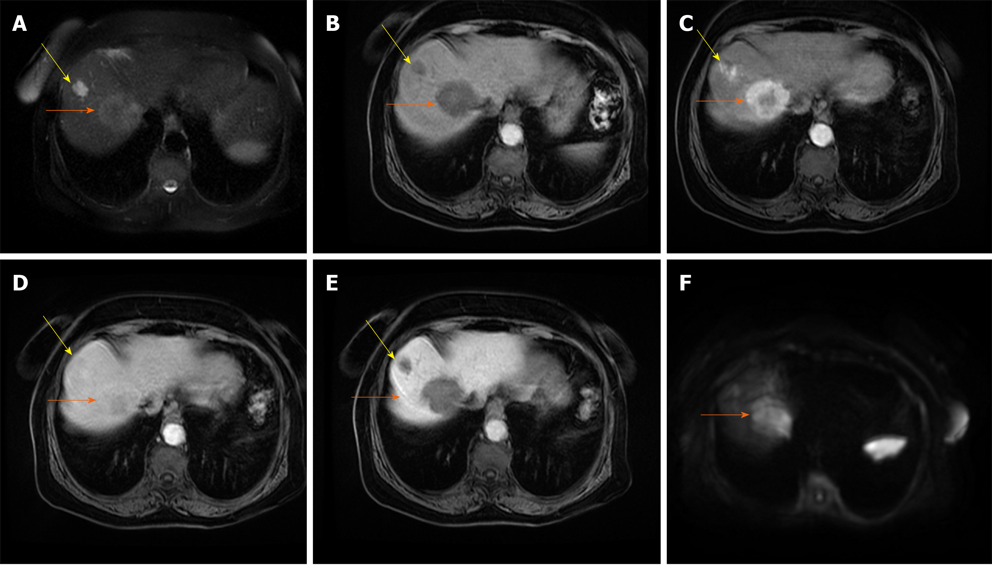
Figure 9 Combined hepatocellular carcinoma and cholangiocarcinoma in 65-year old woman without chronic liver disease.
A: Axial T2-weighted image shows lobulated heterogeneous mass (orange arrow) located centrally in segments VII and VIII; B-D: The tumor is hypointense on T1-weghted fat-saturated image (B), with thick rim of enhancement on arterial phase (C) and progressive enhancement on portal venous phase (D); E and F: The tumor (orange arrows) is hypointense on hepatobiliary phase (E) and hyperintense on diffusion-weighted image (F). Note also hemangioma peripherally in segment VIII (yellow arrow).
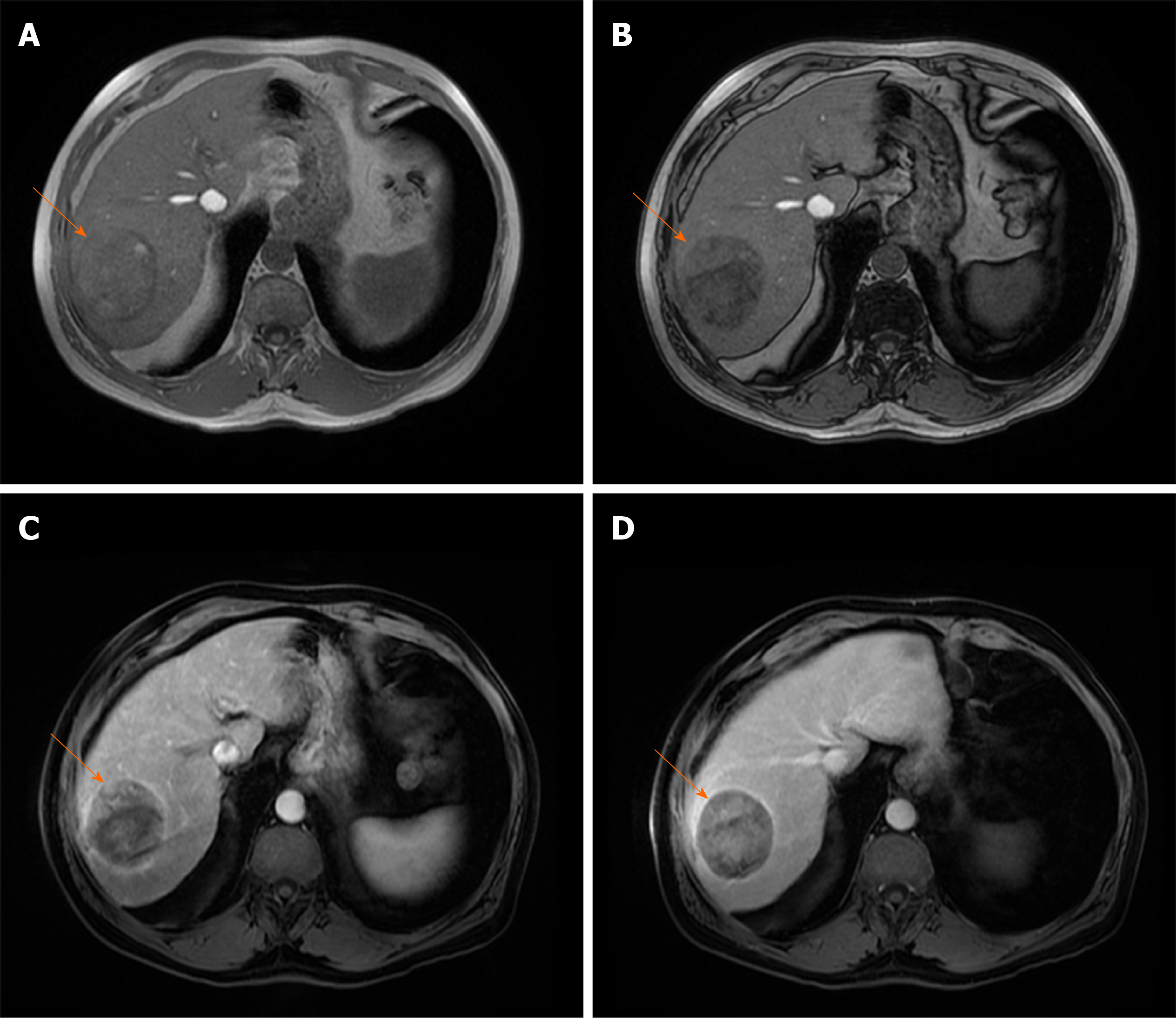
Figure 10 Steatotic hepatocellular carcinoma in 67-year old man with cirrhosis.
A and B: T1-weighted in-phase image shows nodular tumor in segment VII (arrow) which is mostly isointense with surrounding liver parenchyma (A) with signal drop of posterior part of the lesion on opposed-phase image (B); C and D: On arterial phase image hypervascularization is clearly seen for nonstetaotic part of the tumor (C) with washout on portal-venous phase (D). Note relative lack of hypervascularity in steatotic part of the tumor, and extracapsular growth on the lateral border of the lesion.
Figure 11 Scirrhous hepatocellular carcinoma in 68-year old woman with chronic hepatitis C infection.
A: Coronal T2-weighted image shows moderately hyperintense subcapsullary located lesion in segments VI and V (orange arrow); B-D: The tumor (orange arrows) is hypointense on axial T1-weighted FS image (B), hypervascular on arterial phase (C) with only small regions of washout in portal venous phase (D); E: On diffusion-weighted image the lesion is hyperintense. Note also capsular retraction on A (yellow arrow).
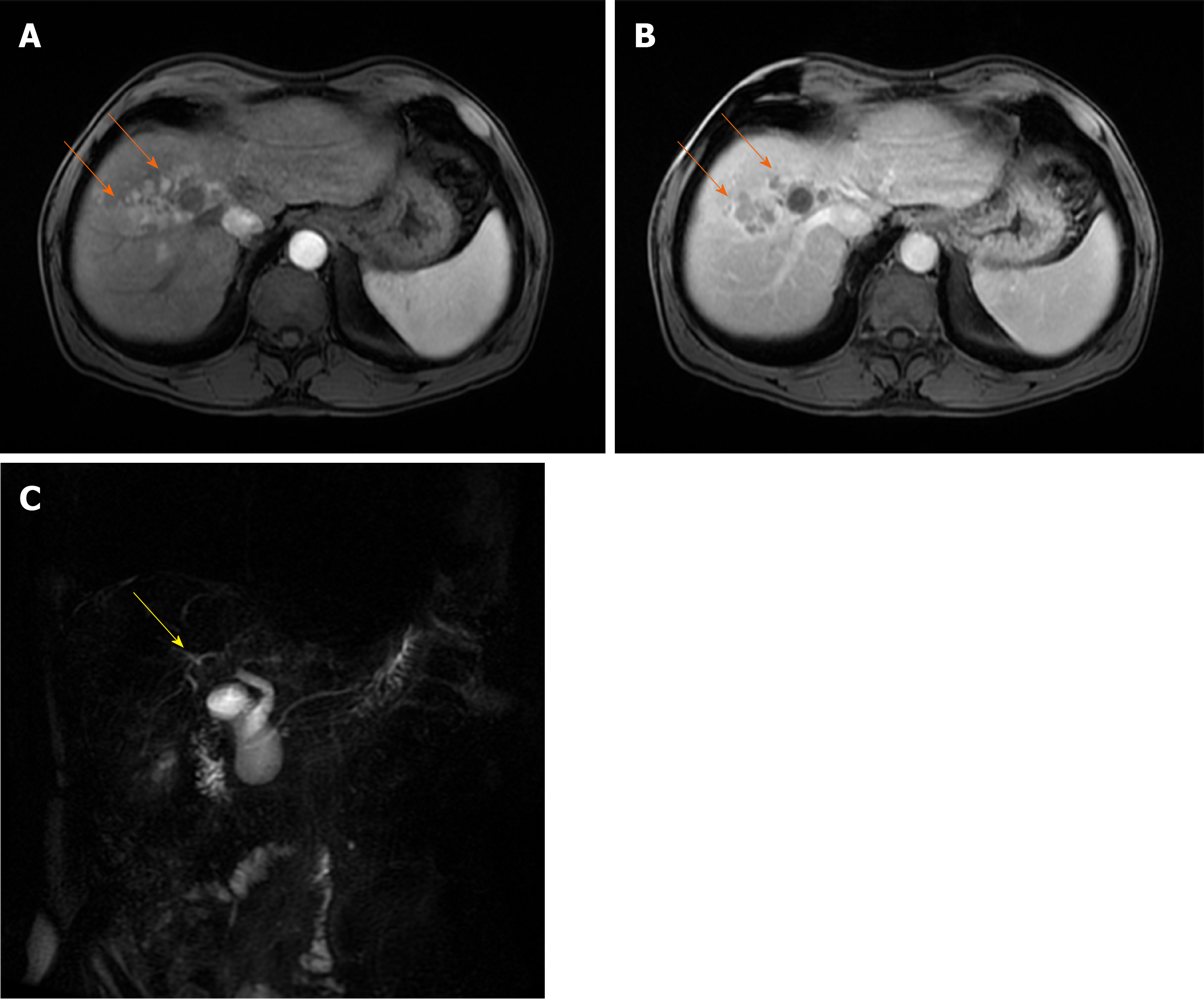
Figure 12 Infiltrative hepatocellular carcinoma in 63-year old man with intra-bile tumor growth.
A and B: On arterial phase image ill-defined hypervascular lesion (arrows) in segment VIII is seen (A) with washout in portal venous phase (B); C: Intra-bile tumor growth is better depicted on coronal magnetic resonance cholangiopancreatography image seen as defect in the lumen right hepatic duct and its posterior branch (yellow arrow).
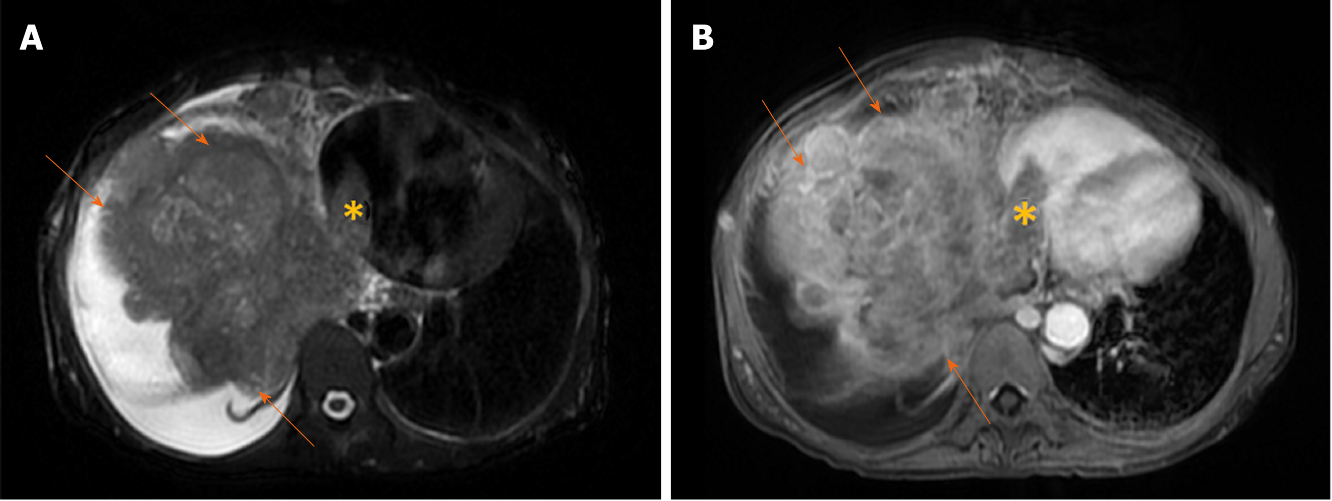
Figure 13 Massive hepatocellular carcinoma in 69-year old woman with cirrhosis.
A: Axial T2-weighted image shows a large mass (arrows) predominantly located in right liver lobe with infiltration of vena cava and tumor thrombus seen in left atrium (asterix); B: Axial image in portal venous phase shows washout in hepatocellular carcinoma (arrows) and thrombus (asterix) in left atrium.
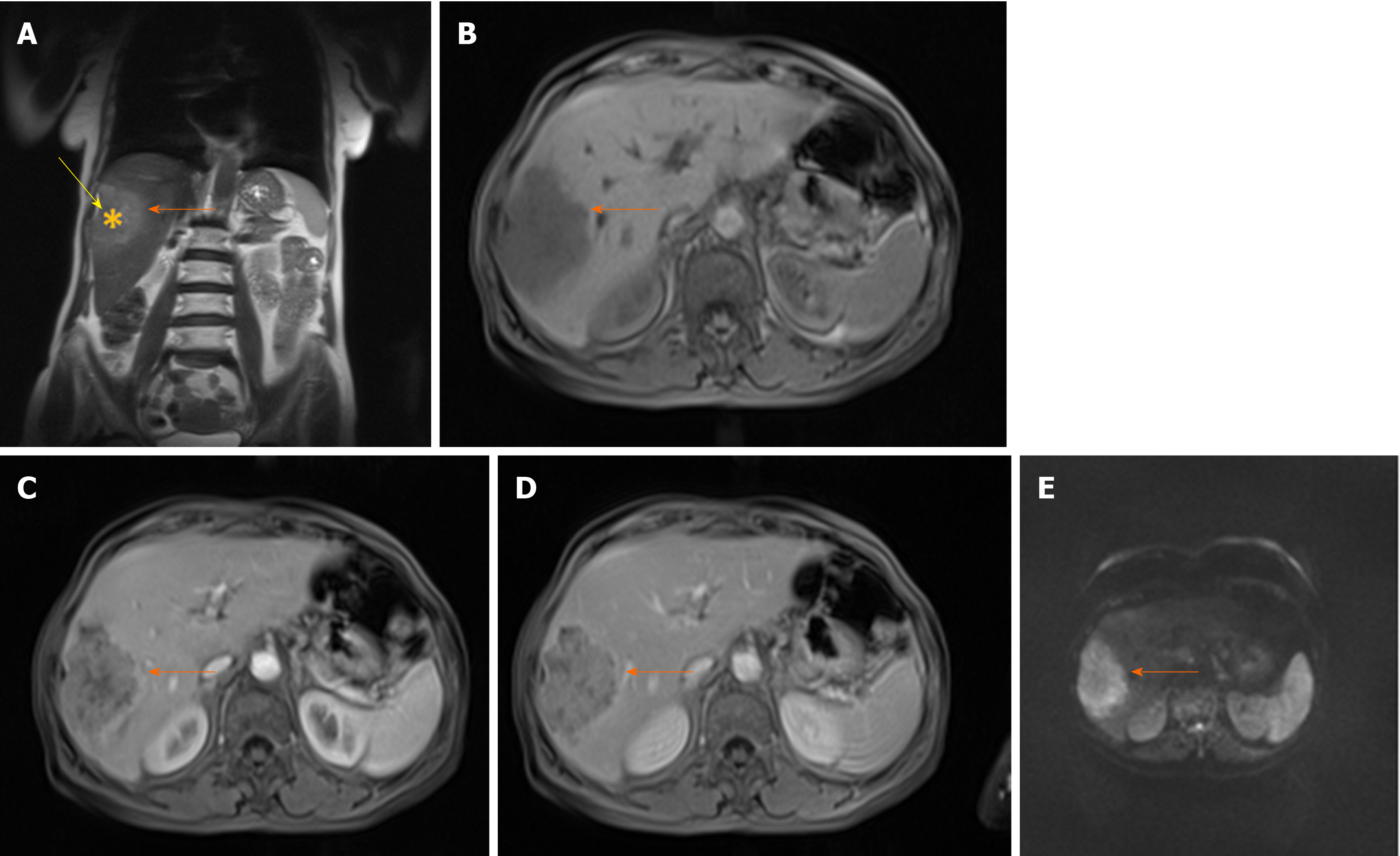
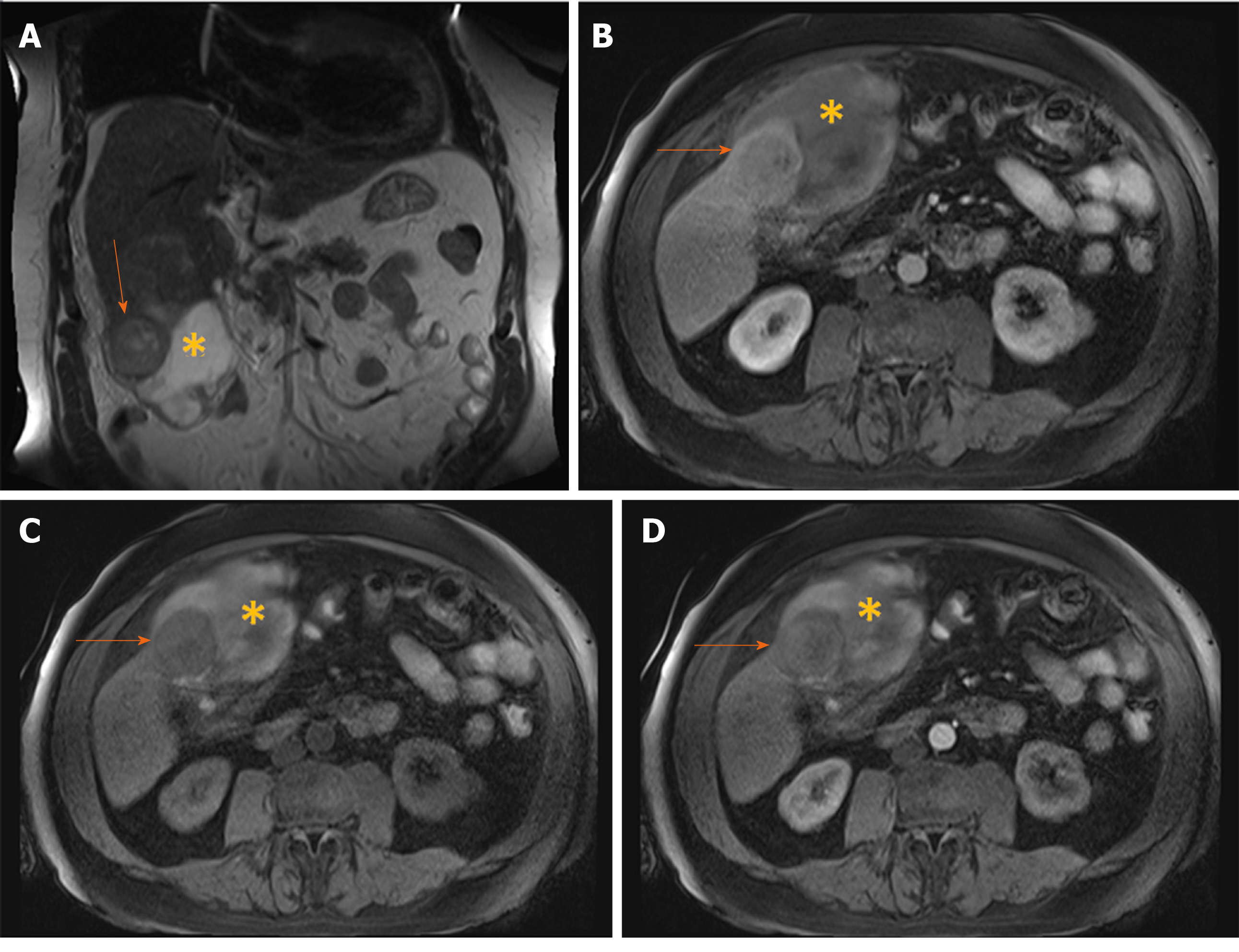
Figure 14 Hemorrhagic hepatocellular carcinoma in 70-year old man with cirrhosis presenting with acute abdominal pain.
A: Coronal T2-weighted image shows a nodular tumor (arrows) in the subcapsular location in segment V, and subhepatic hematoma (asterix); B: Axial T1-weighted FS image shows hypointense tumor (arrows) and hyperintense content of the hematoma (asterix); C and D: Arterial phase displays only subtle hypervascularity in the part of the tumor (arrows) depicted on this section (C) with washout in portal venous phase (D), and hematoma (asterix) located anteriorly.
- Citation: Kovac JD, Milovanovic T, Dugalic V, Dumic I. Pearls and pitfalls in magnetic resonance imaging of hepatocellular carcinoma. World J Gastroenterol 2020; 26(17): 2012-2029
- URL: https://www.wjgnet.com/1007-9327/full/v26/i17/2012.htm
- DOI: https://dx.doi.org/10.3748/wjg.v26.i17.2012


