©The Author(s) 2020.
World J Gastroenterol. Apr 28, 2020; 26(16): 1847-1860
Published online Apr 28, 2020. doi: 10.3748/wjg.v26.i16.1847
Published online Apr 28, 2020. doi: 10.3748/wjg.v26.i16.1847
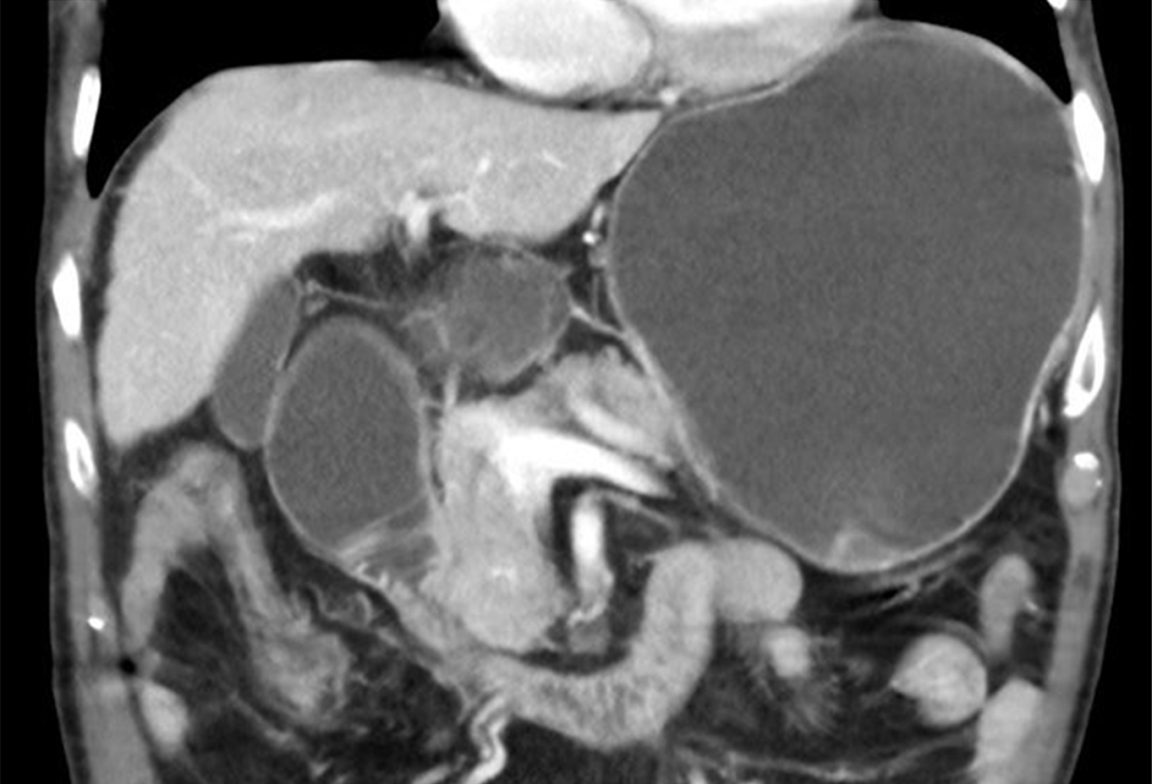
Figure 1 Computed tomography scan appearance of malignant duodenal stricture with gastric distension due to pancreatic cancer.
Figure 2 Graphic representation of the main approaches applied to manage malignant gastric outlet obstruction.
A: Surgical gastrojejunostomy; B: Endoscopic enteral stenting with self-expanding metal stents; C: Endoscopic ultrasound-guided gastroenterostomy.
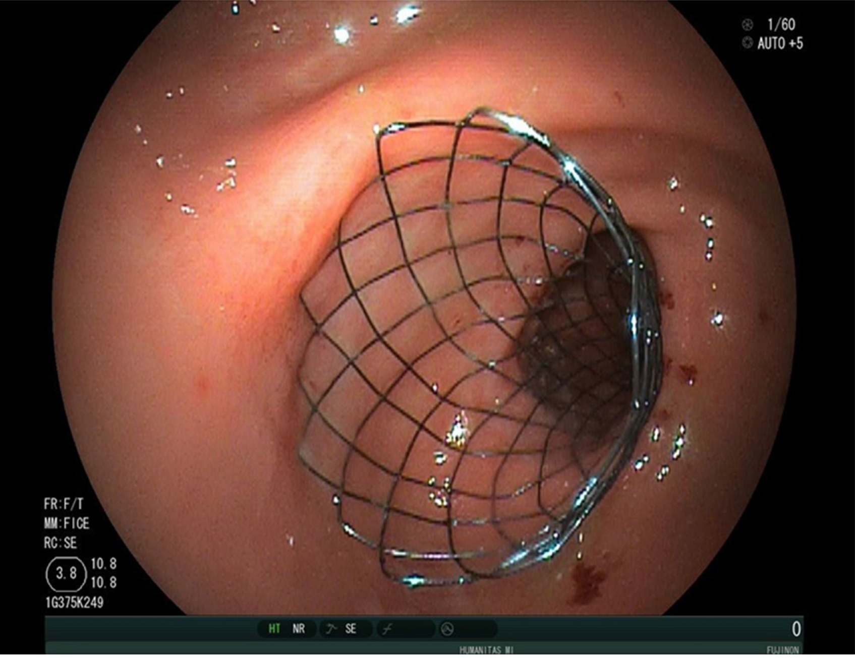
Figure 3 Final endoscopic appearance of a duodenal uncovered self-expanding metal stent deployed across duodenal stricture, in a patient with gastric outlet obstruction due to pancreatic cancer.
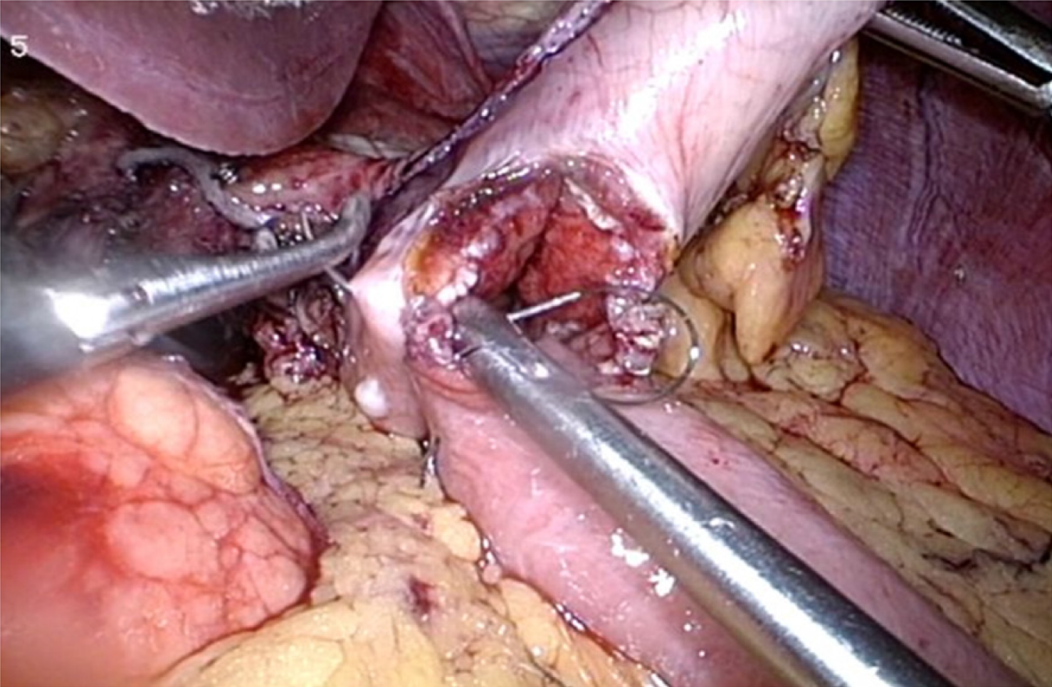
Figure 4 Intra-operative image of laparoscopic gastrojejunostomy.
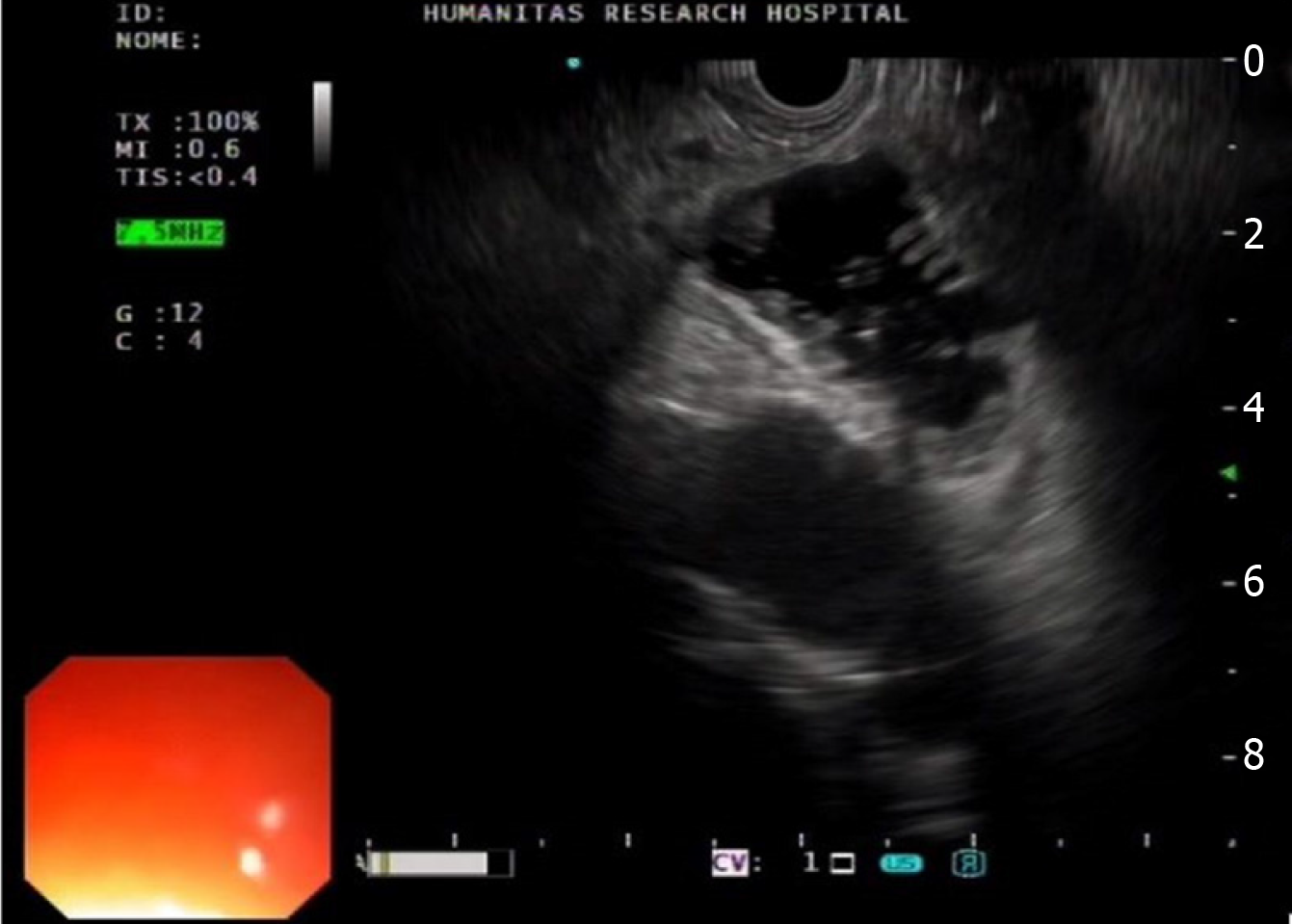
Figure 5 Endoscopic ultrasound view of the distended jejunal loop.
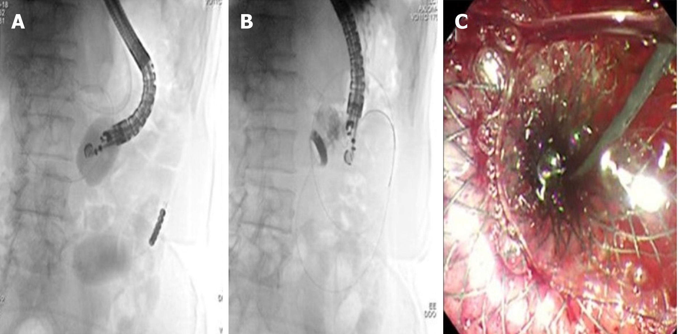
Figure 6 Endoscopic ultrasound-guided gastroenterostomy with the double balloon occluder.
A: The double balloon occluder in place distending the small bowel in between; B: Endoscopic ultrasound-guided placement of lumen-apposing metal stent between the stomach and jejunum; C: Final endoscopic appearance of lumen-apposing metal stent.
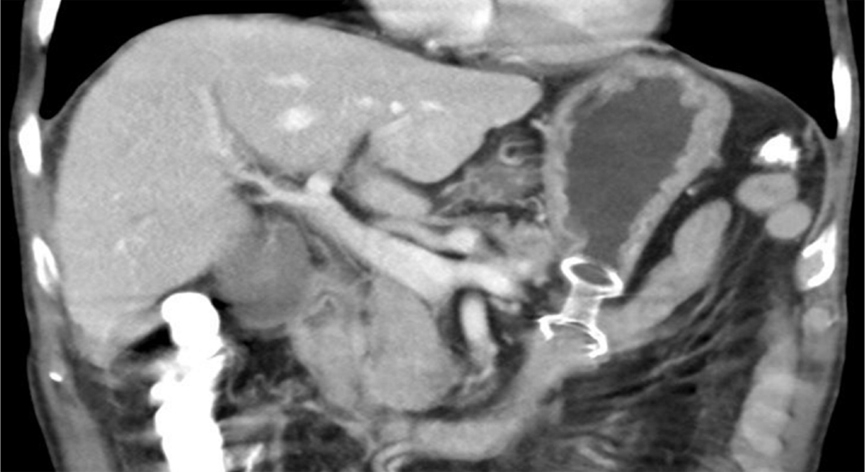
Figure 7 Computed tomography scan appearance of endoscopic ultrasound-guided gastroenterostomy with lumen-apposing metal stent placed between the stomach and jejunum.
- Citation: Troncone E, Fugazza A, Cappello A, Blanco GDV, Monteleone G, Repici A, Teoh AYB, Anderloni A. Malignant gastric outlet obstruction: Which is the best therapeutic option? World J Gastroenterol 2020; 26(16): 1847-1860
- URL: https://www.wjgnet.com/1007-9327/full/v26/i16/1847.htm
- DOI: https://dx.doi.org/10.3748/wjg.v26.i16.1847


