©The Author(s) 2019.
World J Gastroenterol. Feb 21, 2019; 25(7): 824-836
Published online Feb 21, 2019. doi: 10.3748/wjg.v25.i7.824
Published online Feb 21, 2019. doi: 10.3748/wjg.v25.i7.824
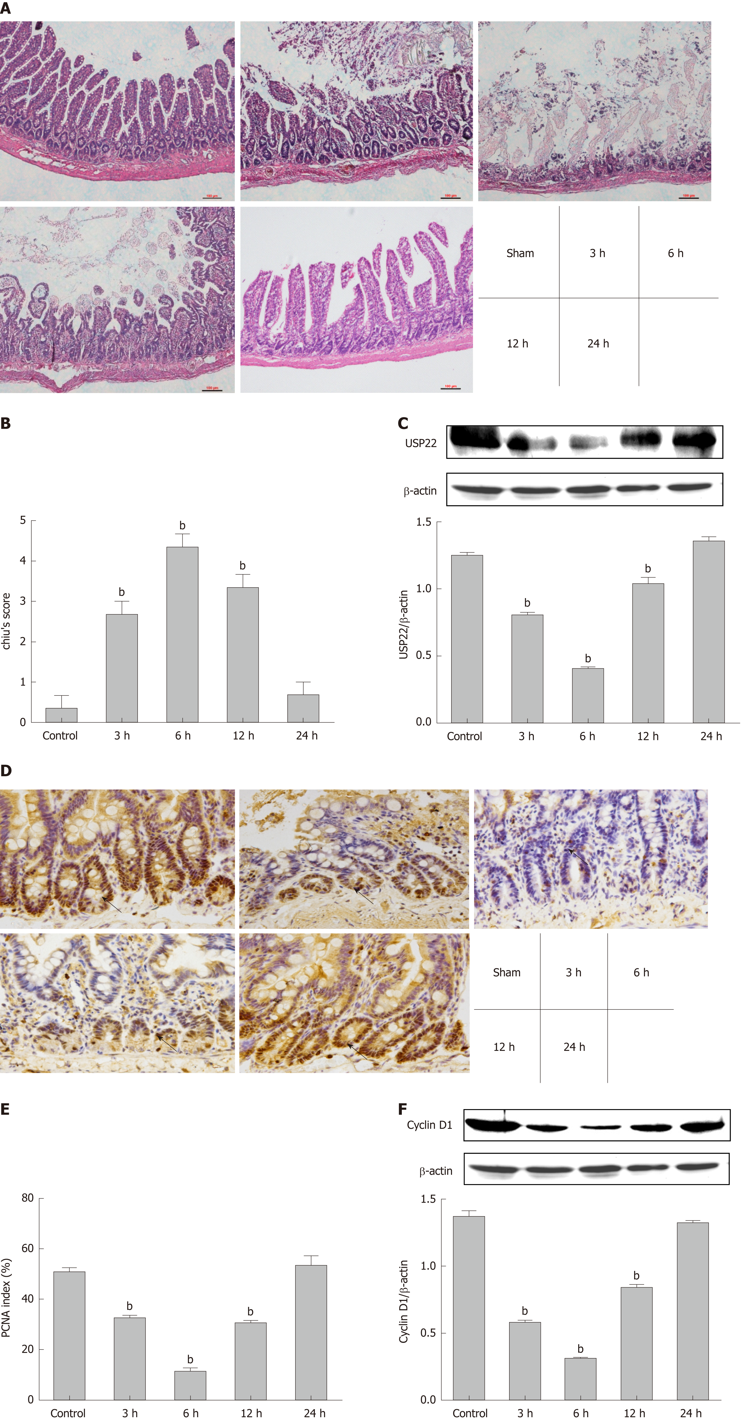
Figure 1 Ubiquitin-specific protease 22 is positively correlated with cell proliferation and tissue regeneration after ischemia reperfusion injury.
Anesthetized rats were subjected to 60 min of intestinal ischemia followed by 3, 6, 12, or 24 h of reperfusion and tissues were harvested at the end of reperfusion. A: Intestinal tissue sections of the experimental groups were stained with HE (200×, bars represent 100 μm); B: Intestinal tissues in injury was scored histopathologically according to a scoring system (Chiu’s score); C: Representative Western blot demonstrating the expression of USP22 in tissues from the experimental groups (n = 3); D: Immunohistochemical staining for PCNA was used for intestinal tissue sections of the experimental groups (400×, bars represent 50 μm); E: Quantitative analyses for immunohistochemical staining. The arrows show brown positive nuclei of proliferating intestinal cells in the crypt area of the intestinal gland; F: Representative protein levels of Cyclin D1 (n = 3) were demonstrated by Western blot. The results are presented as the mean ± SD. bP < 0.01 vs sham. USP22: Ubiquitin-specific protease 22; PCNA: Proliferating cell nuclear antigen.
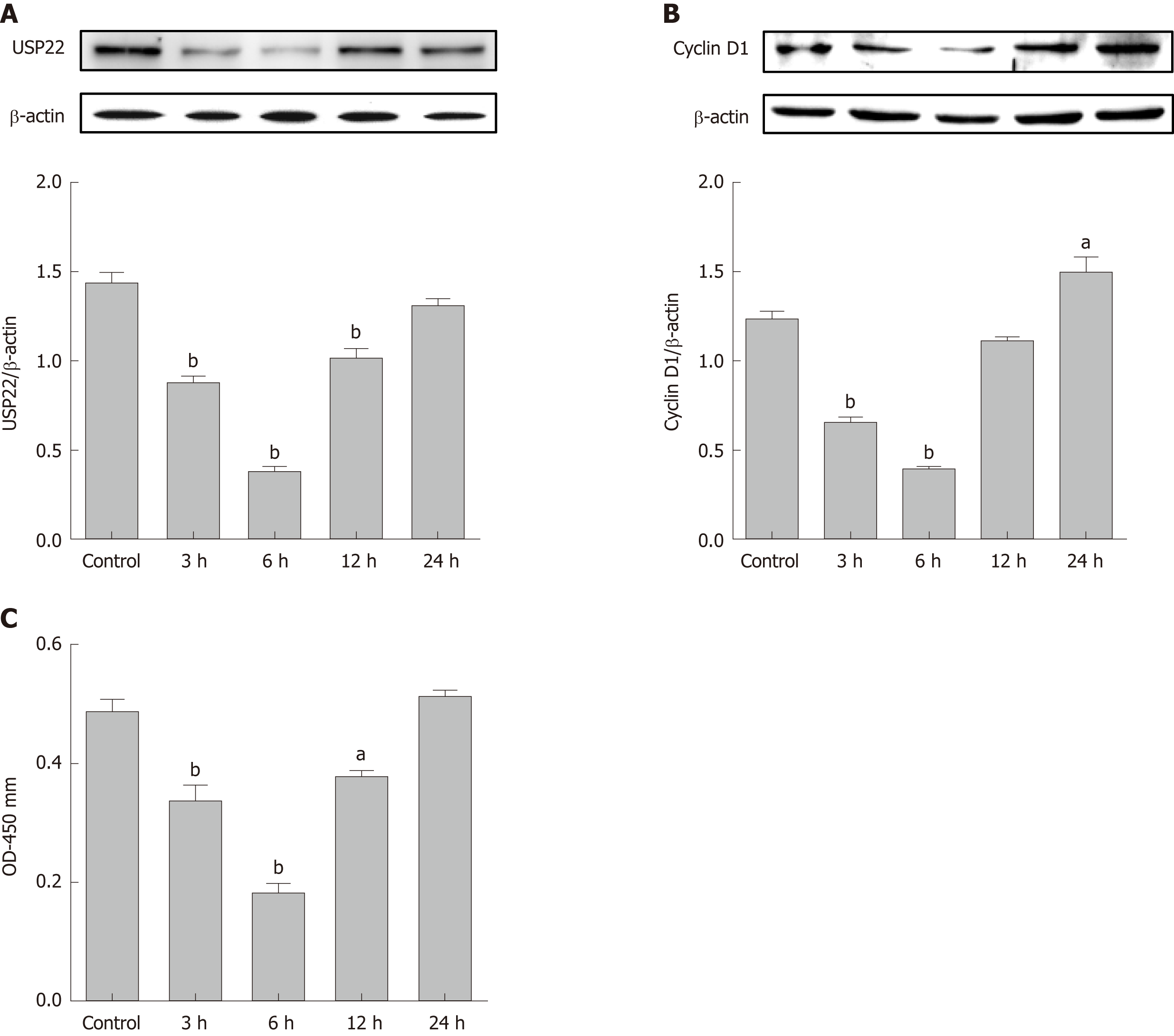
Figure 2 Ubiquitin-specific protease 22 correlates with the proliferative activity of IEC-6 cells after hypoxia/reoxygenation.
IEC-6 cells were subjected to 6 h of hypoxia followed by 3, 6, 12, or 24 h of reoxygenation. A: Representative Western blot demonstrating the expression of USP22 in tissues from the experimental groups (n = 3); B: Representative Western blot demonstrating the expression of Cyclin D1 in tissues from the experimental groups (n = 3); C: CCK-8 was used to examine cell proliferation and viability at the indicated time points (n = 6). The results are presented as the mean ± SD. aP < 0.05 vs the control group; bP < 0.01 vs the control group. USP22: Ubiquitin-specific protease 22; CCK-8: Cell Counting Kit-8.
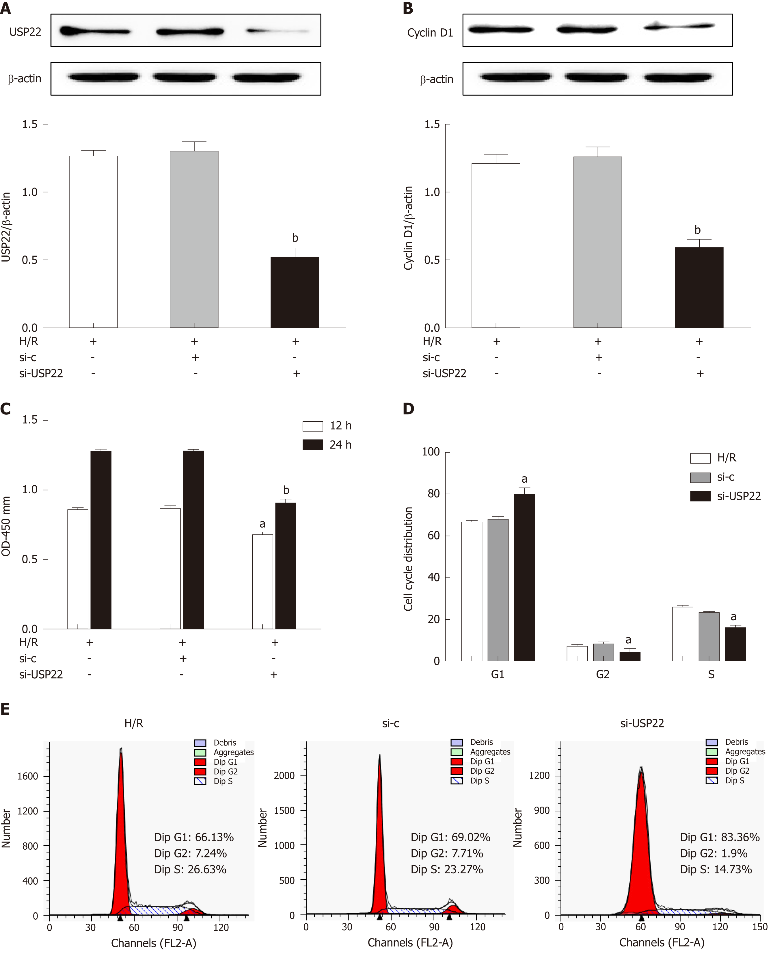
Figure 3 Ubiquitin-specific protease 22 knockdown inhibits cell proliferation and induces cell cycle arrest in IEC-6 cells after hypoxia/reoxygenation.
IEC-6 cells were transfected with USP22 siRNA or unspecific scrambled siRNA and then subjected to hypoxia/reoxygenation or left untreated. A: Western blot analysis for USP22 and β-actin in ICE-6 cells transfected with USP22 siRNA and control siRNA (n = 3); B: Western blot analysis for Cyclin D1 and β-actin in ICE-6 cells transfected with USP22 siRNA and control siRNA (n = 3); C: CCK-8 was used to examine cell viability at the indicated time points (n = 6); D and E: FACSCalibur analysis of the percentages of cells in the G1, G2, and S phases at 24 h of reoxygenation after 6 h of hypoxia (n = 3). The results are presented as the mean ± SD. aP < 0.05 vs the control group; bP < 0.01 vs the control group. USP22: Ubiquitin-specific protease 22; CCK-8: Cell Counting Kit-8.
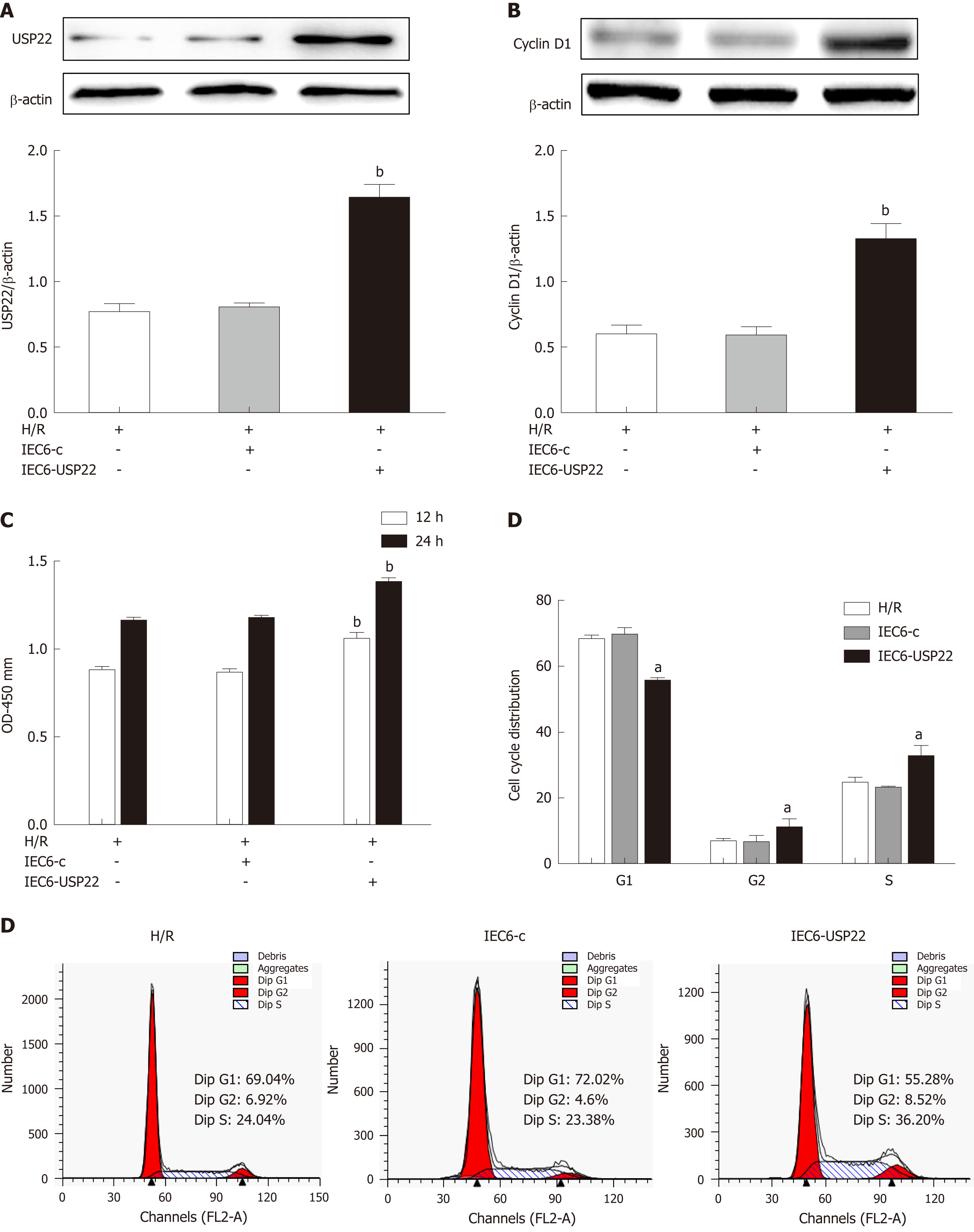
Figure 4 Ubiquitin-specific protease 22 overexpression promotes cell proliferation in IEC-6 cells after hypoxia/reoxygenation.
IEC-6 cells were transfected with a USP22 overexpression plasmid or a negative control plasmid and then subjected to hypoxia/reoxygenation or left untreated. A: Western blot analysis for USP22 and β-actin in ICE-6 cells transfected with IEC6-USP22 and IEC6-Control (n = 3); B: Western blot analysis for Cyclin D1 and β-actin in IEC-6 cells transfected with IEC6-USP22 and IEC6-Control (n = 3); C: CCK-8 assay was used to examine cell viability at the indicated time points (n = 6); D and E: FACSCalibur analysis of the percentages of cells in the G1, G2, and S phases at 24 h of reoxygenation after 6 h of hypoxia (n = 3). The results are presented as the mean ± SD. aP < 0.05 vs the control group; bP < 0.01 vs the control group. USP22: Ubiquitin-specific protease 22; CCK-8: Cell Counting Kit-8.
- Citation: Ji AL, Li T, Zu G, Feng DC, Li Y, Wang GZ, Yao JH, Tian XF. Ubiquitin-specific protease 22 enhances intestinal cell proliferation and tissue regeneration after intestinal ischemia reperfusion injury. World J Gastroenterol 2019; 25(7): 824-836
- URL: https://www.wjgnet.com/1007-9327/full/v25/i7/824.htm
- DOI: https://dx.doi.org/10.3748/wjg.v25.i7.824


