©The Author(s) 2019.
World J Gastroenterol. Feb 7, 2019; 25(5): 608-621
Published online Feb 7, 2019. doi: 10.3748/wjg.v25.i5.608
Published online Feb 7, 2019. doi: 10.3748/wjg.v25.i5.608
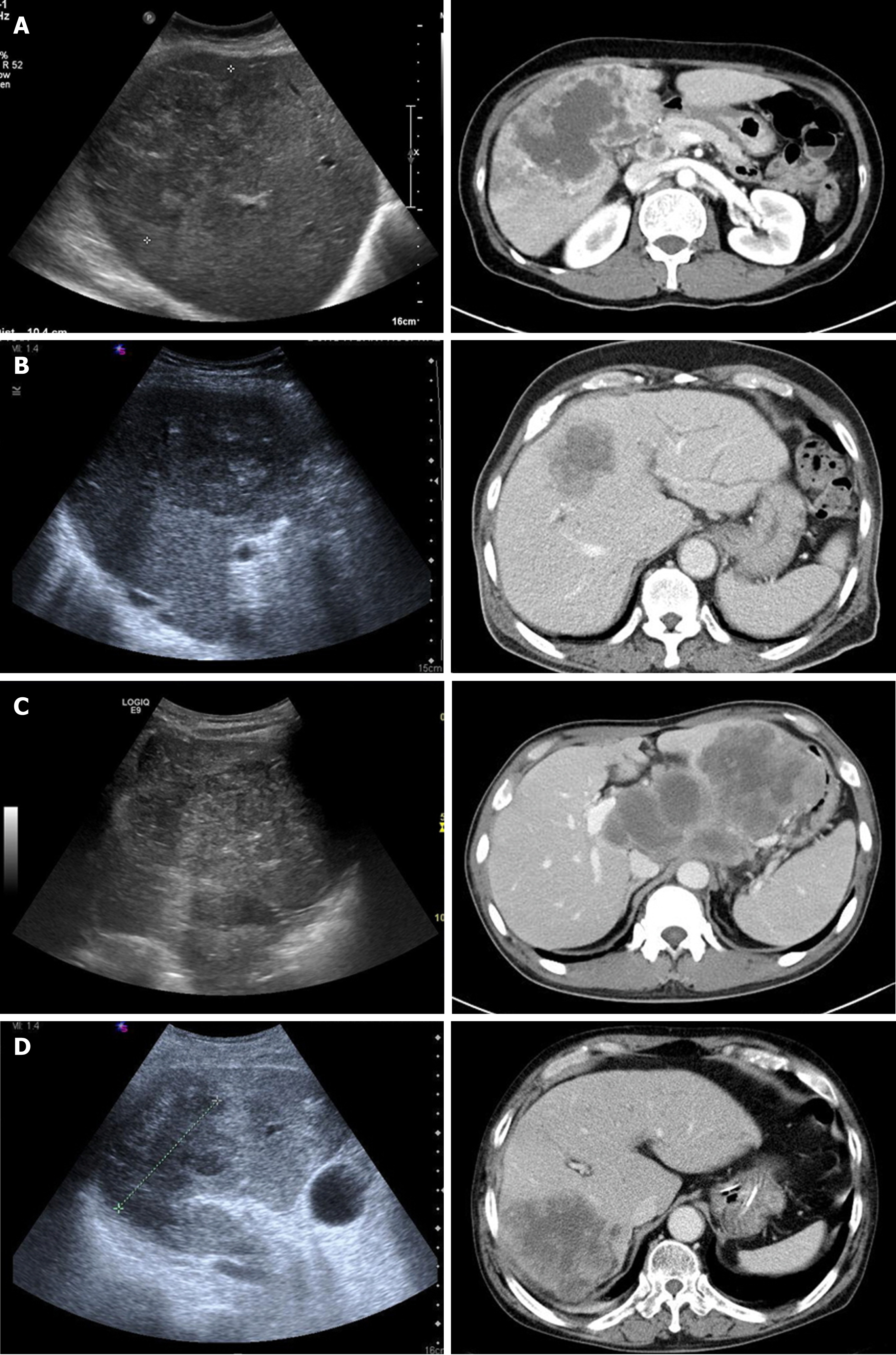
Figure 1 Imaging findings of the patients with various initial diagnoses based on radiologic findings.
A: The initial radiologic impressions were intrahepatic cholangiocarcinoma; B: Hepatocellular carcinoma; C: Lymphoma; D: Hepatic abscess, respectively.
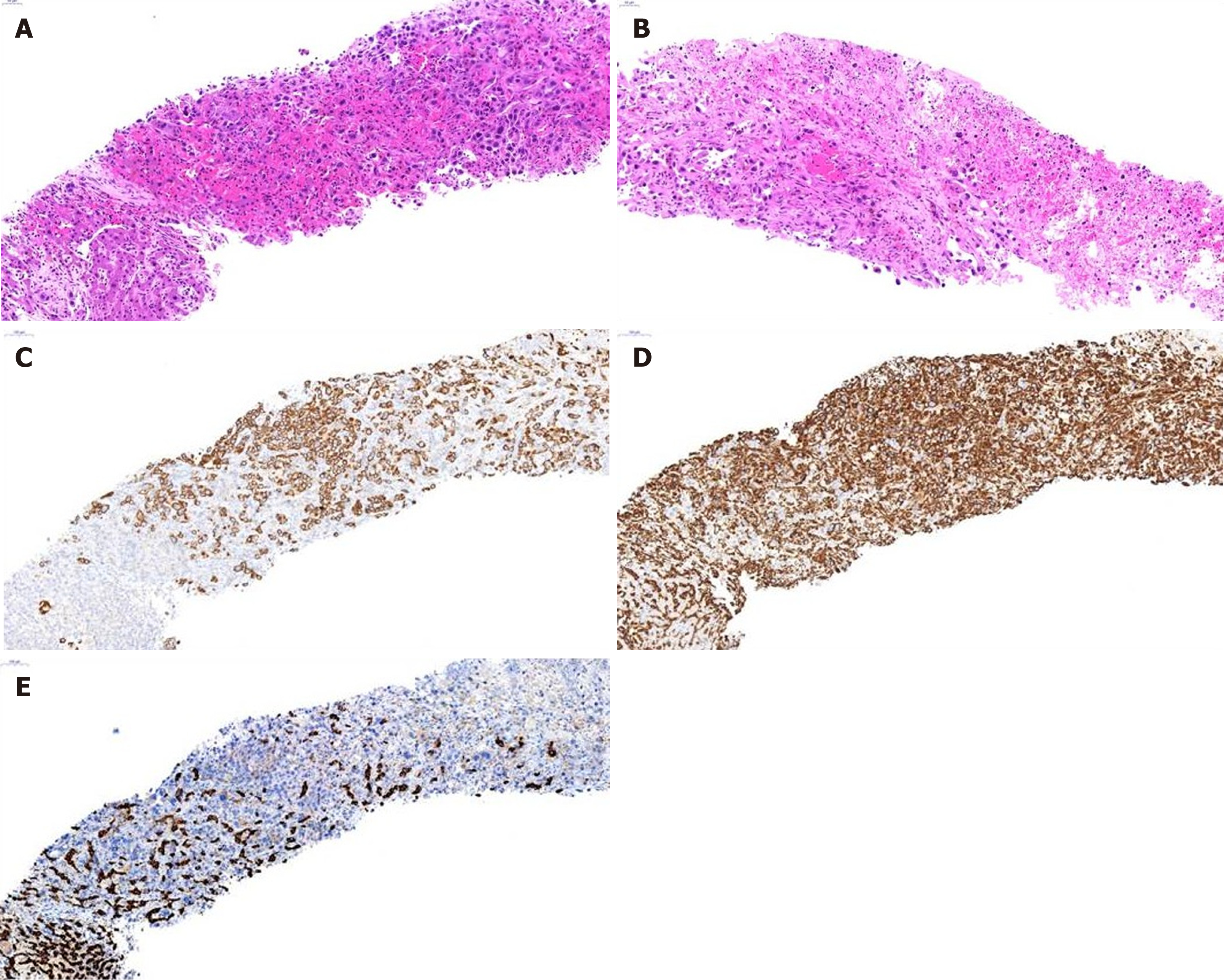
Figure 2 Histologic findings of liver biopsy of case 9.
A and B: Hematoxylin and eosin stain, × 100; C-E: Immunohistochemistry, × 50; C: Cytokeratin 19; D: Vimentin; E: Hepatocyte specific antigen. The liver shows a mass composed for proliferating anaplastic tumor cells with ovoid or rather spindle shape and no organoid structures (A), and necrosis (B). The tumor cells express CK19 (C) and vimentin (D), not HSA (E).
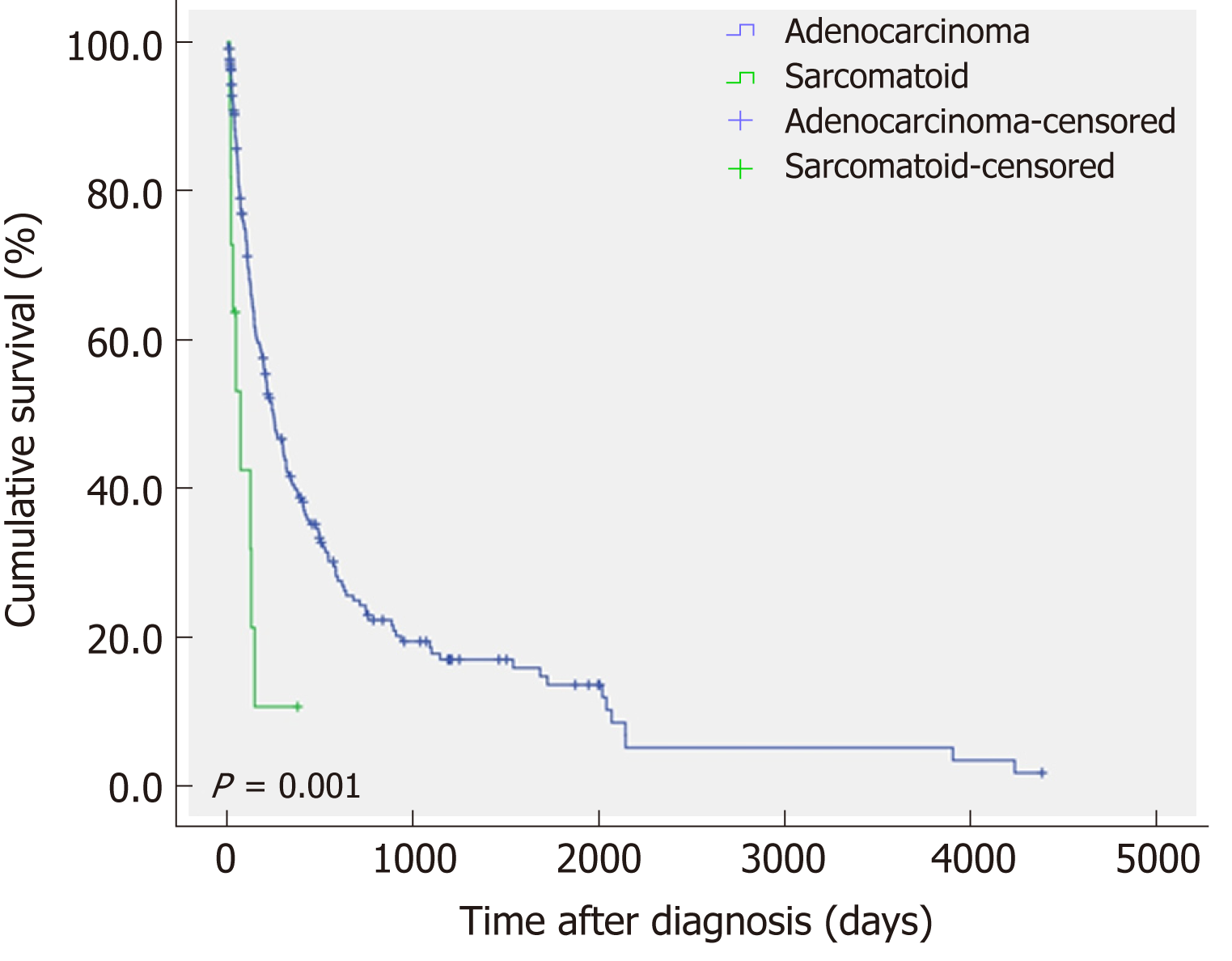
Figure 3 Comparison of cumulative survival rates.
Survival rate of intrahepatic bile duct adenocarcinoma group was better than that of sarcomatoid cholangiocarcinoma group.
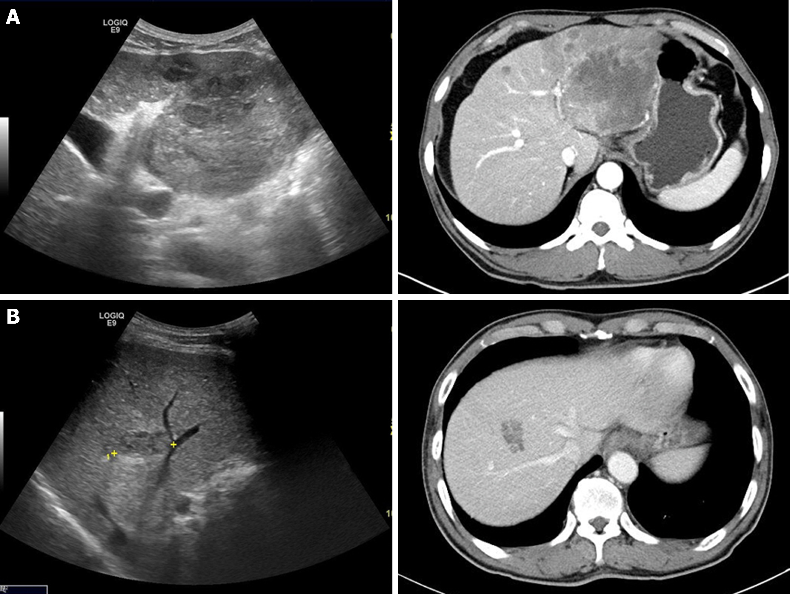
Figure 4 Imaging finding of the patient with mixed pathological findings of hepatocellular carcinoma and intrahepatic cholangiocarcinoma.
A: Case 1; B: Case 2.
- Citation: Kim DK, Kim BR, Jeong JS, Baek YH. Analysis of intrahepatic sarcomatoid cholangiocarcinoma: Experience from 11 cases within 17 years. World J Gastroenterol 2019; 25(5): 608-621
- URL: https://www.wjgnet.com/1007-9327/full/v25/i5/608.htm
- DOI: https://dx.doi.org/10.3748/wjg.v25.i5.608


