©The Author(s) 2019.
World J Gastroenterol. Oct 28, 2019; 25(40): 6077-6093
Published online Oct 28, 2019. doi: 10.3748/wjg.v25.i40.6077
Published online Oct 28, 2019. doi: 10.3748/wjg.v25.i40.6077
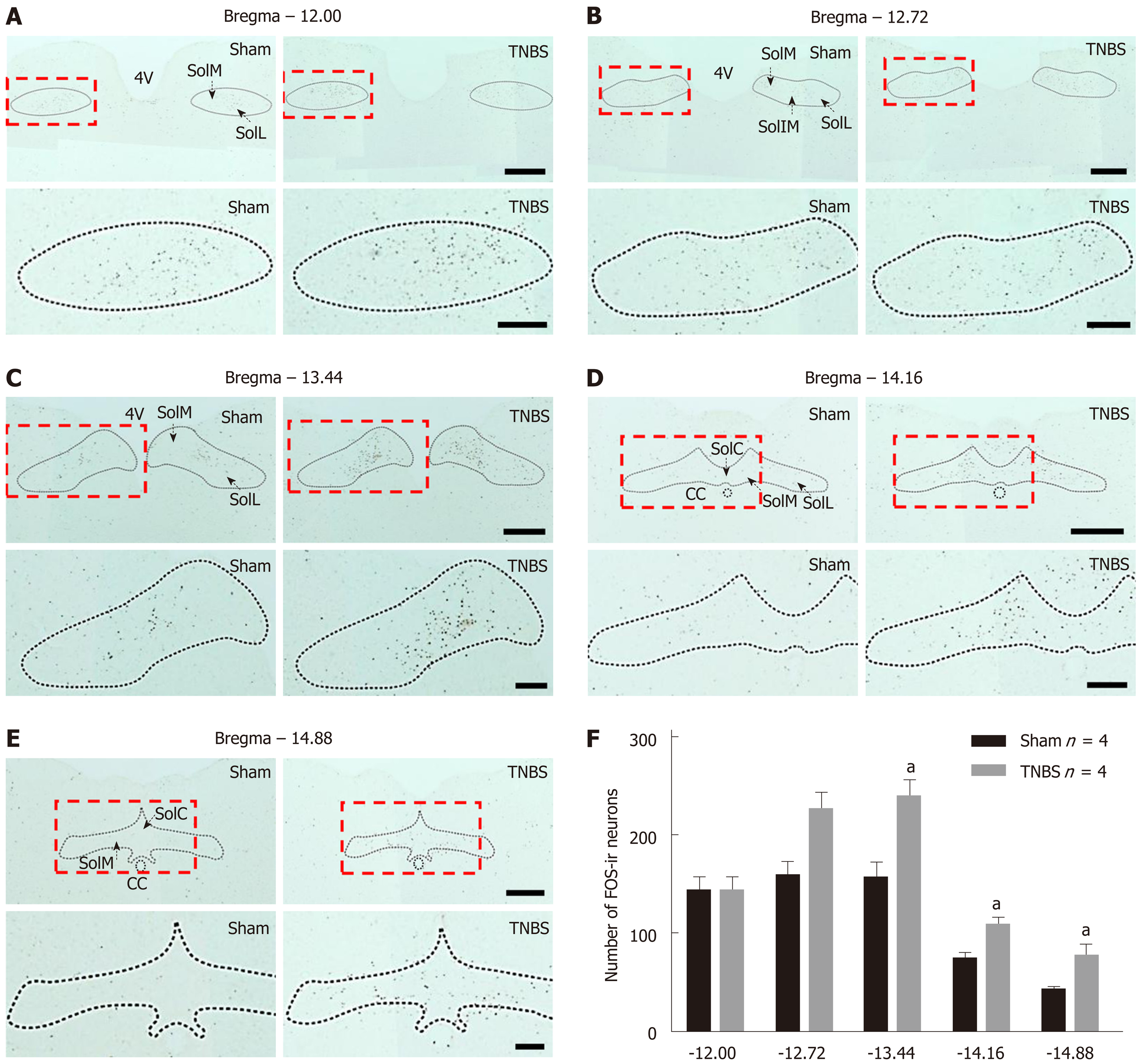
Figure 1 The number of Fos-expressing neurons is upregulated within the nucleus tractus solitarius in rats with chronic pancreatitis.
A-E: Immunochemical staining for Fos within different coronal sections of nucleus tractus solitarius (NTS) in sham (left) and chronic pancreatitis (right) rats. The rectangles in the top panels are further magnified in the bottom panels. Bars = 500 μm in top panels and 200 μm in bottom panels; F: Histogram showing the qualification of Fos-expressing neurons within different parts of NTS in saline and trinitrobenzene sulfonic acid (TNBS)-treated rats. n = 4 rats per each group, unpaired t-test, aP < 0.05, TNBS vs sham. 4V: 4th ventricle; CC: Central canal; SolIM: Nucleus of the solitary tract, intermedial part; SolM: Nucleus of the solitary tract, medial part; SolL: Nucleus of the solitary tract, lateral part; SolC: Nucleus of the solitary tract, commissural part; TNBS: Trinitrobenzene sulfonic acid.
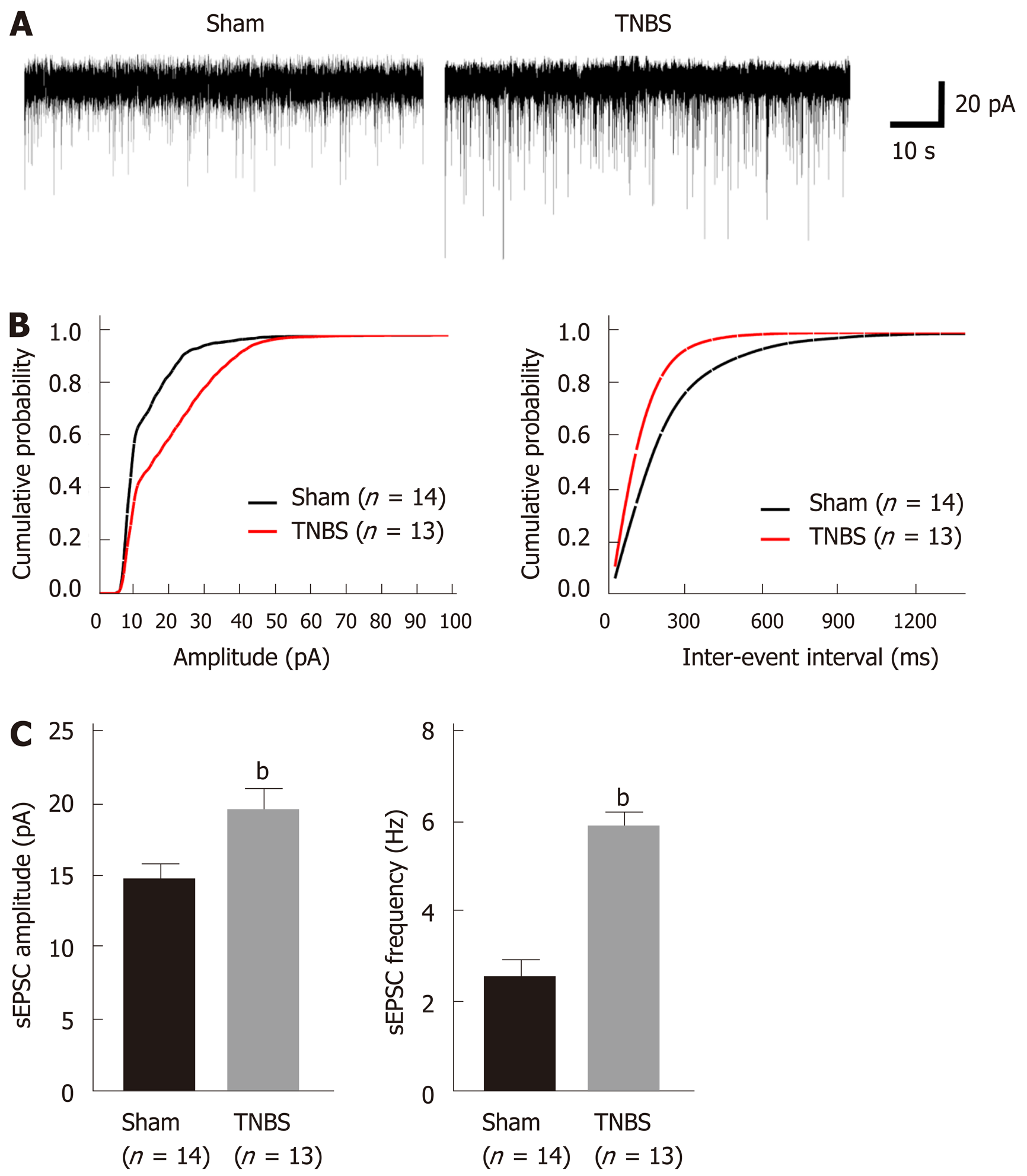
Figure 2 Rats with chronic pancreatitis have increased amplitude and frequency of spontaneous excitatory postsynaptic currents in the caudal nucleus tractus solitarius.
A: Representative spontaneous excitatory postsynaptic currents (sEPSCs) recorded from a sham (left) rat and a trinitrobenzene sulfonic acid (TNBS)-treated (right) rat holding at -60 mV; B: Cumulative amplitude (left) and inter-event interval (right) histograms of sEPSCs recorded from sham and chronic pancreatitis (CP) rats; C: Summary plots showing the frequency (left) and amplitude (right) of sEPSCs from sham and CP rats. Unpaired t-test, bP < 0.01, TNBS vs sham. sEPSC: Spontaneous excitatory postsynaptic currents; TNBS: Trinitrobenzene sulfonic acid.
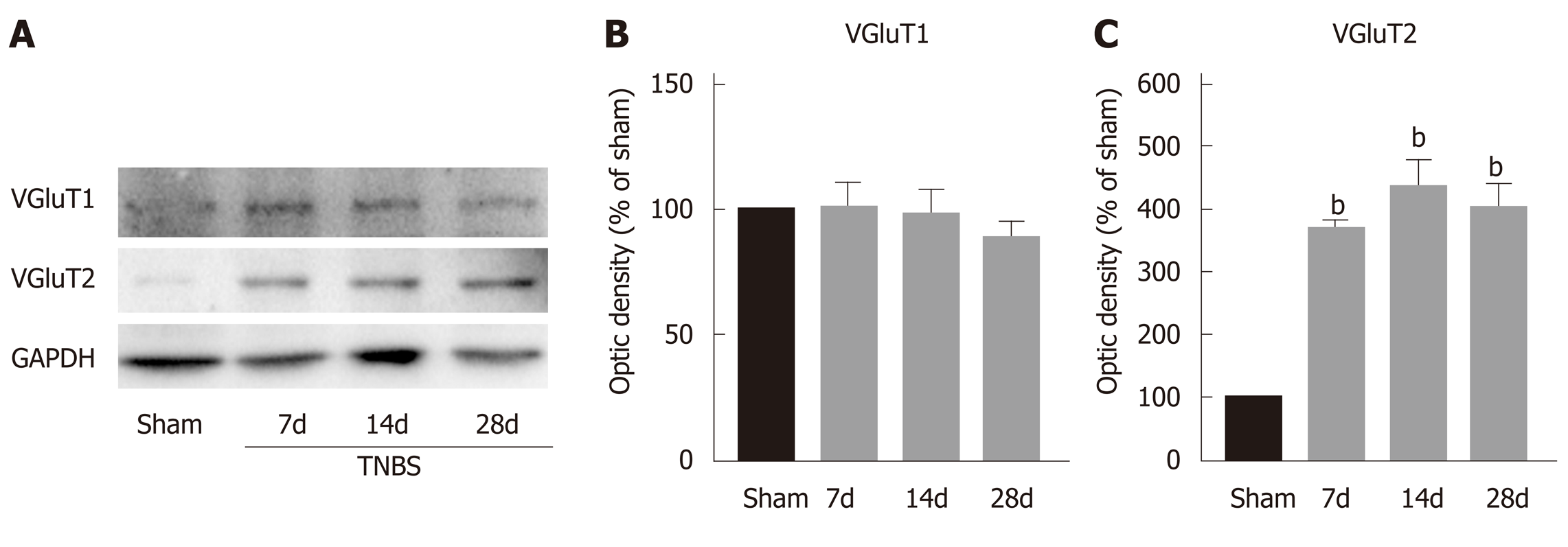
Figure 3 Trinitrobenzene sulfonic acid treatment upregulates the expression of vesicular glutamate transporter 2 within the caudal nucleus tractus solitarius.
A: Representative Western blots for VGluT1 and VGluT2 within the caudal nucleus tractus solitarius (NTS) on postoperative days (POD) 7, 14, and 28; B: VGluT1 amount within the caudal NTS showed no changes after trinitrobenzene sulfonic acid (TNBS) treatment; C: VGluT2 amount within the caudal NTS was significantly increased on POD 7, 14, and 28 after TNBS treatment. n = 3 rats per each group, one-way ANOVA, aP < 0.05, bP < 0.01, TNBS vs sham. TNBS: Trinitrobenzene sulfonic acid; VGluT1: Vesicular glutamate transporter 1; VGluT2: Vesicular glutamate transporter 2.
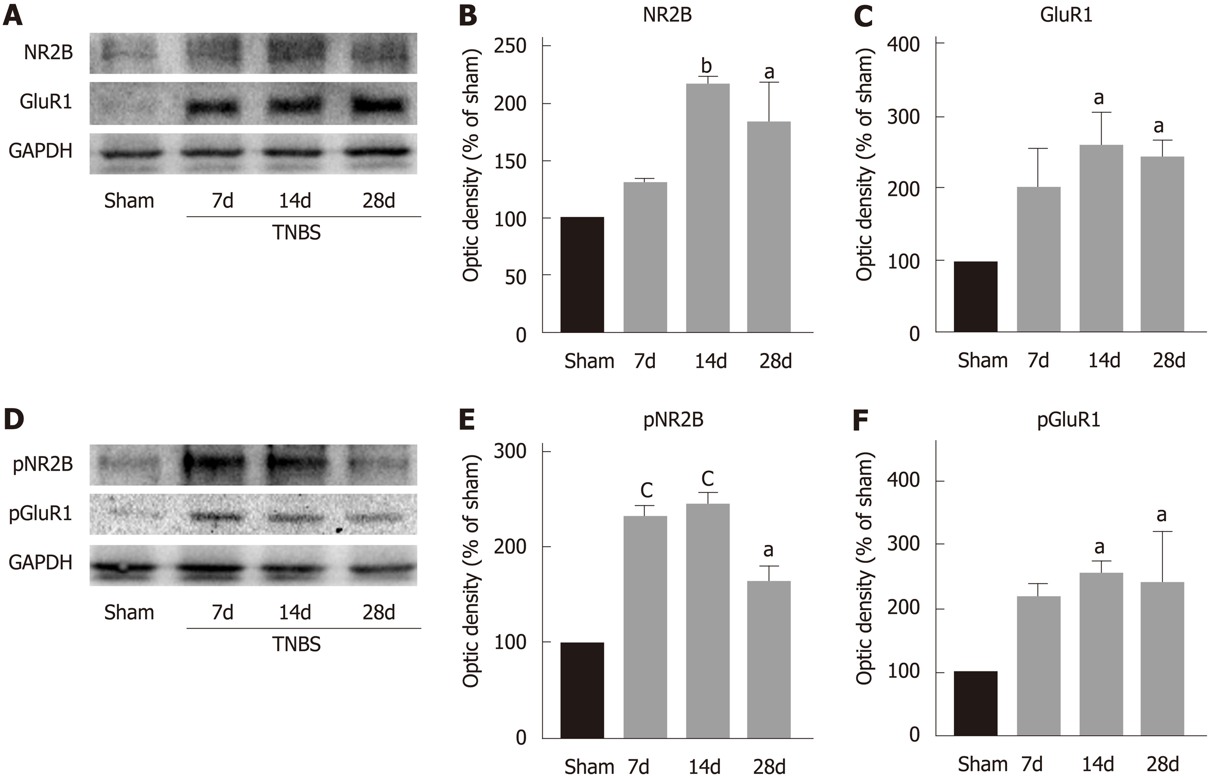
Figure 4 Trinitrobenzene sulfonic acid treatment upregulates the expression of N-methyl D-aspartate receptor subtype 2B and α-amino-3-hydroxy-5-methyl-4-isoxazolepropionic acid receptor subtype 1 within the caudal nucleus tractus solitarius.
A: Representative Western blots for NR2B and GluR1 within the caudal nucleus tractus solitarius (NTS) on postoperative days (POD) 7, 14, and 28; B: The amount of NR2B within the caudal NTS was significantly increased on POD 7, 14, and 28 after trinitrobenzene sulfonic acid (TNBS) treatment; C: The amount of GluR1 within the caudal NTS was significantly increased on POD 7, 14, and 28 after TNBS treatment; D: Representative Western blots for pNR2B and pGluR within the caudal NTS after TNBS treatment; E: The amount of pNR2B within the caudal NTS was significantly increased after TNBS treatment; F: The amount of pGluR1 within the caudal NTS was significantly increased after TNBS treatment. n = 3 rats per each group, one-way ANOVA, aP < 0.05, bP < 0.01, cP < 0.001, TNBS vs sham. GluR1: α-amino-3-hydroxy-5-methyl-4-isoxazolepropionic acid receptor subtype 1; NR2B: N-methyl-D-aspartate receptor subtype 2B; TNBS: Trinitrobenzene sulfonic acid.
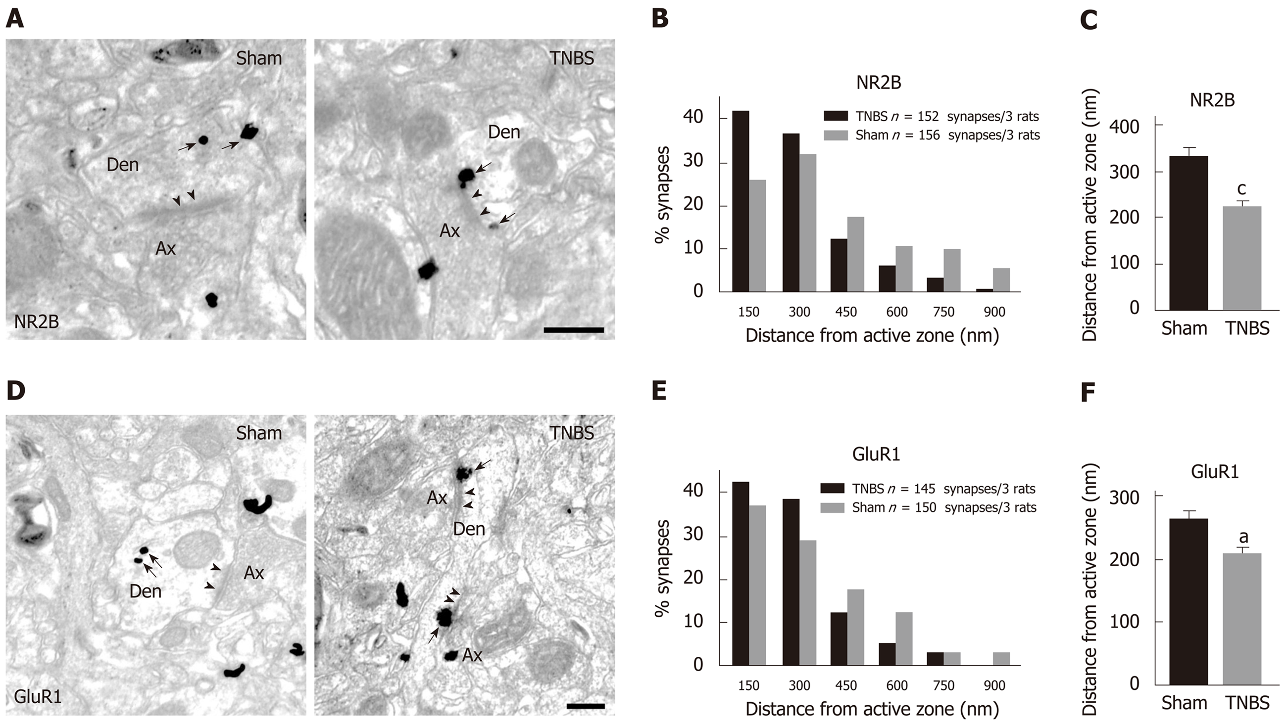
Figure 5 Trinitrobenzene sulfonic acid treatment results in accumulation of postsynaptic N-methyl D-aspartate receptor subtype 2B and α-amino-3-hydroxy-5-methyl-4-isoxazolepropionic acid receptor subtype 1 in the caudal nucleus tractus solitarius.
A: Representative electron microscopy images showing immune-gold particles labeled N-methyl-D-aspartate receptor subtype 2B (NR2B) distributed in an asymmetric synapse within the caudal nucleus tractus solitarius (NTS) of sham (left) and chronic pancreatitis (CP) (right) rats; B: Percentage of NR2B particles in 150 nm bins as a function of distance from the nearest edge of synapses; C: Averaged distances of NR2B from the edge of synapses of sham rats and CP rats; D: Representative electron microscopy images showing immune-gold particles labeled GluR1 distributed in an asymmetric synapse within caudal NTS of sham (left) and CP (right) rats; E: Percentage of α-amino-3-hydroxy-5-methyl-4-isoxazolepropionic acid receptor subtype 1 (GluR1) particles in 150 nm bins as a function of distance from the nearest edge of synapses; F: Averaged distances of GluR1 from the edge of synapses of sham rats and CP rats. Bar = 250 nm in A and D. aP < 0.05, cP < 0.001, TNBS vs sham. Ax: Axon; Den: Dendrite; GluR1: α-amino-3-hydroxy-5-methyl-4-isoxazolepropionic acid receptor subtype 1; NR2B: N-methyl-D-aspartate receptor subtype 2B; TNBS: Trinitrobenzene sulfonic acid.
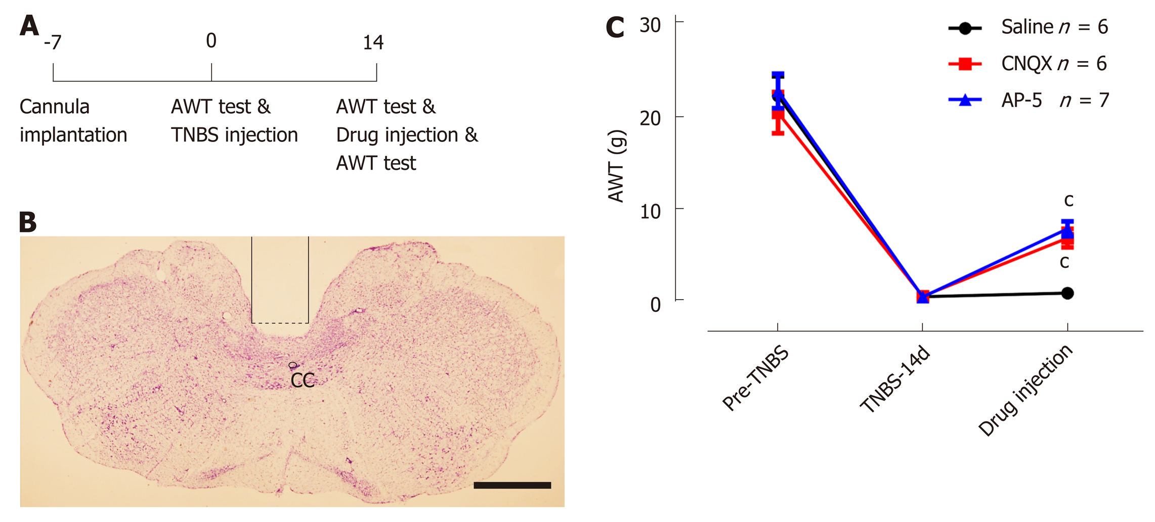
Figure 6 Administration of 6-cyano-7-nitroquinoxaline-2,3-dione and amino-5-phosphonopentaoic acid into the caudal nucleus tractus solitarius alleviates chronic pancreatitis induced visceral hypersensitivity.
A: Schematic diagram of behavioral test; B: A representative section showing the site of cannula implantation within the caudal nucleus tractus solitarius. Bar = 1 mm; C: Microinjection of 6-cyano-7-nitroquinoxaline-2,3-dione (CNQX) and amino-5-phosphonopentaoic acid (AP-5), instead of saline, increases the abdominal withdraw threshold in chronic pancreatitis rats on postoperative day 14. n = 6 rats in saline and CNQX-treated groups and 7 rats in AP5-treated group, one-way ANOVA, cP < 0.001, CNQX or AP5 groups vs saline group. AWT: Abdominal withdraw threshold; CC: Central canal; CNQX: 6-cyano-7-nitroquinoxaline-2,3-dione; AP-5: Amino-5-phosphonopentaoic acid; TNBS: Trinitrobenzene sulfonic acid.
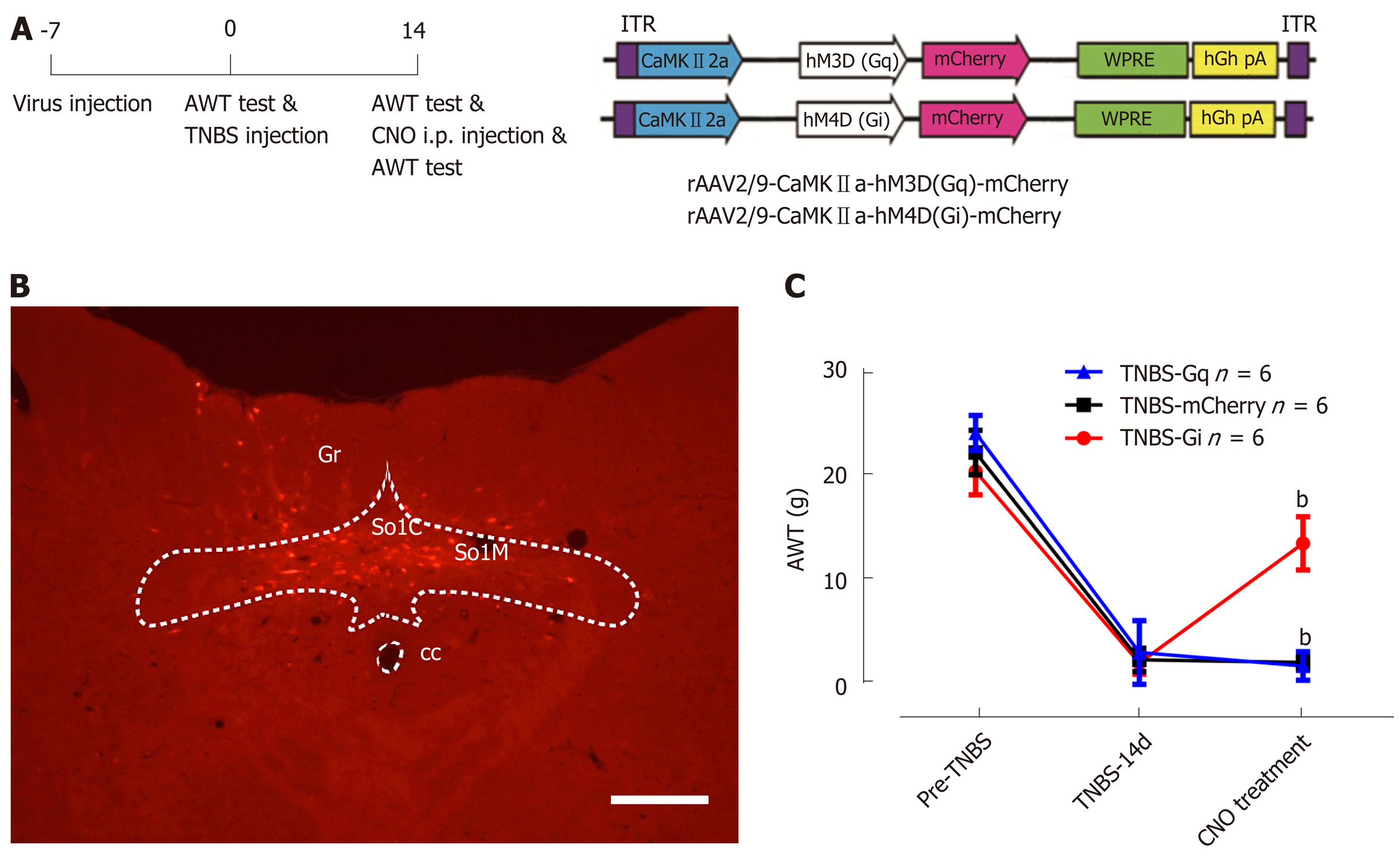
Figure 7 Inactivating excitatory neurons within the caudal nucleus tractus solitarius alleviates visceral hypersensitivity in rats with chronic pancreatitis.
A: The schematic diagram of behavioral experiment (left) and rAAV2/9-CaMKIIa-hM4Di/hM3Dq-mCherry construct (right); B: A representative image showing that the expression of hM4Di-mCherry was confined within the commissure and medial nuclei of the caudal nucleus tractus solitarius (NTS). Bar = 0.4 mm. C: Inhibiting caudal NTS excitatory neurons via i.p. clozapine-N-oxide injection significantly increases the abdominal withdraw threshold (AWT) in chronic pancreatitis rats, while activating these neurons does not influence the AWT. n = 6 rats in mCherry, Gq, and Gi groups, one-way ANOVA. bP < 0.01, Gi vs mCherry. AWT: Abdominal withdraw threshold; CC: Central canal; CNO: Clozapine-N-oxide; SolM: Nucleus of the solitary tract, medial part; SolC: Nucleus of the solitary tract, commissural part; Gr: Gracile nucleus; TNBS: Trinitrobenzene sulfonic acid.
- Citation: Bai Y, Chen YB, Qiu XT, Chen YB, Ma LT, Li YQ, Sun HK, Zhang MM, Zhang T, Chen T, Fan BY, Li H, Li YQ. Nucleus tractus solitarius mediates hyperalgesia induced by chronic pancreatitis in rats. World J Gastroenterol 2019; 25(40): 6077-6093
- URL: https://www.wjgnet.com/1007-9327/full/v25/i40/6077.htm
- DOI: https://dx.doi.org/10.3748/wjg.v25.i40.6077


