©The Author(s) 2019.
World J Gastroenterol. Jan 28, 2019; 25(4): 498-508
Published online Jan 28, 2019. doi: 10.3748/wjg.v25.i4.498
Published online Jan 28, 2019. doi: 10.3748/wjg.v25.i4.498
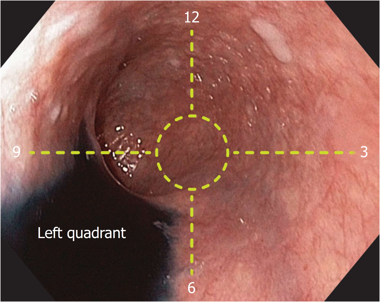
Figure 1 Identification of the left esophageal quadrant.
After pouring Indigo Carmine (Chromoendoscopia, Colombia) into the esophageal lumen and utilizing gravity, the pooled water identified the left quadrant of the esophageal circumference.
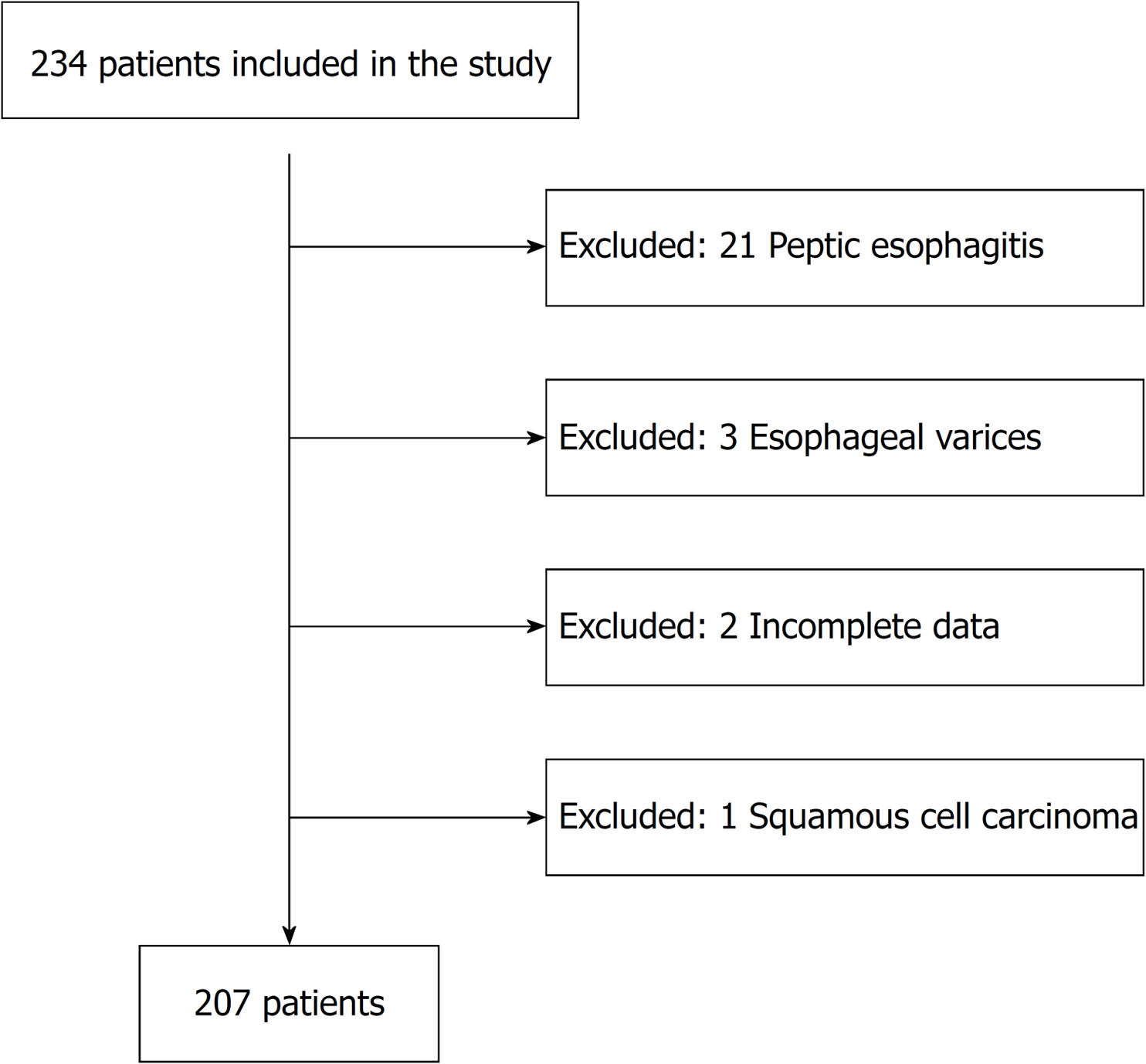
Figure 2 Patient selection.
The chart shows the enrolled, excluded and final number of patients for analysis.
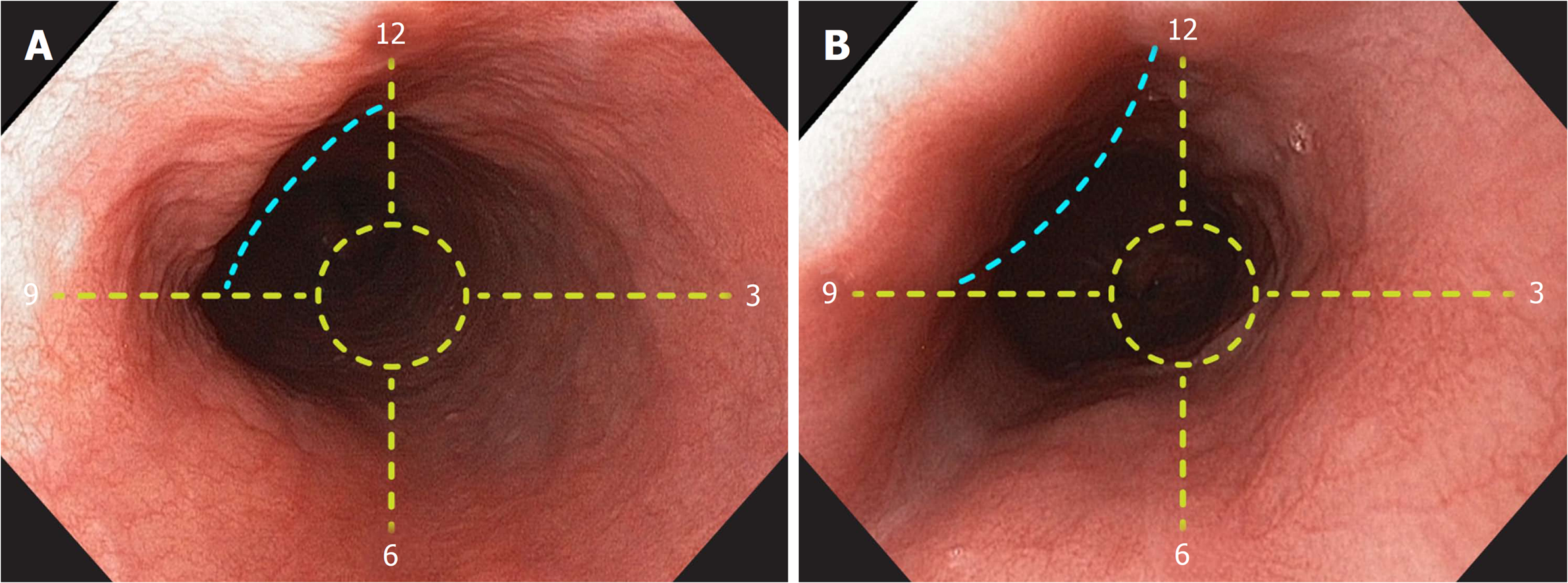
Figure 3 Identification of the left main bronchus and left atrium landmarks.
A: A concave, non-pulsatile, extrinsic esophageal compression (blue lines) is observed 25 cm from the incisors. The landmark is radially oriented at the anterior esophageal quadrant, between 9 and 12 o’clock. B: A convex, pulsatile, extrinsic esophageal compression (blue lines) is observed 31 cm from the incisors. The landmark is radially oriented at the anterior esophageal quadrant, between 9 and 12 o’clock.
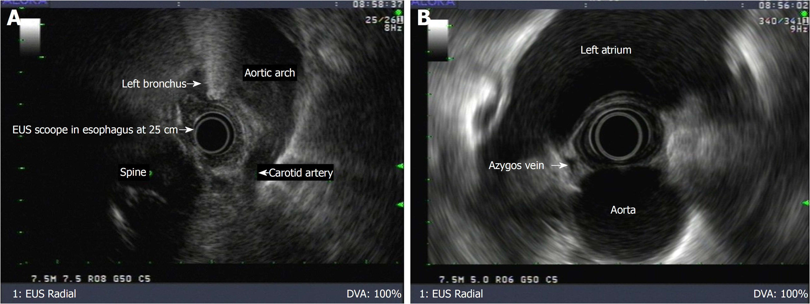
Figure 4 Identification of landmarks by endoscopic ultrasound.
A: Endoscopic ultrasound (EUS) image showing the left main bronchus located on the anterior esophageal quadrant at a distance of 25 cm from the incisors. B: EUS image showing the left atrium located on the anterior esophageal quadrant at a distance of 30 cm from the incisors.
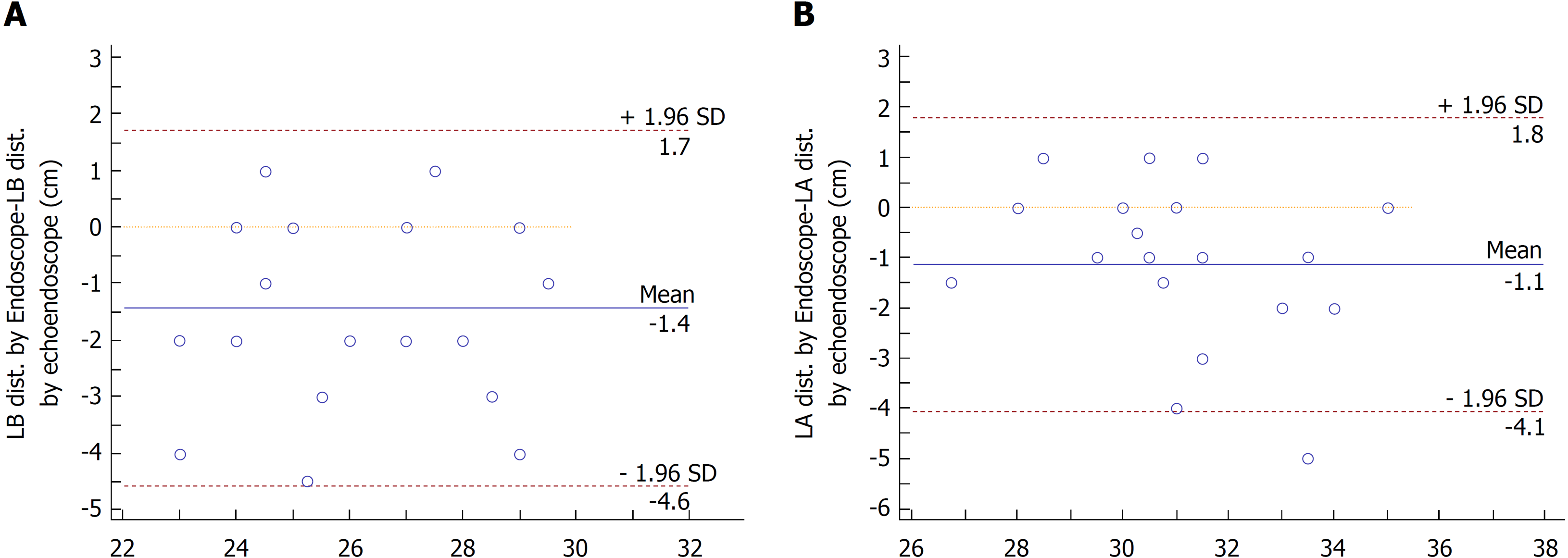
Figure 5 The Bland-Altman plot.
High agreement (IC 95%) is observed between white light and EUS measurements in the sub-study. A: Left main bronchus. B: Left atrium.
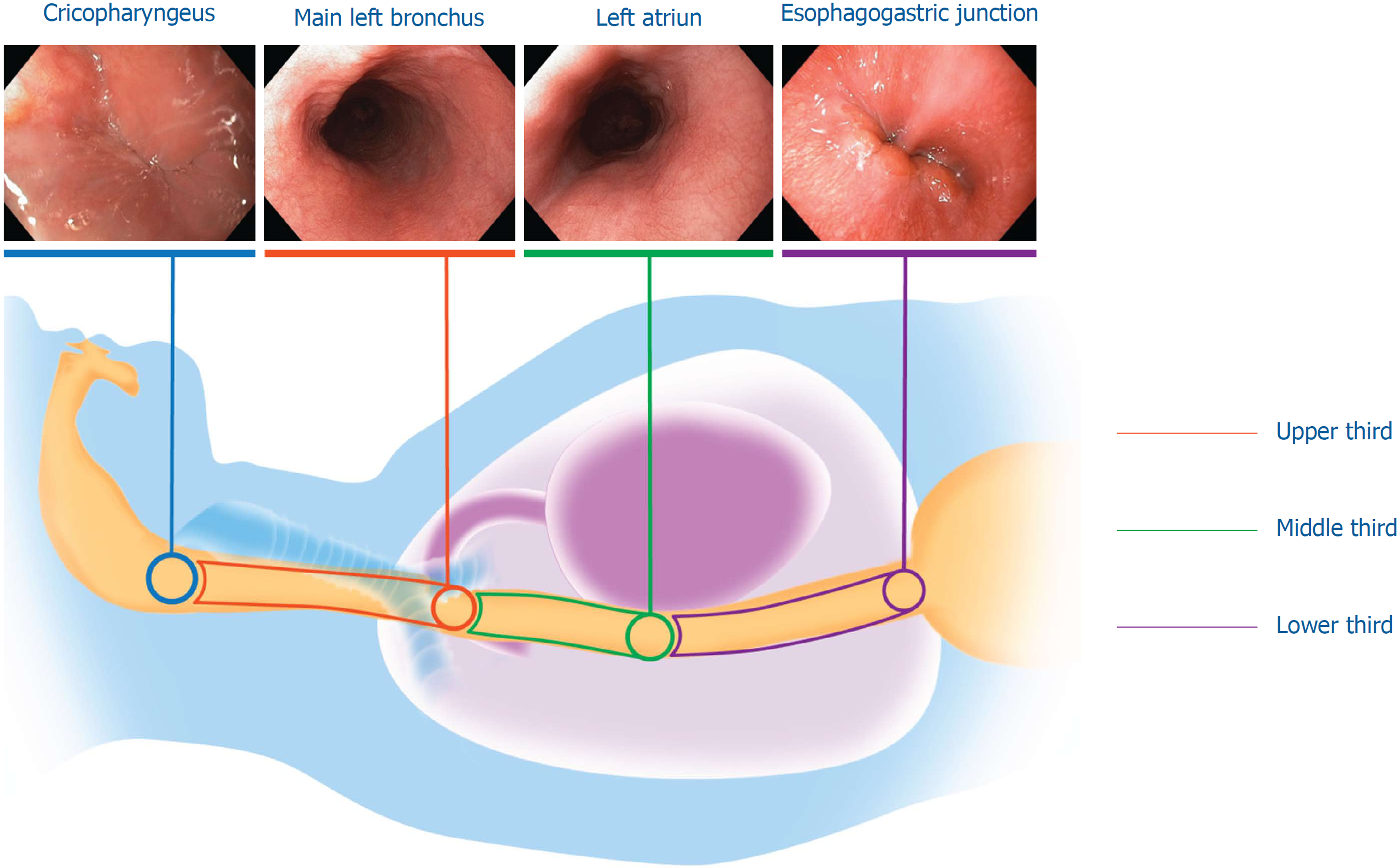
Figure 6 Usefulness of the landmarks for longitudinal orientation.
Esophageal landmarks are represented in colored circles: cricopharyngeus (blue circle), left main bronchus (red circle), left atrium (green circle) and the esophagogastric junction (purple circle); esophageal thirds are represented as colored segments: upper third (red segment), middle third (green segment) and lower third (purple segment).
- Citation: Emura F, Gomez-Esquivel R, Rodriguez-Reyes C, Benias P, Preciado J, Wallace M, Giraldo-Cadavid L. Endoscopic identification of endoluminal esophageal landmarks for radial and longitudinal orientation and lesion location. World J Gastroenterol 2019; 25(4): 498-508
- URL: https://www.wjgnet.com/1007-9327/full/v25/i4/498.htm
- DOI: https://dx.doi.org/10.3748/wjg.v25.i4.498


