©The Author(s) 2019.
World J Gastroenterol. Jun 14, 2019; 25(22): 2720-2733
Published online Jun 14, 2019. doi: 10.3748/wjg.v25.i22.2720
Published online Jun 14, 2019. doi: 10.3748/wjg.v25.i22.2720
Figure 1 Representative images of angioectasia.
A: Video capsule endoscopy confirmed multifocal jejunal angioectasias in patients with chronic anemia; B: Double balloon endoscopy identified an angioectasia classified into Yano-Yamamoto classification Type 1b; C: Argon plasm coagulation cauterization was successfully performed.
Figure 2 Representative images of Dieulafoy’s lesion.
A: Video capsule endoscopy confirmed active bleeding from unknown origin in patient with ongoing overt obscure gastrointestinal bleeding; B: Arterial bleeding occurred from a jejunal punctuate lesion classified into Yano-Yamamoto classification Type 2a; C: Successful hemostasis was achieved by combination of argon plasm coagulation cauterization and endoclips.
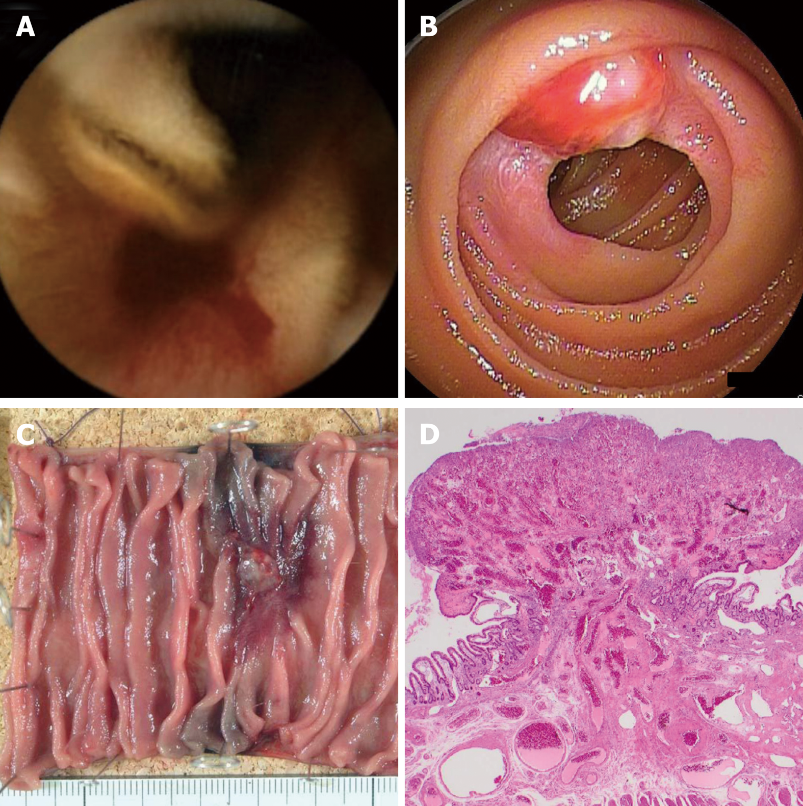
Figure 3 Representative images of arteriovenous malformation.
A: Video capsule endoscopy confirmed active bleeding from jejunum in young patient with ongoing overt obscure gastrointestinal bleeding; B: Double balloon endoscopy identified pulsating subepithelial tumor classified into Yano-Yamamoto classification Type 4; C: Subsequent to endoscopic tattooing, surgical resection was performed; D: Pathological examination revealed a vascular malformation in the submucosa.
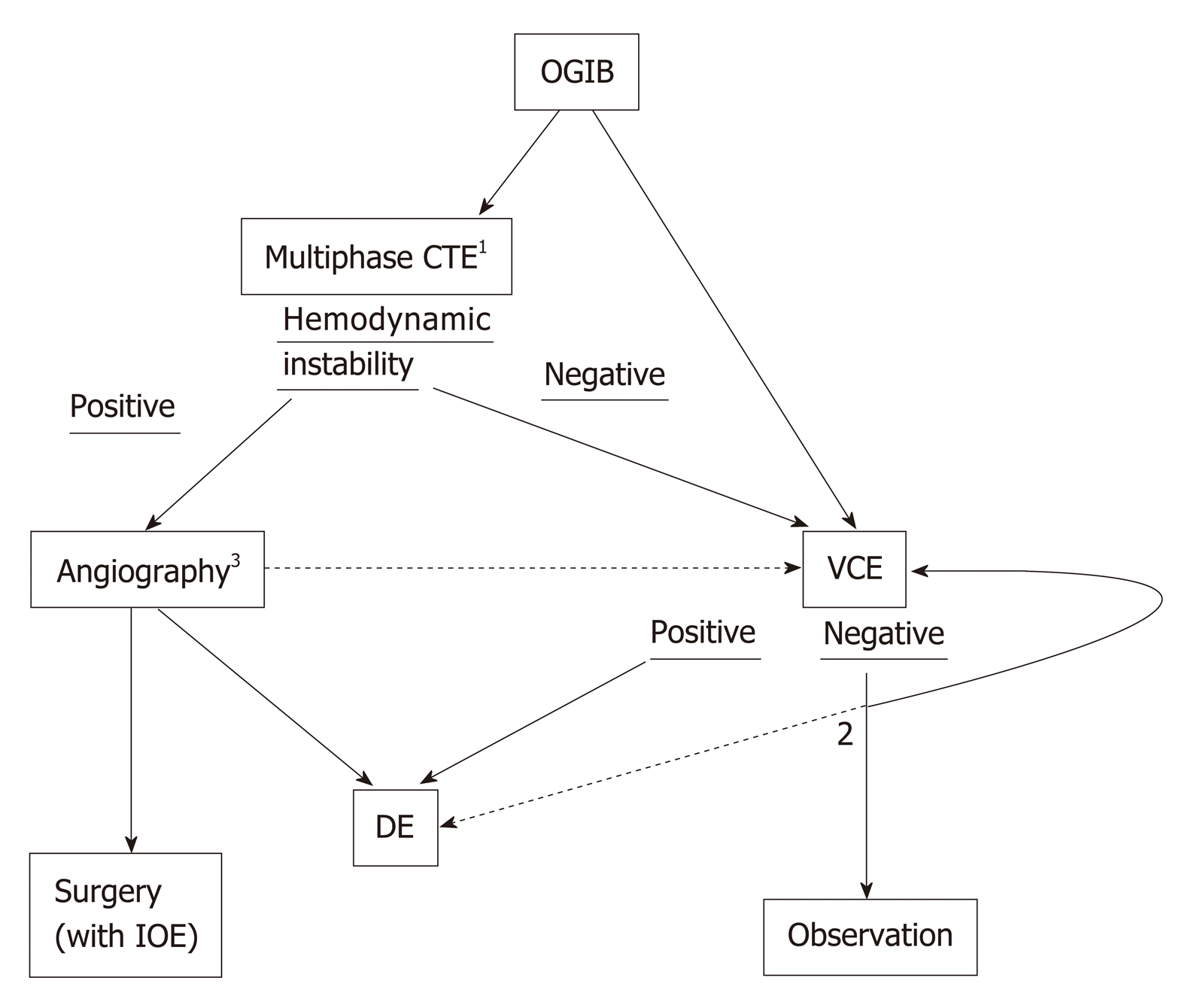
Figure 4 Diagnostic algorithm for small bowel vascular lesions.
Note: 1Computed tomography (CT) scan is especially recommended for patients with ongoing overt obscure gastrointestinal bleeding (OGIB). CT enterography can be replaced to multiphase CT or radionuclide scanning, considering patients general condition; 2Repeated video capsule endoscopy is recommended if the disease presentation changes from occult to overt or if a rapid decrease in the serum hemoglobin level is confirmed. Urgent deep enteroscopy may be useful to reveal the bleeding source in patients with recurrent overt OGIB; 3Surgical intervention with intra-operative endoscopy will be conducted when superselective transcatheter embolization was failed. Meanwhile subsequent endoscopic examination is recommended to reveal bleeding origin, even if hemodynamic instability was relieved. OGIB: Obscure gastrointestinal bleeding; CTE: Computed tomography enteroscopy; VCE: Video capsule endoscopy; DE: Deep enteroscopy; IOE: Intra-operative endoscopy; CT: Computed tomography.
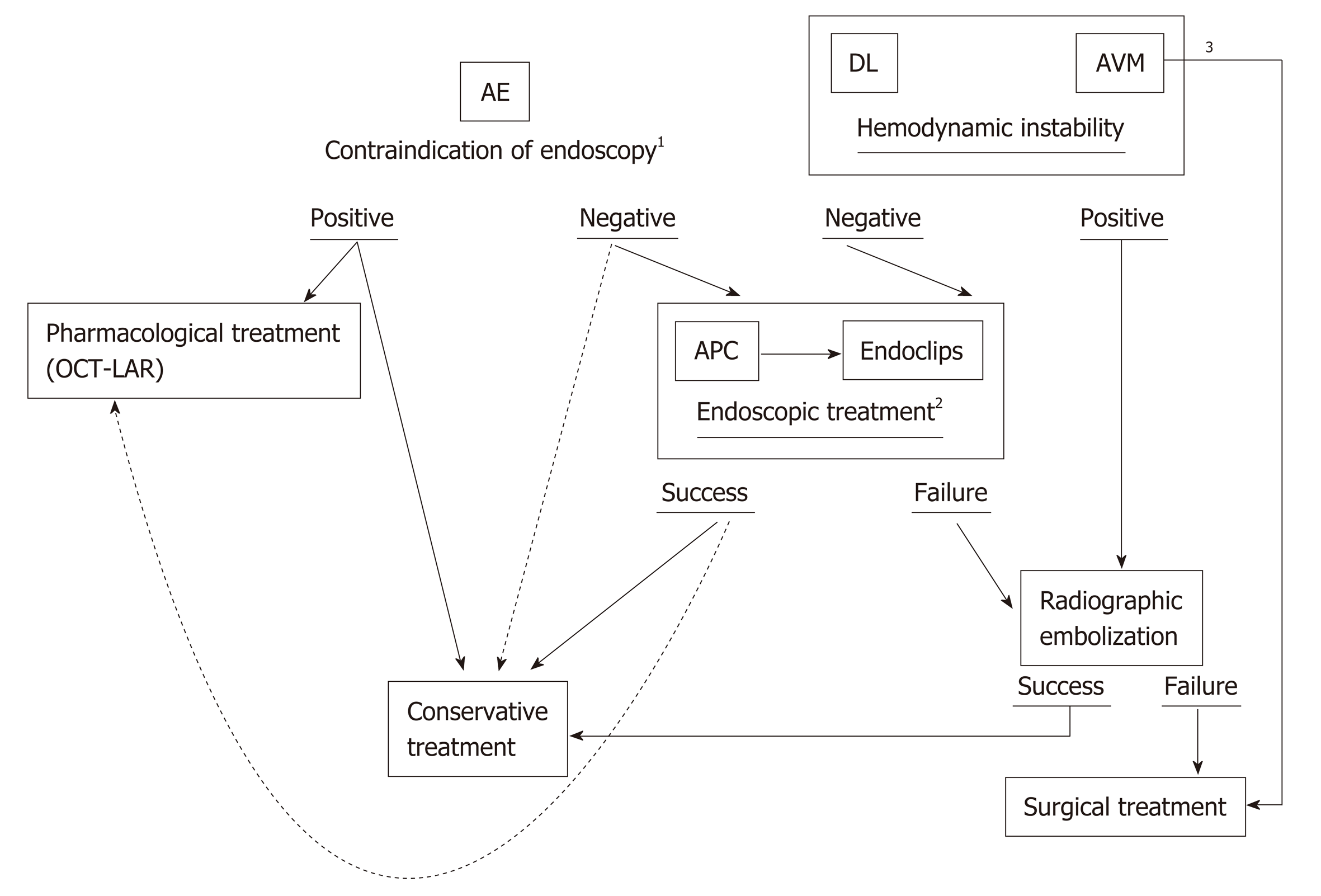
Figure 5 Therapeutic algorithm for small bowel vascular lesions.
Note: 1Pharmacological treatment is recommended for patients whom endoscopy is contraindicated. Meanwhile conservative approach with iron supplementation remains an option for patients with mild anemia; 2Subsequent pharmacological treatment after successful endoscopic treatment may be useful as an adjunct therapy, especially for patients with a high risk of re-bleeding; 3Arteriovenous malformation usually requires surgical resection because of their relatively large size and tendency to re-bleed. AE: Angioectasia; DL: Dieulafoy’s lesion; AVM: Arteriovenous malformation; APC: Argon plasm coagulation; OCT-LAR: Long-acting release octreotide.
- Citation: Sakai E, Ohata K, Nakajima A, Matsuhashi N. Diagnosis and therapeutic strategies for small bowel vascular lesions. World J Gastroenterol 2019; 25(22): 2720-2733
- URL: https://www.wjgnet.com/1007-9327/full/v25/i22/2720.htm
- DOI: https://dx.doi.org/10.3748/wjg.v25.i22.2720


