©The Author(s) 2018.
World J Gastroenterol. Oct 28, 2018; 24(40): 4578-4585
Published online Oct 28, 2018. doi: 10.3748/wjg.v24.i40.4578
Published online Oct 28, 2018. doi: 10.3748/wjg.v24.i40.4578
Figure 1 Delayed gastric emptying fourteen days after subtotal gastrectomy.
A: Food is retained in the remnant stomach because of passage delay at the anastomotic site; B: An endoscope is passed through the anastomotic site; C: On gastroscopy, a patent lumen with edematous mucosa is observed on the efferent loop side; D: Despite the patency of the E-loop, delayed gastrojejunal passage is seen on upper gastrointestinal series.
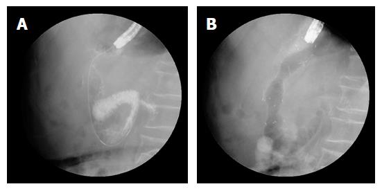
Figure 2 Placement of self-expandable metal stent.
A: A catheter is placed through the lumen; B: A self-expandable metal stent (10-mm in length) is released following adjustment for the suitable position.
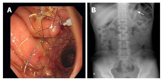
Figure 3 Inserted self-expandable metal stent.
A: A stent is deployed at the anastomotic site with endoscopic clips; B: The stent is identified on an abdominal radiograph (arrow).
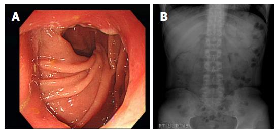
Figure 4 Patent anastomotic site on follow-up gastroscopy.
A: Anastomotic lumen is patent; B: The stent is not identified on a follow-up abdominal radiograph.
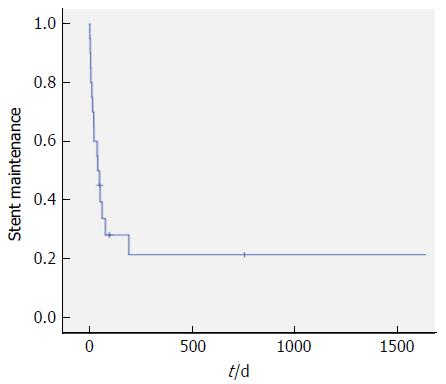
Figure 5 Kaplan–Meier estimates of the stent maintenance period.
The Kaplan–Meier curve is shown. At 30 d, the estimated stent maintenance rate is 58.8%. Inserted stents were passed per rectum spontaneously in 14 of 20 patients (70%) with no significant complications.
- Citation: Kim SH, Keum B, Choi HS, Kim ES, Seo YS, Jeen YT, Lee HS, Chun HJ, Um SH, Kim CD, Park S. Self-expandable metal stents in patients with postoperative delayed gastric emptying after distal gastrectomy. World J Gastroenterol 2018; 24(40): 4578-4585
- URL: https://www.wjgnet.com/1007-9327/full/v24/i40/4578.htm
- DOI: https://dx.doi.org/10.3748/wjg.v24.i40.4578


