©The Author(s) 2018.
World J Gastroenterol. Oct 28, 2018; 24(40): 4527-4535
Published online Oct 28, 2018. doi: 10.3748/wjg.v24.i40.4527
Published online Oct 28, 2018. doi: 10.3748/wjg.v24.i40.4527
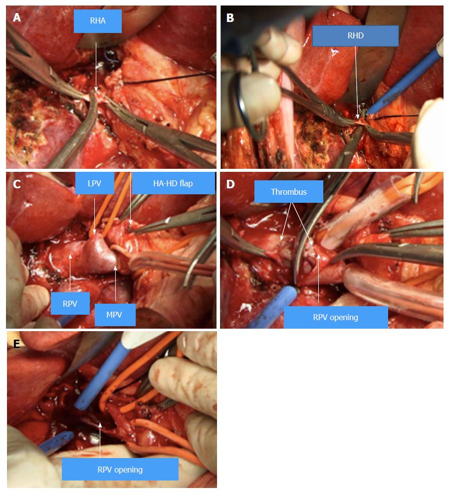
Figure 1 The “thrombectomy first” approach.
A: The right hepatic artery (RHA) was isolated and dissected. B: The right hepatic duct (RHD) was isolated and dissected. C: The left portal vein (LPV), right portal vein (RPV) and the main portal vein (PV) were clearly revealed by dissecting the hepatic artery-hepatic duct (HA-HD) flap. D: The tumor thrombus in the RPV and main portal vein (MPV) was extracted. E: The tape of LPV and PV were both released to flush the remnant tumor thrombus. RHA: Right hepatic artery; RHD: Right hepatic duct; LPV: Left portal vein; RPV: Right portal vein; PV: Portal vein; MPV: Main portal vein; HA-HD: Hepatic artery-hepatic duct.
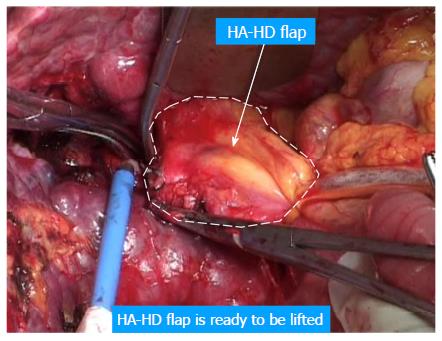
Figure 2 The hepatic artery-hepatic duct flap was ready to be lifted.
HA-HD: Hepatic artery-hepatic duct.
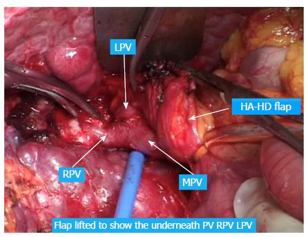
Figure 3 The hepatic artery-hepatic duct flap was lifted to show the portal vein, right portal vein, and left portal vein.
RPV: Right portal vein; LPV: Left portal vein; MPV: Main portal vein; HA-HD: Hepatic artery-hepatic duct.
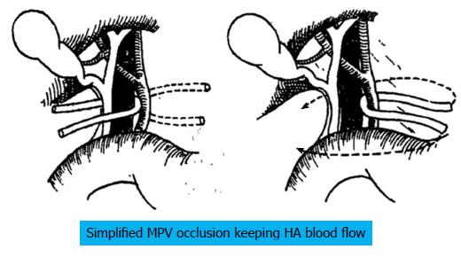
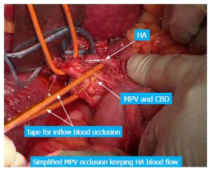
Figure 4 A simplified method to occlude the main portal vein while keeping hepatic artery blood flow.
MPV: Main portal vein; HA: Hepatic artery.
Figure 5 A simplified method to occlude the main portal vein while keeping hepatic artery blood flow.
HA: Hepatic artery; MPV: Main portal vein; CBD: Common bile duct.
Figure 6 Case 1.
A: The computed tomography scan showed tumor thrombus in the right portal vein. B: Transcatheter arterial chemoembolization was performed before the operation. C: The thrombus was extracted from the right portal vein opening. PV: Portal vein; PVTT: Portal vein tumor thrombus.
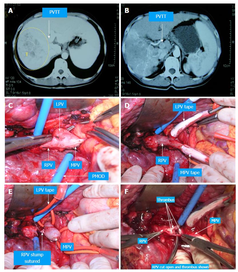
Figure 7 Case 2.
A: The computed tomography scan showed tumor thrombus in the right portal vein (RPV) and portal vein (PV). B: Transcatheter arterial chemoembolization was performed before the operation. C and D: The left portal vein (LPV), RPV and PV were clearly revealed by dissecting the hepatic artery-hepatic duct flap. E: The LPV and PV were taped. F: Tumor thrombus in the RPV and PV was extracted. G: The tumor thrombus in the LPV was extracted, and the RPV stump was closed. H: Live transection was performed by the anterior approach. PV: Portal vein; PVTT: Portal vein tumor thrombus; RPV: Right portal vein; LPV: Left portal vein; MPV: Main portal vein; HA-HD: Hepatic artery-hepatic duct.
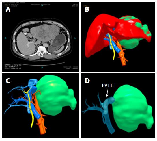
Figure 8 Case 3.
A: The computed tomography scan showed the left hepatocellular carcinoma (HCC) and tumor thrombus in the left portal vein (LPV) and portal vein (PV). B and C: 3D reconstruction of the left HCC. D: 3D reconstruction of tumor thrombus in the LPV and PV. PVTT: Portal vein tumor thrombus.
- Citation: Peng SY, Wang XA, Huang CY, Li JT, Hong DF, Wang YF, Xu B. Better surgical treatment method for hepatocellular carcinoma with portal vein tumor thrombus. World J Gastroenterol 2018; 24(40): 4527-4535
- URL: https://www.wjgnet.com/1007-9327/full/v24/i40/4527.htm
- DOI: https://dx.doi.org/10.3748/wjg.v24.i40.4527


