©The Author(s) 2018.
World J Gastroenterol. Jan 28, 2018; 24(4): 543-548
Published online Jan 28, 2018. doi: 10.3748/wjg.v24.i4.543
Published online Jan 28, 2018. doi: 10.3748/wjg.v24.i4.543
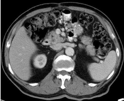
Figure 1 Pre-treatment abdominal contrast-enhanced computed tomography images.
These reveal thickening of the stomach wall above the gastrojejunostomy site without enlarged perigastric lymph nodes. There is no evidence of lesion extension into the serosa or surrounding soft tissues.
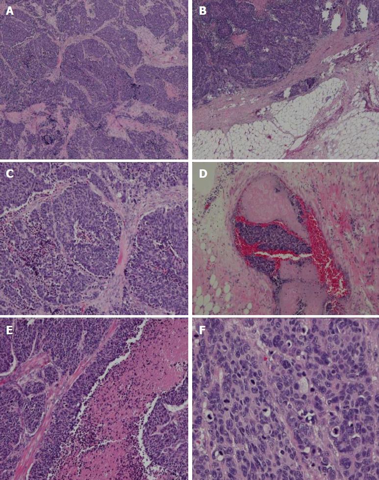
Figure 2 Histological findings.
A: A low-power histological view. Tumor cells show infiltrative growth from the muscularis propria to the subserosa (HE, × 40). B: Large-cell carcinoma showing invasion into the subserosa. C: High-power view shows monotonous large tumor cells with abundant cytoplasm and large irregular nuclei with prominent nucleoli (HE, × 100). D and E: Angiolymphatic invasion and carcinoma cell embolus. F: Mitotic figures were also observed (60 per 10 high-power fields). HE: Hematoxylin and eosin.
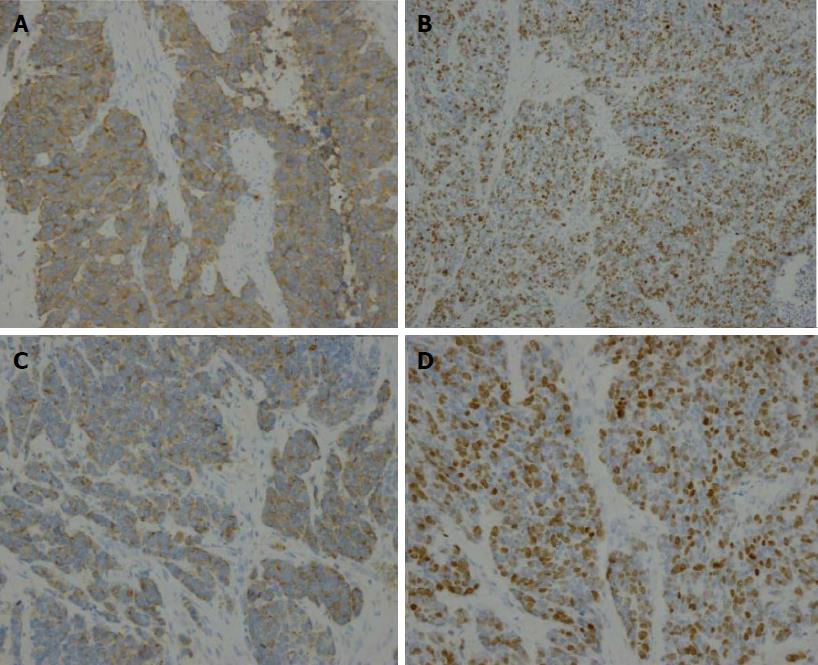
Figure 3 Immunohistochemical staining.
Positive immunohistochemical staining for (A) synaptophysin (× 200), (B) CD56 (× 200), and (C) chromogranin A (× 200). D: The Ki-67 index is about 60% (× 200).
Figure 4 Computed tomography scan 7 mo after the operation.
This image reveals lymph node metastasis in the region around the gastrojejunal anastomosis in the abdominal cavity.
- Citation: Ma FH, Xue LY, Chen YT, Xie YB, Zhong YX, Xu Q, Tian YT. Neuroendocrine carcinoma of the gastric stump: A case report and literature review. World J Gastroenterol 2018; 24(4): 543-548
- URL: https://www.wjgnet.com/1007-9327/full/v24/i4/543.htm
- DOI: https://dx.doi.org/10.3748/wjg.v24.i4.543


