Copyright
©The Author(s) 2018.
World J Gastroenterol. Jan 21, 2018; 24(3): 360-370
Published online Jan 21, 2018. doi: 10.3748/wjg.v24.i3.360
Published online Jan 21, 2018. doi: 10.3748/wjg.v24.i3.360
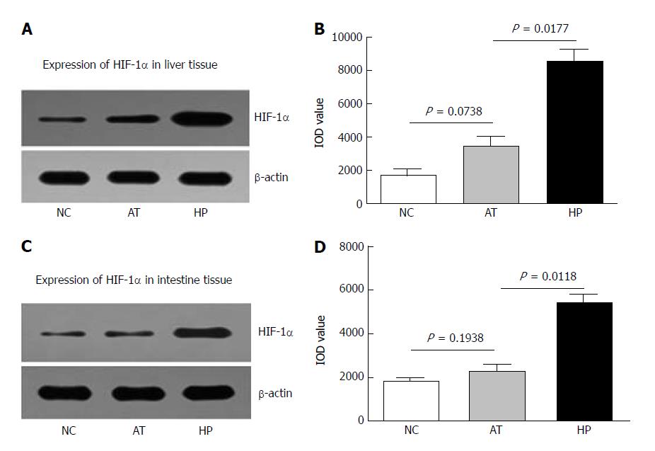
Figure 1 Expression of HIF-1α in liver and intestinal tissues in a rat autologous orthotopic liver transplantation model.
A: Western blot assay showed that the level of HIF-1α protein expression induced by hypoxia preconditioning was increased in rat liver tissue at 12 h postoperatively; B: The chart shows that the level of HIF-1α in the HP group was higher at different postoperative time points. At 12 h postoperatively, HIF-1α expression in liver tissue in the HP group was significantly higher than in the AT group and NC group; C: Western blot assay showed that HIF-1α expression was increased in rat intestinal tissue after hypoxia preconditioning at 12 h postoperatively; D: The chart shows that HIF-1α expression induced by hypoxia preconditioning in intestinal tissue was elevated at different postoperative time points. HIF-1α expression was significantly higher in the HP group compared with the AT group and NC group at 12 h postoperatively.
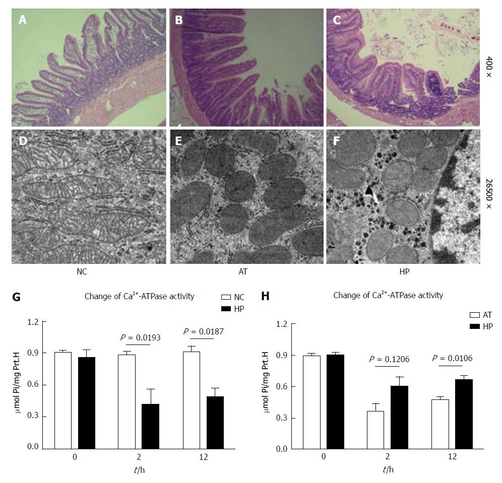
Figure 2 Hypoxia-induced HIF-1α expression increases intestinal cellular mitochondrial integrity and Ca2+-ATPase activity after I/R injury.
A-C: Pathological changes in intestinal cells are shown (HE, 10 × 20). A: The normal morphology of intestinal cells in the NC group; B: The intestinal cells in the AT group exhibited oedema and red blood cell deposition and blood clots in capillary vessels; the villus epithelial cells were shedding, had severely damaged glands, and had been infiltrated by inflammatory cells; C: The intestinal cells in the HP group exhibited slight oedema and fewer inflammatory cells. The villus epithelial cells were less damaged. D-F: The mitochondrial changes in intestinal cells are shown (× 46000). D: The normal morphology of mitochondria in the NC group; E: The mitochondria in the AT group appeared swollen, round, and degenerated. The visible mitochondrial cristae appeared less fractured or disappeared; F: The mitochondria in the HP group showed less swelling and fewer cristae. The endoplasmic reticulum structure had survived; G: The Ca2+-ATPase activity in the AT group was significantly lower compared with the NC group, which decreased to the lowest level at postoperative 2 h (AT group vs NC group: P < 0.05) and then recovered; H: The Ca2+-ATPase activity in the HP group was lower after the operation but was significantly higher than that in the AT group at postoperative 2 h (HP group vs AT group: P < 0.05).
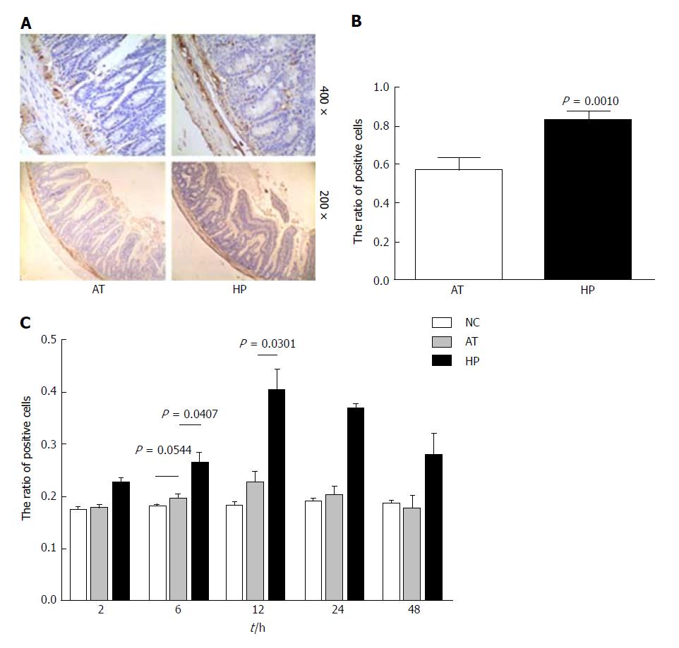
Figure 3 Hypoxia preconditioning promotes BCL2 expression in rat intestinal tissues.
A: Immunohistochemistry revealed the expression of BCL2 in rat intestinal tissue in the AT and HP groups at postoperative 12 h; B: The ratio of BCL2 expression in the HP group was significantly higher than that in the AT group at postoperative 12 h (HP vs AT P = 0.001); C: The ratio of positive BCL2 expression was detected at several time points postoperatively (2, 6, 12, 24 and 48 h) in the NC, AT, and HP groups. The expression of BCL2 in the HP group was increased after the operation, peaked at postoperative 2 h, and was maintained at a high level. At postoperative 12 h, BCL2 expression in the HP group was significantly higher than that the NC group and the AT group. No significant difference in BCL2 expression was found in the AT group and NC group.
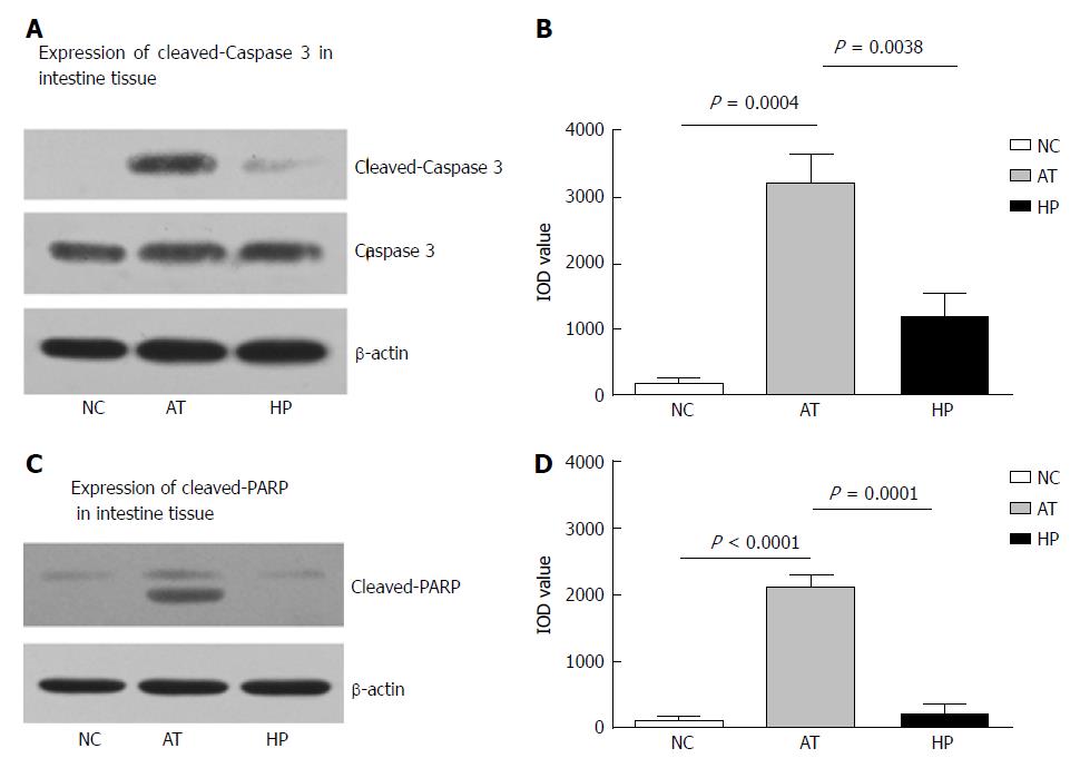
Figure 4 Changes of Caspase 3 and PARP expression in rat intestinal tissue at postoperative 24 h.
A: The apoptosis was detected by Western blot assay, which showed that the expression of cleaved Caspase 3 as increased in rat intestinal tissue with ischemia and reperfusion injury at postoperative 24 h; B: The bar-table shows the level of cleaved Caspase 3 expression in AT group was significantly higher than that in the NC group. The cleaved Caspase 3 expression in the HP group was lower than that in the AT group at postoperative 24 h; C: The expression of cleaved PARP was increased in rat intestinal tissue with ischemia and reperfusion injury at postoperative 24 h; D: The bar-table shows that the level of cleaved PARP expression in the AT group was significantly higher than that in the NC group. The cleaved PARP expression in the HP group was lower than that in the AT group at postoperative 24 h.
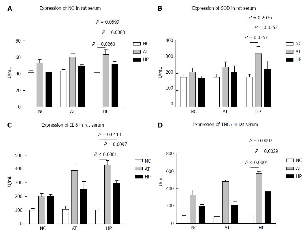
Figure 5 Hypoxia preconditioning decreases the expression of inflammatory factors in rat serum after postoperative I/R injury of intestinal tissue.
A: In the HP group, NO levels in the serum were lower, especially at 12 h and 24 h after the operation, compared with the AT group; B: The SOD levels in the HP group 12 h and 24 h after the operation were significantly lower than those in the AT group; C: After the operation, the IL-6 levels in rat liver tissues that had undergone ischaemia-reperfusion injury were significantly higher in the AT group compared with the HP group and the NC group; D: TNF-α levels in the HP group were slightly decreased compared with those in the AT group.
- Citation: Ji ZP, Li YX, Shi BX, Zhuang ZN, Yang JY, Guo S, Xu XZ, Xu KS, Li HL. Hypoxia preconditioning protects Ca2+-ATPase activation of intestinal mucosal cells against R/I injury in a rat liver transplantation model. World J Gastroenterol 2018; 24(3): 360-370
- URL: https://www.wjgnet.com/1007-9327/full/v24/i3/360.htm
- DOI: https://dx.doi.org/10.3748/wjg.v24.i3.360


