Copyright
©The Author(s) 2018.
World J Gastroenterol. May 7, 2018; 24(17): 1839-1858
Published online May 7, 2018. doi: 10.3748/wjg.v24.i17.1839
Published online May 7, 2018. doi: 10.3748/wjg.v24.i17.1839
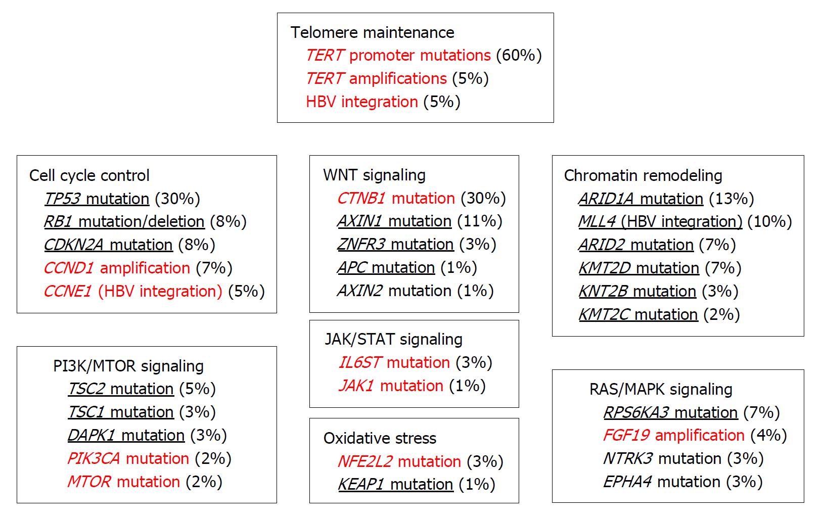
Figure 1 Mutational landscape of hepatocellular carcinoma.
The figure was made by modifying the original figure in Ref. 7. Gain and loss of function events are indicated by red color and with underlines, respectively.
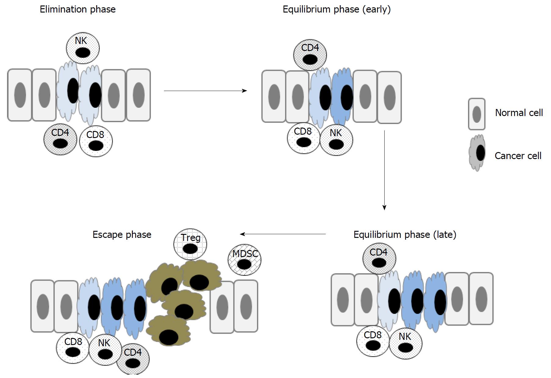
Figure 2 Cellular mechanisms underlying immunoediting.
At the elimination phase, newly-appearing cancer cells can be recognized and killed by a number of immune cells, particularly natural killer (NK) cells, and CD8+ and CD4+ T cells. At the equilibrium phase, variant cancer cells arise that are less immunogenic, and consequently more resistant to being killed by immune cells. Over time, a variety of different cancer variants develop. At the escape phase, one variant may finally escape the killing mechanism or recruit immunosuppressive cells such as Tregs and MDSCs, and eventually form an appreciable tumor mass. MDSC: Myeloid-derived suppressive cell.
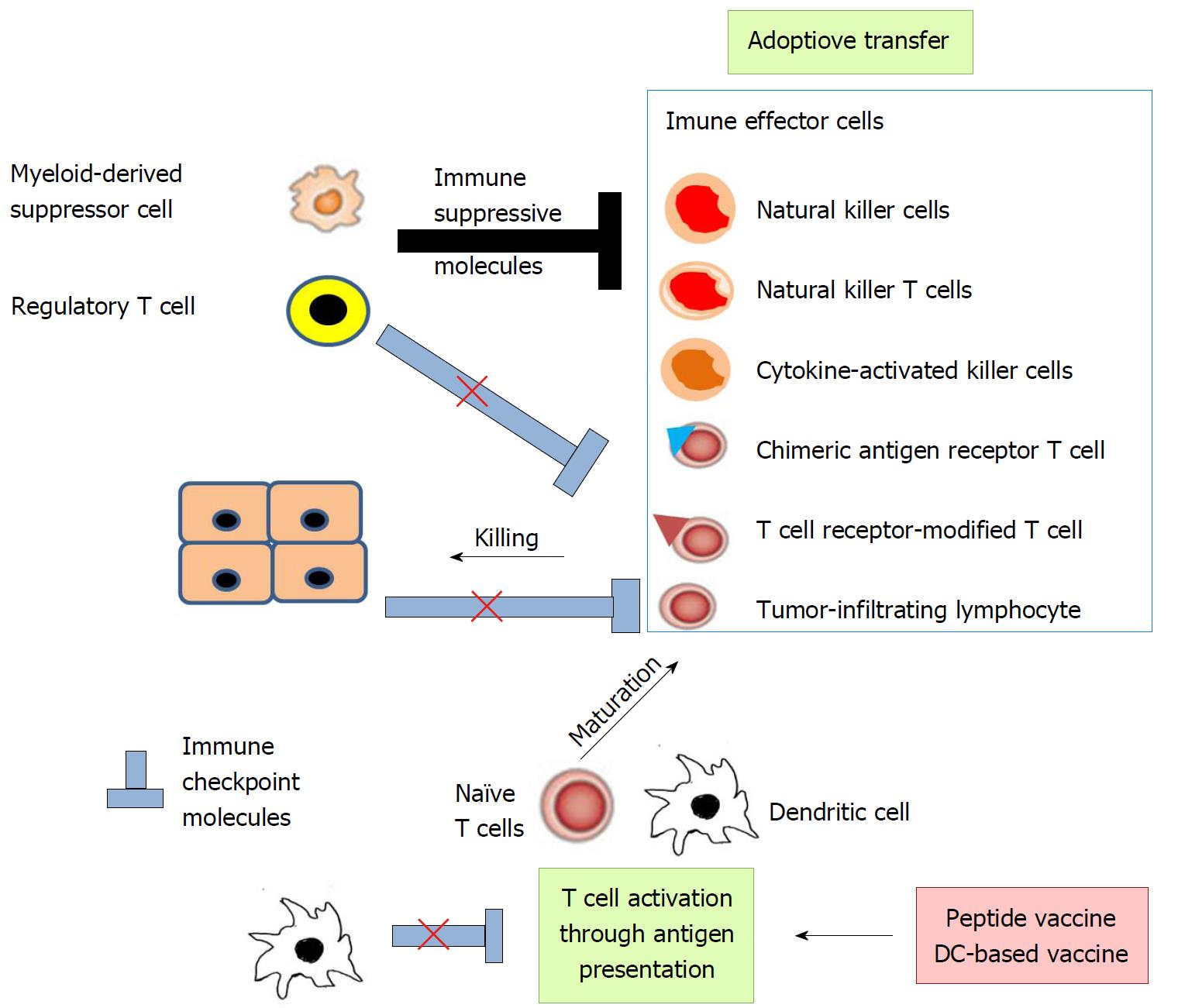
Figure 3 Strategies of immune therapy.
Immune therapy can be classified to two types, promotion of immune effector cell function and reversal of depressed anti-tumor immunity. Immune effector cell function can be enhanced by peptide vaccine, DC-based vaccine, and adoptive transfer of effector cells including tumor-infiltrating lymphocytes (TILs), T cell receptor (TCR)-modified T cells, chimeric antigen receptor (CAR) T cells, natural killer (NK) cells, NKT cells, and cytokine-activated killer cells (CIKs). Depressed anti-tumor immunity can be reversed by the blockade of immune checkpoint pathways and immune suppressor cells including regulatory T cells (Tregs) and myeloid-derived suppressor cells (MDSCs).
Figure 4 Tumor-associated antigen presentation of antigen-presenting cells to T cells.
Endogenous antigens are degraded to peptides and loaded on MHC class II on APCs to be presented to the CD4+ T cells, while exogenous antigens are degraded to peptides and loaded on MHC class I to be presented to the CD8+ T cells. These pathways deliver activation signals to corresponding T cells. However, full activation and subsequent survival require the co-stimulatory signals delivered by several pathways including the CD80/CD86-CD28 pathway. In the absence of co-stimulatory signals, T cells become unresponsive to the antigen, a condition called anergy. TAA: Tumor-associated antigen; APCs: Antigen-presenting cells.
Figure 5 Mechanism underlying immune functions of CTLA-4 and PD-1-PD-L pathways.
A: CTLA-4 has a higher binding affinity to CD80/CD86 than the co-stimulatory signal molecule CD28. As a consequence, CTLA-4 competitively antagonizes the stimulatory signal, which the interaction between CD80/86 and CD28 generates at the priming phase of T cells. B: The PD-L1/L2-PD-1 interaction interferes with T cell activation signals in the effector phase.
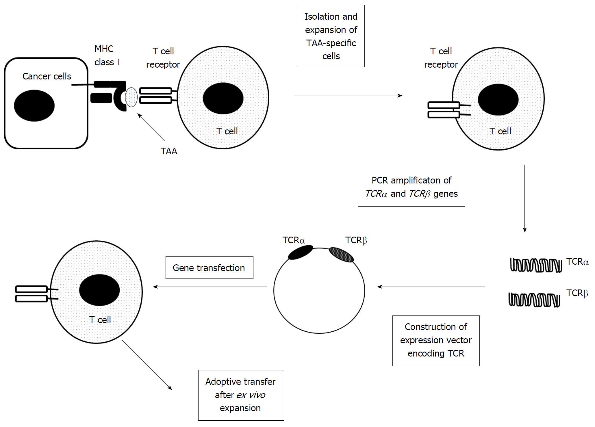
Figure 6 Preparation of genetically modified TCR-expressing T cells.
TCRα and TCRβ genes are cloned and amplified with the use of PCR from TAA-specific T cells, which are isolated and expanded ex vivo. The obtained TCRα and TCRβ genes are cloned into an expression vector, which is used for transfection into T cells. The transfected cells are adoptively transferred after ex vivo expansion.TAA: Tumor-associated antigen.
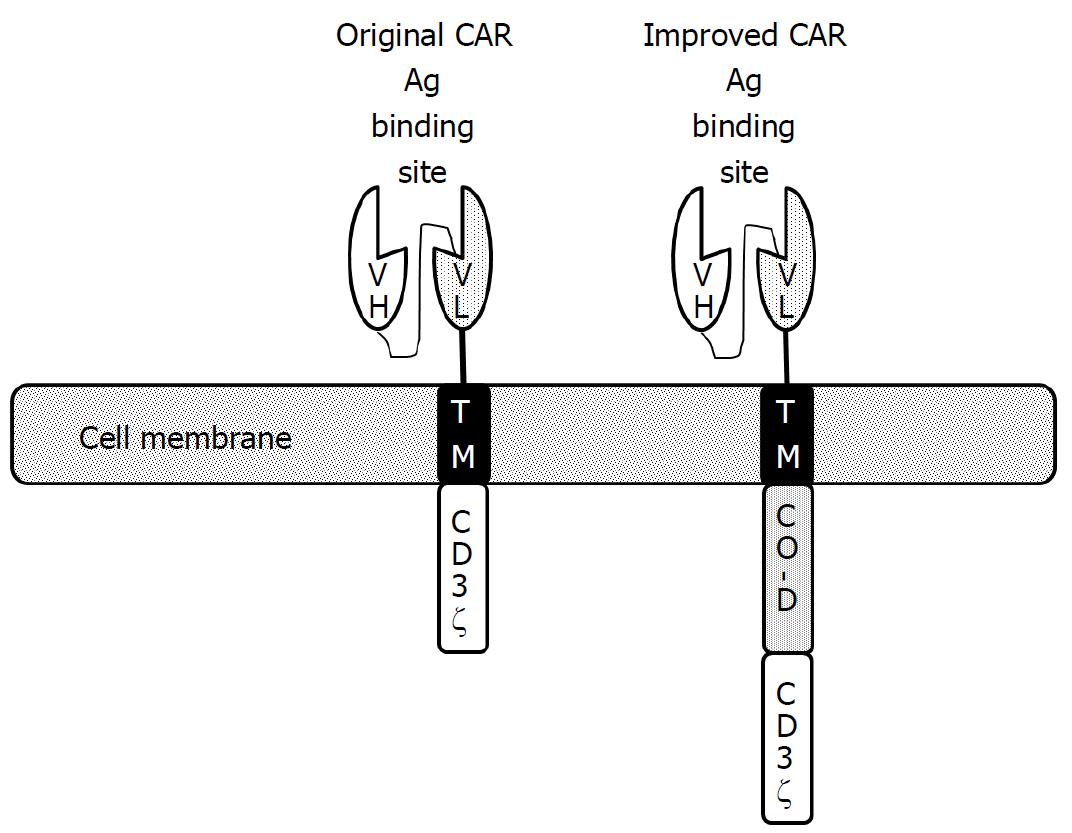
Figure 7 Schematic structure of chimeric antigen receptors.
Chimeric antigen receptors (CARs) are composed of a single-chain fragment variable (scFv) containing the heavy chain variable (VH) and the light chain variable domain (VL) of a monoclonal antibody. ScFv is attached to a transmembrane (TM) domain and CD3ζ chain in the case of the first-generation CARs. Improved CARs additionally contain one or two more costimulatory domains. VH, heavy chain variable domain; VL, light chain variable domain; TM, transmembrane domain; CO-D, domain derived from co-stimulatory molecules.
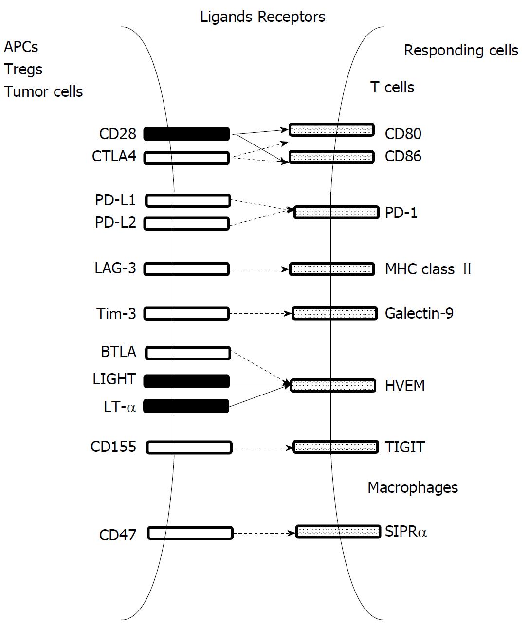
Figure 8 Interaction of major immune co-stimulatory and inhibitory molecules, and their cognate receptors.
Co-stimulatory and co-inhibitory molecules are indicated by closed and open boxes, respectively. Co-stimulatory and inhibitory signals are indicated by filled and hatched arrows, respectively.
- Citation: Mukaida N, Nakamoto Y. Emergence of immunotherapy as a novel way to treat hepatocellular carcinoma. World J Gastroenterol 2018; 24(17): 1839-1858
- URL: https://www.wjgnet.com/1007-9327/full/v24/i17/1839.htm
- DOI: https://dx.doi.org/10.3748/wjg.v24.i17.1839


